




No CrossRef data available.
Article contents
Ebstein's malformation in the setting of Down's syndrome
Published online by Cambridge University Press: 24 May 2005
Abstract
We report a patient with Down's syndrome referred to our Department of Echocardiography for evaluation after an episode of tachyarrhythmia. We diagnosed Ebstein's malformation. As far as we know, this association has been reported previously on only three occasions. We review these previous cases, describe our echocardiographic techniques, and discuss our experience.
Information
- Type
- Brief Report
- Information
- Copyright
- © 2003 Cambridge University Press
In almost all cases, Down's syndrome is a genetic abnormality caused by trisomy of chromosome 21. It was Seguin, and John Langdon Down, who published the first clinical descriptions, in 1846 and 1866, respectively. It is the most common genetic cause of mental retardation, with prevalence in the United States of America of 9.2 per 10,000 live births.1
In two-fifths of cases, there are associated cardiac defects resulting from maldevelopment of the mesenchymal cushions of the atrioventricular canal and outflow tract, along with the myocardium of the atrial and ventricular septums. Thus, the resulting congenital defects include atrial, atrioventricular, and ventricular septal defects, patency of the arterial duct, and Fallot's tetralogy. Associated abnormal ventriculo-arterial connections, or malformation of the aortic arch, such as coarctation, are uncommon.2
Ebstein's malformation is a rare malformation, with an incidence in children between 0.03 and 1%. Its association with Down's syndrome is extremely unusual, with only three cases, to the best of our knowledge, being previously reported.3–5 We now describe another case of Ebstein's malformation seen in association with Down's syndrome, in which the principal clinical manifestation was a tachyarrhythmia producing hemodynamic compromise.
Case report
The patient was a 20-year-old, male, and known to have Down's syndrome. An atrial septal defect had been documented by cardiac catheterization 20 days after birth. On this occasion, he was hospitalized after an episode of syncope lasting 5 min, with loss of sphincteric control, but without a postictal state or other neurologic sequels. Over the previous four months, he had often had oppressive chest pain, with shortness of breath and diaphoresis.
When seen in the emergency room, he was perspiring profusely, and complaining of dizziness and generalized weakness. His heart rate was 170 beats/min, and there was systemic hypotension. On physical examination, epicanthic folds typical for Down's syndrome were noted (Fig. 1). The chest was symmetrical, with the point of maximum impulse found in the fifth intercostal space on the left mid-axillary line. A thrill could be palpated over the sternum. The second heart sound was widely split, without respiratory changes, and third and fourth sounds were also audible. The peripheral pulses were present and symmetrical. There was no evidence of cyanosis, nor osteoarthropathy.

Figure 1. The facial characteristics of Down's syndrome as seen in our patient. Note the broad base of the nose and the epicanthic folds.
The electrocardiogram revealed right bundle branch block and wide QRS complexes, and the tachyarrhythmia failed to respond to vagal maneuvers. Electrical cardioversion, at 150 J, returned the patient to sinus rhythm at a rate of 70 beats/min, whereupon the tracings showed left bundle branch block, with delta waves seen in leads DI, aVL, V5 and V6.
The frontal chest X-ray revealed significant cardiomegaly, with increase in the silhouettes of both the right and left heart, a prominent pulmonary arch, and evidence of increased pulmonary flow.
A transthoracic echocardiogram confirmed a left-sided heart, and revealed usual atrial arrangement with concordant segmental connections. Systemic and pulmonary venous returns were normal. The atrial septum was grossly deficient, with an aneurysm of the flap valve. Severe Ebstein's malformation was also found, with extensive displacement of the septal leaflet of the tricuspid valve, restricted movement of the antero-superior leaflet, and marked atrialization of the right ventricle. Mild tricuspid regurgitation and mild pericardial effusion were also present.
A transesophageal echocardiogram, with three-dimensional reconstruction, optimized the visualization of the cardiac defects, and permitted comparison with the anticipated morphology as seen in autopsied specimens (Fig. 2).

Figure 2. We compared the three-dimensional reconstruction of the transesophageal images from our patient (b) with an autopsied specimen from our archive (a). The specimen shows the characteristic displacement of the septal leaflet of tricuspid valve (arrows). The black arrows show the limit between the atrialized right ventricle and the functional chamber. The dotted line represents the atrioventricular groove. Note also a co-existing atrial septal defect is visualized (black arrowhead). The three-dimensional reconstruction similarly shows the displacement of the septal leaflet (white arrowhead), and abnormal tethering of the antero-superior leaflet (white arrow). aRV: atrialized right ventricle; RV: right ventricle; RA: right atrium; LA: left atrium; LV: left ventricle.
Five days after admission to the hospital, he was taken to the electrophysiology laboratory, where an inferior paraseptal pathway was found. An attempt was made to ablate the pathway, but it failed. Because of the persisting preexcitation, propafenone hydrochloride was started in an attempt to avoid recurrences.
Discussion
Ebstein's malformation, first described by Wilhelm Ebstein in 1866, is characterized by abnormal displacement of the hingepoints of the septal and inferior leaflets of the tricuspid valve, along with diminution in the size of the functional right ventricle, and fibrosis of the thinned and atrialised inlet component. The antero-superior leaflet, although retaining its hinge from the atrioventricular junction, is always redundant and thickened, and sometimes has a fibromuscular structure.6, 7
The abnormalities of the tricuspid valve can produce either regurgitation or stenosis, although the former is more frequent. In half the cases, it is associated with an atrial septal defect, and less often with pulmonary stenosis or atresia. Clinical manifestations vary from severe heart failure seen during fetal or neonatal stages, to life-long absence of symptoms, with discovery on routine examination,8 or because of arrhythmias, as in our patient. The tachyarrhythmias seen in these patients can adversely affect their functional class, increase cyanosis, and even cause syncope. The association with an atrial septal defect adds the risk of paradoxical embolisation and its consequences. Preexcitation occurs in about one-fifth of patients with Ebstein's anomaly, and in our patient it's related to the congenital anomaly and not to the Down syndrome. Catheter ablation of the accessory tract has less success than in structurally normal hearts, and recurrence is common.9
The association with Down's syndrome is very rare. As mentioned before, as far as we can establish, only three cases have previously been reported. The first was a 17-year-old young woman, who was admitted with heart failure and respiratory distress secondary to bronchopneumonia that was subsequently fatal. Diagnosis was established at autopsy.3 The second case involved an asymptomatic 55-year-old man, whose malformation was discovered during a routine cardiologic examination.4 The last case was diagnosed by fetal echocardiography, examination of amniotic fluid revealing trisomy 21.5
In our patient, the history of frequent episodes of tachyarrhythmia made it obligatory to look for structural abnormalities of the heart. Echocardiography then played a decisive role in confirming the diagnosis, especially when the transesophageal interrogation was supported by three-dimensional reconstruction. We are now aware that our patient continues to have a functioning accessory muscular atrioventricular connection as the source of the tachyarrhythmias, so careful follow-up, and appropriate medication, will be needed for his future care.
References
Epstein CJ. Down syndrome (trisomy 21). In: Scriver CR, Beaudet AL, Sly WS, Valle D (eds). The Metabolic and Molecular Bases of Inherited Disease. McGraw-Hill, New York, 1995, pp 749–794.
Liu MC, Corlett K. A study of congenital heart defects in mongolism. Arch Dis Child 1959; 34: 410–419.Google Scholar
Johnson CD, Ortiz-Colom PM, Sainz de la Peña H, Barroso E. Ebstein's anomaly in a patient with Down's syndrome. Bol Assoc Med PR 1989; 81: 221–222.Google Scholar
Venturi E, Musaio L, Strazzeri R, Baroni F. Ebstein's tricuspid anomaly and Down's syndrome. A clinical case report. Recenti Prog Med 1992; 83: 556–558.Google Scholar
Silvia SR, Bruner JP, Moore CA. Prenatal diagnosis of Down's syndrome in the presence of isolated Ebstein's anomaly. Fetal Diagn Ther 1999; 14: 149–151.Google Scholar
Anderson KR, Zuberbuhler JR, Anderson RH, Becker AE, Lie JT. Morphologic spectrum of Ebstein's anomaly of the heart. A review. Mayo Clin Proc 1979; 54: 174–180.Google Scholar
Muñoz-Castellanos L, Barros W, García F, Salinas CH, Kuri M. Estudio patológico de la displasia y el adosamiento valvulares en la anomalía de Ebstein. Arch Inst Cardiol Méx 1993; 63: 101–109.Google Scholar
Celermajer DS, Bull C, Till AJ, et al. Ebstein's anomaly: presentation and outcome from fetus to adult. J Am Coll Cardiol 1994; 23: 170–176.Google Scholar
Cappato R, Schluter M, Weiss C, et al. Radiofrequency current catheter ablation of accessory atrioventricular pathways in Ebstein's anomaly. Circulation 1996; 94: 376–383.Google Scholar

The facial characteristics of Down's syndrome as seen in our patient. Note the broad base of the nose and the epicanthic folds.
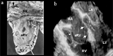
We compared the three-dimensional reconstruction of the transesophageal images from our patient (b) with an autopsied specimen from our archive (a). The specimen shows the characteristic displacement of the septal leaflet of tricuspid valve (arrows). The black arrows show the limit between the atrialized right ventricle and the functional chamber. The dotted line represents the atrioventricular groove. Note also a co-existing atrial septal defect is visualized (black arrowhead). The three-dimensional reconstruction similarly shows the displacement of the septal leaflet (white arrowhead), and abnormal tethering of the antero-superior leaflet (white arrow). aRV: atrialized right ventricle; RV: right ventricle; RA: right atrium; LA: left atrium; LV: left ventricle.
 You have
Access
- 9
- Cited by