Introduction
Recent studies using oocytes, which are ideally suited for observing the dynamic changes of the nuclear envelope (NE) structure in vivo, have revealed general and specific features of oocytes that both resembled and differed from those of somatic mammalian cells (Lenart & Ellenberg, Reference Lenart and Ellenberg2003). Most biochemical knowledge about the NE comes from meiotic in vitro systems using Xenopus eggs, but our understanding of NE dynamics is still limited (Lenart & Ellenberg, Reference Lenart and Ellenberg2003). The NE generates a unique structural and functional environment for chromatin in the nucleus. The structural integrity of the NE is essential for nuclear functions such as DNA replication, RNA transcription and RNA processing (Stuurman et al., Reference Stuurman, Heins and Aebi1998; Gotzmann & Foisner, Reference Gotzmann and Foisner1999; Gruenbaum et al., Reference Gruenbaum, Wilson, Harel, Goldberg and Cohen2000; Nickerson, Reference Nickerson2001). There is increasing evidence that the NE is involved in the organization of interphase chromatin, but the molecular mechanisms are not yet understood (Foisner & Gerace, Reference Foisner and Gerace1993; Ye & Worman, Reference Ye and Worman1994; Furukawa, 1998; Furukawa et al., Reference Furukawa1999; Zheng et al., Reference Zheng, Ghirlando, Lee, Mizuuchi, Krause and Craigie2000).
The nuclear lamina is one of the key structural elements of the nucleus. It is comprised of two major types of lamin proteins: constitutively expressed lamin B and developmentally regulated lamin A/C (Gruenbaum et al., Reference Gruenbaum, Wilson, Harel, Goldberg and Cohen2000). Lamin B associates predominantly with the membrane vesicle through combination with lamin-binding proteins throughout the cell cycle, whereas lamin A/C becomes soluble and is dispersed into the cytosol during mitosis (Hozak et al., Reference Hozak, Sasseville, Raymond and Cook1995; Foisner, Reference Foisner1997; Moir et al., Reference Moir, Yoon, Khuon and Goldman2000). LAP2β proteins are a family of related nuclear integral membrane proteins that may fulfil related functions in nuclear architecture and cell-cycle-dependent nuclear dynamics (Foisner & Gerace, Reference Foisner and Gerace1993; Maison et al., Reference Maison, Pyrpasopoulou, Theodoropoulos and Georgatos1997) and that bind to lamin B and chromosomes (Foisner & Gerace, Reference Foisner and Gerace1993; Furukawa et al., Reference Furukawa, Pante, Aebi and Gerace1995, Reference Furukawa, Fritze and Gerace1998). These three nuclear membrane proteins gather at the chromatin periphery during nuclear formation in somatic cells, and it has been reported that the assembly of LAP2β at late anaphase is earlier than that of lamin A/C and B (Foisner & Gerace, Reference Foisner and Gerace1993; Maison et al., Reference Maison, Pyrpasopoulou, Theodoropoulos and Georgatos1997; Dechat et al., Reference Dechat, Gotzmann, Stockinger, Harris, Talle, Siekierka and Foisner1998; Bodoor et al., Reference Bodoor, Shaikh, Salina, Raharjo, Bastos, Lohka and Burke1999). The assembly of LAP2β near the chromatin may be involved in the chromatin targeting of other nuclear-membrane proteins such as lamin proteins (Dechat et al., Reference Dechat, Gotzmann, Stockinger, Harris, Talle, Siekierka and Foisner1998).
There have been few detailed reports about the process of pronucleus (PN) formation in mammalian oocytes after fertilization or activation. In our earlier study, localization of LAP2β was observed in the periphery of oocyte chromatin (oCh) after the extrusion of the second polar body (PB2) before PN formation in some fertilized oocytes, but not in activated ones (Isaji et al., Reference Isaji, Iwata, Harayama and Miyake2004). It was difficult to assess exactly the beginning of chromatin decondensation from DNA images alone. In the mitotic metaphase, specific histone H3 phosphorylation at Ser10 is believed to be essential for the initial chromosome assembly and recruitment of condensins (de la Barre et al., Reference de la Barre, Gerson, Gout, Creaven, Allis and Dimitrov2000; Giet & Glover, Reference Giet and Glover2001; Murnion et al., Reference Murnion, Adams, Callister, Allis, Earnshaw and Swedlow2001). Histone H3 phosphorylation at Ser10 is related to chromosome condensation (Goto et al., Reference Goto, Tomono, Ajiro, Kosako, Fujita, Sakurai, Okawa, Iwamatsu, Okigaki, Takahashi and Inagaki1999; Wei et al., Reference Wei, Lanlan, Josephine, Gorovsky and Allis1999; Prigent & Dimitrov, Reference Prigent and Dimitrov2003). In the late G2 phase, only histone H3 on the pericentromeric chromatin is phosphorylated (Hendzel et al., Reference Hendzel, Wei, Mancini, Van Hooser, Ranalli, Brinkley, Bazett-Jones and Allis1997; Sauve et al., Reference Sauve, Anderson, Ray, James and Roberge1999; Van Hooser et al., Reference Van Hooser, Goodrich, Allis, Brinkley and Mancini1998). Then, the phosphorylation spreads along the chromosomes and is complete at prophase (Hendzel et al., Reference Hendzel, Wei, Mancini, Van Hooser, Ranalli, Brinkley, Bazett-Jones and Allis1997; Van Hooser et al., Reference Van Hooser, Goodrich, Allis, Brinkley and Mancini1998; Sauve et al., Reference Sauve, Anderson, Ray, James and Roberge1999). At the end of mitosis, histone H3 is dephosphorylated (Hendzel et al., Reference Hendzel, Wei, Mancini, Van Hooser, Ranalli, Brinkley, Bazett-Jones and Allis1997; Van Hooser et al., Reference Van Hooser, Goodrich, Allis, Brinkley and Mancini1998; Sauve et al., Reference Sauve, Anderson, Ray, James and Roberge1999). Oocytes resume meiosis for PN formation after fertilization and activation. As the oocytes of most mammalian species are arrested at the metaphase of the second meiosis (Met II), they must undergo chromatin decondensation before PN formation, which must be preceded by the dephosphorylation of histone H3 at Ser10 (Wei et al., Reference Wei, Lanlan, Josephine, Gorovsky and Allis1999; Goto et al., Reference Goto, Tomono, Ajiro, Kosako, Fujita, Sakurai, Okawa, Iwamatsu, Okigaki, Takahashi and Inagaki1999).
We previously observed LAP2β assembly around oocyte-derived chromatin during the Tel II/G1 transition in fertilized oocytes, but not in activated oocytes after treatment with the calcium ionophore A23187 and cycloheximide (Isaji et al., Reference Isaji, Iwata, Harayama and Miyake2004). In the present study, we investigated the onset of LAP2β assembly and histone H3 dephosphorylation at Ser10 in fertilized and activated oocytes to examine the differences in LAP2β assembly between fertilized and activated oocytes, and to determine the precise state of the oocyte chromatin at LAP2β assembly.
Materials and methods
Collection and maturation of oocytes
Because of the necessity for bovine spongiform encephalopathy (BSE) screening, maturation culture took place at a local slaughterhouse. The bovine ovaries after BSE screening were brought back to our laboratory within an hour. Cumulus–oocyte complexes (COCs) were aspirated from antral follicles 4–6 mm in diameter with an 18 gauge needle and a 5 ml syringe, and stored in modified synthetic oviduct fluid (mSOF) supplemented with 1 mM glutamine and 5 mM taurine (Sigma Chemical, St Louis, MO) in a polystyrene dish (60 × 15 mm; Becton Dickinson Labware, Lincoln Park, NJ). The collected COCs were then washed twice with fresh mSOF and three times with a specified maturation medium. The maturation medium consisted of mSOF containing 1 mg/ml glucose, the essential amino acids solution for Eagle's minimal essential medium (Sigma), the non-essential amino acids solution for basal Eagle's medium (Sigma), 10 μg follicle stimulating hormone (FSH) (Antrin, Denka Pharmaceutical, Kawasaki, Japan), 1 μg/ml β-estradiol (Sigma) and 10% (v/v) heat-treated fetal calf serum (FCS; Biocell, Carson, CA).
Before maturation culture, the COCs were morphologically assessed under a stereomicroscope. Only COCs surrounded by compact layers of cumulus cells and with homogeneously distributed cytoplasm were considered to be of good quality. Some of the COCs were subjected to removal of cumulus cells and fixed as described below. The other COCs were cultured for 22 h in 100 μl droplets of maturation medium covered with paraffin oil (analytical grade for amino acids; Nacalai Tesque, Kyoto, Japan) in a polystyrene culture dish (60 × 15 mm) that had previously been placed in a CO2 incubator for more than 3 h to equilibrate the gas phase and temperature. The incubation environment consisted of 5% CO2 and humidified air at 38.5 °C. After maturation culture for 20–21 h the oocytes in the maturation medium were kept at 38.5 °C and transported to the laboratory within 1 h.
Collection and culture of cumulus cells
Glass slides were sterilized by immersion in 70% (v/v) alcohol for several days, followed by exposure to flame from a Bunsen burner, prior to use. Cumulus cells were collected from the medium that was used for washing of bovine oocytes and deposited in a 15 ml conical tube. A small aliquot of the medium from the bottom was dropped on a glass slide (76 × 26 mm) in a culture dish and cultured in a CO2 incubator for 3–4 days.
In vitro fertilization, oocyte activation and culture
The mSOF supplemented with 20 μg/ml heparin (H-3393; Sigma), 5 mM taurine, 3 mg/ml fatty-acid-free bovine serum albumin (BSA, 012-79; Nacalai), 50 μg/ml penicillin GK and 100 μg/ml streptomycin was used for in vitro fertilization (IVF). Frozen semen from a Japanese Black bull was thawed at 37 °C, overlaid on a 45% and 60% (v/v) discontinuous Percoll (Pharmacia, Uppsala, Sweden) gradient in a 15 ml conical tube and centrifuged at 540 g for 10 min. The sperm pellet was collected and diluted to a concentration of 2.5 × 10⁶ sperm/ml with IVF medium. Matured oocytes were washed with IVF medium, and each group of 10 COCs was transferred to a 100 μl droplet of sperm suspension for fertilization and then cultured for 8 h in a CO2 incubator at 38.5 °C.
At the end of the maturation period, COCs were placed in 0.1% (w/v) hyaluronidase (Type I-S; Sigma) in phosphate-buffered saline including 0.1% (w/v) polyvinylalcohol (PBS-PVA) for 3–5 min. The oocytes were then transferred to 100 μl droplets of mSOF in a culture dish (60 × 15 mm) under paraffin oil and were freed from almost all of the cumulus cells by gentle pipetting. Denuded oocytes with a polar body were activated by three different methods. After maturation culture for 22 h, oocytes were exposed to 5 μM calcium ionophore A23187 (CaA; Sigma) for 5 min, followed by incubation with 10 μg/ml cycloheximide (CH; Sigma) alone (CaA + CH) or in combination with 2.5 μg/ml cytochalasin D (CD; Sigma) (CaA + CH + CD) for 6 h. Some oocytes were exposed to CaA for 5 min after maturation culture for 28 h to activate them without CH treatment (CaA). The CaA stock was prepared by dissolving 5 mM CaA in dimethylsulfoxide (Sigma) and diluting the mixture to the prescribed concentration with mSOF with 0.1% (w/v) PVA (mSOF-PVA). All stock solutions were kept at −20 °C until use. After the treatment with CaA, CaA + CH or CaA + CH + CD, the oocytes were washed with mSOF and then cultured in freshly prepared mSOF for 6–8 h in a CO2 incubator.
Detection of lamin-associated polypeptide 2β (LAP2β) and phosphorylation of histone H3
Mouse monoclonal anti-LAP2β (BD Transduction, Lexington, KY) diluted 1:200 in PBS with 1% (w/v) BSA (PBS-B) and rabbit polyclonal anti-phosphorylated histone H3 at Ser10 (p-H3) antibodies (1:200 dilution in PBS-B; Cell Signaling Technology, Wilmington, MA) were used as the first antibodies. Then, anti-LAP2β and anti-p-H3 antibodies were detected by Alexa Fluor 488-labelled goat-anti-mouse IgG antibody (1:200; Molecular Probes, Eugene, OR) and Alexa Fluor 594-labelled goat-anti-rabbit IgG antibody (1:200; Molecular Probes), respectively. In the negative control, the oocytes were treated with normal rabbit serum and mouse IgG instead of rabbit polyclonal anti-p-H3 at Ser10 and mouse monoclonal anti-LAP2β antibody, respectively.
Immunofluorescence microscopy
Cumulus cells and cumulus-free oocytes were fixed in 3.5% paraformaldehyde in PBS-PVA containing 0.5% (v/v) Triton X-100 for 40 min and then washed twice in PBS-PVA. All specimens were washed twice in PBS containing 1% (w/v) BSA and 10% (v/v) FCS (PBS-BF) for 15 min each, and then stored in PBS-BF at 4 °C until use. They were blocked with PBS-BF for 2 h at room temperature and then incubated with the first antibody at 4 °C overnight. After being washed three times in PBS-BF for 15 min each, the oocytes were incubated with the second antibody for 1 h at room temperature under light-shielded conditions. After being washed three times in PBS-B for 15 min each, the DNA was stained with 2.0 μg/ml 4′,6-diamidino-2-phenylindole (DAPI; DAKO, Glostrup, Denmark) in PBS-B for 20 min. The cumulus cells and oocytes were washed three times in PBS-B for 15 min each. The cumulus cells and oocytes were whole-mounted in a drop of mounting solution (Vectashield; Vector Laboratories, Burlingame, CA) to prevent optical fading.
The samples were observed under an epifluorescence microscope (U-LH100HGAPO; Olympus Optical, Tokyo, Japan) using the excitation mirror sets U-MWIB2 for Alexa Fluor 488, U-MWIG2 for Alexa Fluor 594 and U-MWU2 for DAPI.
Results
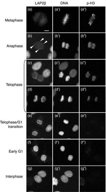
The typical patterns of LAP2β assembly and histone H3 phosphorylation in bovine cumulus cells during the metaphase/interphase transition are shown in Fig. 1. No fluorescence of LAP2β was observed around metaphase chromosomes (Fig. 1a). Several fluorescent granules of LAP2β were detected near the chromatin in some of the anaphase cells (arrowheads, Fig. 1b). LAP2β granules were observed around the chromatin of all telophase cells (Fig. 1c), and then LAP2β clearly surrounded the periphery of the chromatin from late telophase (Fig. 1d) to the telophase II/G1 (Tel II/G1) transition (Fig. 1e). The LAP2β staining was very strong all over the nucleoplasm, and the boundary between the nuclear periphery and the internal LAP2β fluorescence was obscure (Fig. 1f). At the complete interphase with somewhat enlarged pronuclei, LAP2β fluorescence was detected on both the periphery and the inside of the nucleus, and the LAP2β granules were completely lost in the cytoplasm of G1 cells (Fig. 1g), although many LAP2β granules were observed in the cytoplasm from telophase to early G1 (Fig. 1d–f). Histone H3 of chromatin was still strongly phosphorylated at the onset of LAP2β assembly in the anaphase-to-telophase cells (Fig. 1b, b″). Histone H3 phosphorylation was maintained until late telophase (Fig. 1d″), and then histone H3 was almost completely dephosphorylated at the Tel II/G1 transition (Fig. 1e″).
LAP2β assembly and histone H3 phosphorylation in proliferating bovine cumulus cells. The images of LAP2β (a–g), DAPI (a′–g′) and phosphorylated histone H3 at Ser10 (p-H3; a″–g″) are shown. LAP2β assembly was observed as several granules around chromatin at anaphase (arrowheads b) to early telophase (c). A high level of LAP2β assembly around the chromatin was observed beginning in late telophase (d), the telophase/G1 transition, and interphase (e–g). The staining of p-H3 was observed on the chromatin from metaphase to late telophase (a″–d″). The p-H3 became hardly detectable on the chromatin from the telophase/G1 transition to interphase (e″–g″). The scale bar represents 10 μm.

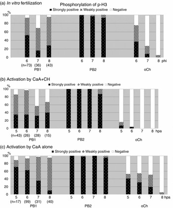
To show the detailed chronology between the assembly of LAP2β around the oocyte chromatin and disaggregation of the chromatin, we examined the phosphorylation of histone H3 at Ser10 and localization of LAP2β. Fig. 2 summarizes the chronological changes in histone H3 phosphorylation in the first (PB1) and second polar bodies (PB2) and oocyte chromatin (oCh) after fertilization and activation. Only oocytes from the extrusion of PB2 to just before pronucleus (PN) formation were examined, and the degree of histone H3 phosphorylation was classified into three categories: strongly positive, weakly positive and negative (Fig. 3a″–d″). The phosphorylation was stronger in PB2 than PB1, while it was maintained at a higher level in both PB1 and PB2 than in the oocyte. Just after telophase II, oCh kept the condensed form until the formation of the pronucleus (Tel II/G1 transition). The histone H3 of oCh only was progressively dephosphorylated in both fertilized and activated oocytes. In fertilized oocytes (Fig. 2a), histone H3 of oCh was still phosphorylated in 75% (55/73) of oocytes at 6 h post-insemination (hpi), and then almost completely dephosphorylated in 95% (45/47) of oocytes at 8 hpi. In the oocytes activated by CaA + CH, the dephosphorylation of histone H3 took place faster than in fertilized oocytes. Histone H3 of oCh was already dephosphorylated in 84% (36/43) of activated oocytes at 5 h post-activation (hpa), and almost all oCh (96%; 25/30) of the activated oocytes until 6 hpa (Fig. 2b). The dephosphorylation of oCh proceeded faster in the case of activation using CaA + CH than in that using CaA alone and in vitro fertilization.
Intensity of histone H3 phosphorylation during the second meiosis in bovine in vitro fertilized and activated oocytes. These graphs show the intensity of the fluorescence against anti-phosphorylated histone H3 at Ser10 (p-H3) in the first (PB1) and second polar body (PB2) and oocyte chromatin (oCh) in oocytes between the late telophase II and early pronuclear stage (Tel II/G1 transition). Oocytes were in vitro inseminated (a, IVF) and activated by treatment with the calcium ionophore A23187 (CaA) followed by cycloheximide (CH) (b, CaA + CH) or by treatment with CaA (c, CaA). Oocytes were fixed at 6–8 h post-insemination (hpi) or at 5–8 h post-activation (hpa), respectively.

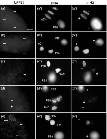
LAP2β assembly and histone H3 phosphorylation in oocyte chromatin during the second meiosis in bovine oocytes after in vitro fertilization. The images of LAP2β (a–e), DAPI (a′–e′) and phosphorylated histone H3 at Ser10 (p-H3; a″–e″) are shown. No LAP2β assembled around oocyte chromatin (oCh; a–b) with strong phosphorylation (a″–b″) in oocytes at telophase II (a, a′ and a″) and the early Tel II/G1 transition stage. Histone H3 of oCh was gradually dephosphorylated (c and c″), but LAP2β did not assemble around oCh (c). Several granules of LAP2β assembled on the periphery of oCh (d) without phosphorylation of histone H3 (d″). The DNA image (d′) show that the oCh was in a condensed condition before the disaggregation of chromatin. The oCh without phosphorylation of histone H3 (e″) was clearly surrounded by many granules of LAP2β (e). Oocytes were fixed at 6–8 h post-insemination (hpi). PB1, first polar body; PB2, second polar body; oCh, oocyte chromatin; ++, strongly positive; +, weakly positive; −, negative. The scale bar represents 10 μm.

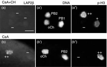
LAP2β assembly was not observed in PB1, PB2 or oCh until telophase II, at which stage histone H3 was intensely phosphorylated in fertilized oocytes (Fig. 3a–a″). LAP2β was also absent around the oCh whose histone H3 was still intensely phosphorylated (Fig. 3b–b″), and was hardly observed around oCh with clear p-H3 staining in oocytes at the beginning of the Tel II/G1 transition (Fig. 3c–c″). A few granules of LAP2β generally appeared around oCh with very weak or no fluorescence of p-H3 (Fig. 3d–d″). Then, LAP2β assembly proceeded and many strong granules surrounded p-H3-negative oCh (Fig. 3e–e″). Both the PB1 and PB2 chromatins were still highly phosphorylated (Fig. 3a″–d″), and LAP2β assembly was always undetectable (Fig. 3a–e). Additionally, the p-H3 intensity of PB2 was generally higher than that of PB1. Typical images of LAP2β assembly and histone H3 phosphorylation in oocytes activated by treatment with CaA + CH and CaA alone are shown in Fig. 4. LAP2β assembly was hardly detectable in any of the activated oocytes using CaA + CH, even around oCh with no p-H3 fluorescence (Fig. 4a–a″). On the other hand, the patterns of LAP2β assembly and histone H3 dephosphorylation in the oocytes activated using CaA alone (Fig. 4b–b″) were more similar to those in fertilized oocytes than those in oocytes activated using CaA + CH.
LAP2β assembly and histone H3 phosphorylation in oocyte chromatin of activated oocytes. The images of LAP2β (a, b), DAPI (a′, b′) and phosphorylated histone H3 at Ser10 (p-H3; a″, b″) are shown. Oocytes were activated by treatment with the calcium ionophore A23187 followed by cycloheximide (CaA + CH; a and a″) or CaA alone (b and b″), and were fixed at 5–8 h post-activation (hpa). In most oocytes activated by CaA + CH, the LAP2β did not assemble around oCh (a) irrespective of oCh dephosphorylation (a″), with a very few exceptions. The oCh without phosphorylation of histone H3 (b″) was clearly surrounded by many granules of LAP2β (b) in the oocytes activated by CaA alone. PB1, first polar body; PB2, second polar body; oCh, oocyte chromatin; ++, strongly positive; +, weakly positive; −, negative. The scale bar represents 10 μm.

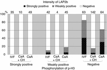
The relationship between the timing of LAP2β assembly and histone H3 phosphorylation in oCh of bovine fertilized and activated oocytes is summarized in Fig. 5. According to the grade of histone H3 phosphorylation, oocytes were divided into three groups, which were further divided into three categories according to the grade of LAP2β assembly in oCh. No LAP2β assembled around oCh with strongly positive p-H3 in spite of fertilization and activation. The total frequencies of oocytes with LAP2β-positive oCh were 41% (58/140), 14% (21/156) and 30% (48/158) in oocytes that were fertilized, activated by CaA + CH and activated by CaA alone, respectively. On the other hand, the total frequencies of oocytes with p-H3 positive oCh were 55% (77/140), 9% (14/156) and 59% (94/158) in oocytes that were fertilized, activated by CaA + CH and activated by CaA alone, respectively. Strong LAP2β assembly was not observed around oCh showing strongly positive staining of p-H3 (Fig. 5; also see Figs 3 and 4), and histone H3 was completely dephosphorylated in 91% (115/127) of LAP2β-positive oCh in the three experimental groups. The pattern of histone H3 dephosphorylation and LAP2β assembly was similar in the fertilized oocytes and oocytes activated by CaA alone, but not in oocytes activated by CaA + CH. On the other hand, LAP2β assembly was observed in only a small proportion of dephosphorylated oCh (15%, 21/142) in oocytes activated by CaA + CH, although the dephosphorylation of histone H3 in these oocytes was much faster than in oocytes after fertilization or treatment with CaA alone (Figs 4a, 5b).
Relationship between intensity of LAP2β expression and histone H3 phosphorylation in oocyte chromatin of bovine in vitro fertilized and activated oocytes around telophase II. Oocytes were in vitro inseminated or activated by treatment with the calcium ionophore A23187 (CaA) followed by cycloheximide (CH) after maturation culture. The oocytes were fixed at 6–8 h post-insemination (hpi) and at 5–8 h post-activation (hpa), respectively. Oocytes around the Tel II/G1 transition were classified into three groups – negative, weakly positive and strongly positive – by the degree of histone H3 dephosphorylation (x-axis). Then, oocytes in each class of p-H3 phosphorylation were further classified into three categories – negative, weakly positive and strongly positive – by the intensity of LAP2β (indicated by the patterns of columns). Figures on each bar show the number of oocytes in the indicated categories. n, total number of oocytes examined.

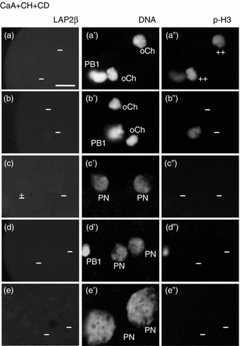
Although oCh was rapidly dephosphorylated and LAP2β assembled around oCh temporarily, the chromatin of PB2 continued to show high phosphorylation of histone H3 and did not show LAP2β assembly. To trace the fate of the counterpart of the oocyte chromatin, we examined the dephosphorylation of histone H3 and LAP2β assembly in oocytes activated by CaA +CH + CD treatment for inhibition of the PB2 extrusion. Both oChs in the ooplasm were strongly phosphorylated during the second meiosis, and then these chromatins were simultaneously dephosphorylated (Fig. 6a, a″, b, b″). However, LAP2β assembly was undetectable around these dephosphorylated oCh (Fig. 6a, b), just as in the oocytes activated by CaA + CH treatment (Fig. 4a). LAP2β assembly was not observed either in small pronuclei with evidence of chromatin decondensation in DNA images (Fig. 6c, c′, d, d′) or in developing pronuclei (Fig. 6e, e′).
LAP2β assembly and histone H3 phosphorylation during pronuclear formation in oocyte chromatin of bovine oocytes activated by treatment with calcium ionophore, cycloheximide and cytochalasin D. The images of LAP2β (a–e), DAPI (a′–e′) and phosphorylated histone H3 at Ser10 (p-H3; a″–e″) are shown. Oocytes were activated by treatment with the calcium ionophore A23187 (CaA) followed by treatment with cycloheximide (CH) and cytochalasin D (CD), and were fixed at 6–8 h post-activation (hpa). The fluorescence of p-H3 was clearly detected in both oocyte-condensed chromatin masses (oChs in a′ and a″). LAP2β was still undetectable around the oChs (b) after complete disappearance of p-H3 from both oChs (b″), and LAP2β did not assemble even immediately after the formation of pronuclei (PN, c–e and c′–e′). PB1, first polar body; PB2, second polar body; oCh, oocyte chromatin. ++, strongly positive; ±, very weak or obscure; −, negative. The scale bar represents 10 μm.

Discussion
We previously reported that LAP2β assembly around oCh was observed in some fertilized oocytes from PB2 extrusion to the beginning of PN formation, i.e., the Tel II/G1 transition (Isaji et al., Reference Isaji, Iwata, Harayama and Miyake2004). The timing of LAP2β assembly was precisely determined and the cause of the lack of LAP2β assembly in some fertilized oocytes was examined here. As there is a strong correlation between histone H3 phosphorylation at Ser10 and chromosome condensation during cell division (Hendzel et al., Reference Hendzel, Wei, Mancini, Van Hooser, Ranalli, Brinkley, Bazett-Jones and Allis1997; Van Hooser et al., Reference Van Hooser, Goodrich, Allis, Brinkley and Mancini1998), we immunohistochemically analysed the phosphorylation of histone H3 at Ser10 as an indicator of oocyte chromatin decondensation.
First, the timing of both histone H3 dephosphorylation and LAP2β assembly were confirmed in somatic cells using bovine cumulus cells. LAP2β firstly assembled around the anaphase chromatin, and the granules accumulated in the vicinity of each telophase chromatin mass that was still highly phosphorylated, supporting previous reports using other somatic cells (Foisner & Gerace, Reference Foisner and Gerace1993; Maison et al., Reference Maison, Pyrpasopoulou, Theodoropoulos and Georgatos1997; Dechat et al., Reference Maison, Pyrpasopoulou, Theodoropoulos and Georgatos1998; Bodoor et al., Reference Bodoor, Shaikh, Salina, Raharjo, Bastos, Lohka and Burke1999). The degree of phosphorylation declined around late telophase, and the chromatin at the Tel II/G1 transition was finally completely dephosphorylated.
In bovine oocytes around the Tel II/G1 transition after fertilization and activation, the level of phosphorylation of histone H3 at Ser10 in PB1, PB2 and oCh was high until telophase II, and it was generally higher in PB2 than in PB1 in spite of fertilization or activation. Histone H3 dephosphorylation in oCh rapidly progressed, while that in PB1 and PB2 was somewhat slower than that in oCh after completion of the second meiosis. The LAP2β, however, did not assemble around any chromatins with a high level of p-H3. A few granules of LAP2β gradually appeared in the vicinity of oCh after the decrease or disappearance of p-H3, and then LAP2β assembled surrounding oCh. No assembly of LAP2β was observed around the chromatin in PB1 or PB2. This may have resulted from the highly phosphorylated state of chromatin histone H3 and/or the lack of LAP2β in the cytoplasm of polar bodies.
Aurora B kinase and protein phosphatase 1 (PP1) are associated with chromosome modification during mitosis in vertebrates (Murnion et al., Reference Murnion, Adams, Callister, Allis, Earnshaw and Swedlow2001). PP1 is a phosphatase of p-H3 in somatic cells (Hsu et al., Reference Hsu, Sun, Li, Reuben, Tatchell, Bishop, Grushcow, Brame, Caldwell, Hunt, Lin, Smith and Allis2000; Scrittori et al., Reference Scrittori, Hans, Angelov, Charra, Prigent and Dimitrov2001), and dephosphorylates Ser10 of histone H3 as suggested in Caenorhabditis elegans and Saccharomyces cerevisiae. Furthermore, it also dephosphorylates and inactivates aurora B itself (Murnion et al., Reference Murnion, Adams, Callister, Allis, Earnshaw and Swedlow2001). The aurora B kinase is also activated by okadaic acid, a PP1/PP2 inhibitor, which indicates that the kinase should interact with the phosphatase, although direct interactions have not been reported (Sugiyama et al., Reference Sugiyama, Sugiura, Hara, Sugimoto, Shima, Honda, Furukawa, Yamashita and Urano2002). The high level of p-H3 in PB1 and PB2 may be a result of the decrease in PP1 activity and/or its amount. As there is a sufficient volume of cytoplasm, the abundance of the activator of PP1 in the ooplasm may immediately raise PP1 activity. This is supported by the fact that p-H3 in both oChs is simultaneously and completely dephosphorylated in activated oocytes in which PB2 extrusion was suppressed by CD treatment. Assessment of the difference in the phosphorylation level of histone H3 enables us to discriminate correctly between oocyte and PB2 chromatin, although it is difficult to distinguish the oCh from the PB2 chromatin during the Tel II/G1 transition by DNA staining alone.
In somatic cells, the sequential phosphorylation by both protein kinase C (PKC) and CDK1 brings about the depolymerization of LAP2β, lamin A/C and lamin B, while the dephosphorylation by PP1 causes their polymerization (Collas, Reference Collas1999; Steen et al., Reference Steen, Martins, Tasken and Collas2000; Steen & Collas, Reference Steen and Collas2001). Both chromatin decondensation by histone H3 dephosphorylation and LAP2β assembly are thought to result from dephosphorylation by PP1 for pronucleus formation. Furthermore, it is inferred that PP1 activity might increase during the metaphase/anaphase transition, since histone H3 phosphorylation and LAP2β assembly proceed simultaneously in somatic cells. The chromosomes of cumulus cells were strongly phosphorylated until the Tel II/G1 transition, while LAP2β assembled around the anaphase chromatin. LAP2β generally assembled around oCh after the disappearance of dephosphorylation of histone H3 in the second meiosis of the bovine oocyte. It is concluded that the timing of LAP2β assembly in the bovine oocyte is clearly different from that in somatic cells from the point of histone H3 dephosphorylation, although the significance of the difference is still unclear. It is considered that both LAP2β assembly and histone H3 dephosphorylation are controlled by the activity of PP1 (Collas, Reference Collas1999; Steen et al., Reference Steen, Martins, Tasken and Collas2000; Steen & Collas, Reference Steen and Collas2001); however, differential timing of the two events was certainly observed between the somatic cell and oocyte. These results suggest that additional factors and/or a different pathway, which may be oocyte-specific, are involved in LAP2β assembly. LAP2β disappeared again from the periphery of oCh at the early PN stage in oocytes after fertilization and activation by CaA, while histone H3 phosphorylation remained at an undetectable level. These results suggest that there is no relevant relationship between the disappearance of LAP2β and the phosphorylation of histone H3 in the oocyte, although there is dephosphorylation of histone H3 followed by LAP2β assembly.
Our previous study showed that LAP2β assembled around oCh in fertilized bovine oocytes, but not in oocytes activated by CaA + CH (Isaji et al., Reference Isaji, Iwata, Harayama and Miyake2004). In the present study, the oocytes activated by CaA alone showed the clear assembly of LAP2β around dephosphorylated oCh as well as fertilized oocytes. Oocyte chromatin was completely dephosphorylated in almost all oocytes activated by CaA + CH treatment; nevertheless, LAP2β did not assemble around oCh. These results suggest that CH, an inhibitor of protein synthesis, may inhibit the synthesis of LAP2β and/or some factors that have a role in LAP2β assembly. Thus, LAP2β assembly is not essential for lamin A/C and B integration at least for PN formation, because the pronucleus was normally formed in either the presence or absence of LAP2β assembly (Isaji et al., Reference Isaji, Iwata, Harayama and Miyake2004).
It is generally difficult to distinguish the detailed stages of fertilized oocytes from PB2 extrusion to early PN formation by DNA staining alone. The boundaries between telophase II and the Tel II/G1 transition with condensed chromatin, and between the Tel II/G1 transition and the beginning of the PN stage could be clarified by the assessment of chromatin phosphorylation and LAP2β assembly combined with DNA images. Both chromatin masses are non-spherical and equally and strongly phosphorylated until the end of telophase II. The starting point of the transition phase is distinguished by the spherical shape of the chromatin masses, and the phosphorylation level begins to decrease in oCh only by the time of complete separation of the PB2 from the oocyte, referred to as the early Tel II/G1 transition or early condensed chromatin stage. Oocytes that have almost completely dephosphorylated histone H3 could be classified into the late Tel II/G1 transition, and several granules of LAP2β assembly appear around the oCh. The successive assembly of LAP2β surrounding oCh without p-H3 is the obvious index for the beginning of the PN stage. The commencement of oCh decondensation, namely when the PN begins growing, is concomitant with the disappearance of the LAP2β assembly from oCh. Of course, lamin A/C and B assemble on the nuclear membrane at this stage (Isaji et al., Reference Isaji, Iwata, Harayama and Miyake2004). Since LAP2β assembles again on the nuclear membrane around the end of DNA synthesis (Isaji et al., Reference Isaji, Iwata, Harayama and Miyake2004), the PN is referred to as the early or growing pronucleus until the reassembly of LAP2β.
From the results of the present study, it is concluded that LAP2β assembles around only oCh after complete dephosphorylation of histone H3 in bovine oocytes after fertilization and activation using CaA alone, and that the timing of histone H3 dephosphorylation and LAP2β assembly in these oocytes is clearly different from that of somatic cells. The results also indicate that CH treatment inhibits LAP2β assembly around oCh but not histone H3 dephosphorylation.
Acknowledgements
The present study was supported by a grant for Creative Scientific Research (13GS0008) and the 21st Century COE Program of the Ministry of Education, Culture, Sports, Science and Technology of Japan. The authors are grateful to the staff of the Nishiharima and Kobe City Meat Inspection Offices for supplying bovine ovaries.