Introduction
Chronic rhinosinusitis is a common disease that significantly affects quality of life (QoL).Reference Fokkens, Lund, Hopkins, Hellings, Kern and Reitsma1 Prevalence has been shown to be between 5 and 10 per cent of the population,Reference Adams and Marano2,Reference Benninger, Ferguson, Hadley, Hamilos, Jacobs and Kennedy3 and effective treatment significantly improves QoL.Reference Fokkens, Lund, Hopkins, Hellings, Kern and Reitsma1
The treatment of chronic rhinosinusitis is usually undertaken in a stepwise process. Surgery is reserved for cases resistant to appropriate medical treatment. There are a wide range of surgical procedures available to treat chronic rhinosinusitis, with the current ‘gold standard’ being endoscopic sinus surgery. This involves opening the sinus ostia whilst preserving as much mucosa as possible under endoscopic vision. The natural drainage pathways are opened or widened to restore mucociliary clearance and allow topical steroid medication to reach the mucosa. Powered instruments are often used. Known complications include heavy bleeding, orbital injury, cerebrospinal fluid leak and significant intranasal post-operative scarring.
In a national, high quality audit, surgery for polyps and chronic rhinosinusitis has been shown to be safe and effective with a follow up of three years.Reference Hopkins, Browne, Slack, Lund, Topham and Reeves4 However, techniques are always being adapted and modified, and it is important to assess and monitor new techniques.
The Sino-Nasal Outcome Test (SNOT)-20 questionnaire was one of the first patient-reported outcome measures that was validated for use in chronic rhinosinusitis.Reference Piccirillo, Merritt and Richards5 Additional symptoms of anosmia and nasal obstruction have now been added to the questionnaire as these form part of the chronic rhinosinusitis diagnostic guidelines in Europe and North America. The patient-reported outcome measures are now referred to as SNOT-22, and this has again been assessed and validated.Reference Gillett, Hopkins, Slack and Browne6,Reference Noon and Hopkins7
Balloon sinuplasty is a relatively new technique. It follows on from the successful use of balloon dilatation in other medical specialties. The balloon technique was first used for paranasal sinus use by the US Food and Drug Administration in the USA in 2005. The ‘CE’ mark for use in Europe was achieved in 2006.
The aim of this study was to use the SNOT-22 patient-reported outcome measures to monitor the outcomes of balloon sinuplasty and, in particular, review if there was a lasting effect for over two years.
Materials and methods
This was a longitudinal cohort study of nine patients who underwent balloon sinuplasty and no other procedure. The majority of the patients (n = 8) underwent surgery between June and December of 2017 and were followed up for over two years. One patient underwent surgery in October 2019. Surgery was undertaken when the patient still had significant symptoms and impaired QoL despite appropriate medical treatment. All patients had chronic rhinosinusitis using the European Position Paper on Rhinosinusitis and Nasal Polyps 2012 definition 1 and had a computed tomography (CT) scan before surgery. Patients with polyps were not included. In this series, balloon sinuplasty was limited to patients whose symptoms were predominantly frontal pain, pressure or headache. Where there was widespread disease based on symptoms and imaging, traditional endoscopic sinus surgery was undertaken.
The primary outcome measure was improvement in symptoms assessed using the SNOT-22 questionnaire. The specific question of facial pain or pressure was analysed individually. Secondary outcomes included complications, failure of the procedure, and need for revision or repeat procedures. Patients completed the SNOT-22 questionnaire in person before their surgery and then at an approximate two month follow-up out-patient appointment. A telephone consultation was undertaken at two years after their original surgery to fill out the final questionnaire. In data analysis, significance was classed as a p < 0.05, and t-test was calculated using Microsoft Excel® spreadsheet software for Mac® 16.35.
Procedure
Patients underwent a general anaesthetic, and the balloon catheter was guided to the correct position under direct vision. There were no local anaesthetic procedures in this series. The XprESS ENT Dilation System was used (Striker, Kalamazoo, USA). The guidewire with a light-emitting diode indicator was gently passed into the frontal sinus. The balloon was then inserted over the wire to sit in the natural ostium. Saline was used to inflate the balloon. After dilatation, the surgical site was reviewed using the endoscope. No packing was used.
Results
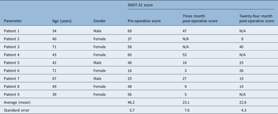
Patient demographics are shown in Table 1. Age ranged from 34 to 71 years, and the mean age was 51 years. All nine patients had successful procedures, but some were more difficult compared with others. This difficulty was often predicable through reviewing the frontal sinus drainage pathway on sagittal CT scans. None of the patients required any instrumentation or uncapping of agger nasi cells.Reference Fokkens, Lund, Hopkins, Hellings, Kern and Reitsma1 There were no complications. One of the nine patients required a repeat procedure. Despite this repeat, there was no significant long-term benefit in symptoms for this single patient.
Patient demographics and average SNOT-22 score

SNOT = Sino-Nasal Outcome Test; N/A = not applicable
Follow up at the 2–3 month point ranged from 7 to 16 weeks with a mean of 11 weeks. Long-term follow up was from 28 to 36 months (mean, 32 months). One patient's long-term follow up was at 17 months and was excluded from the long-term follow up data collection as it was less than 2 years. Before surgery, one patient had a mild SNOT-22 score, three had a moderate score and four had a severe score using the system described by Toma and Hopkins in 2016.8
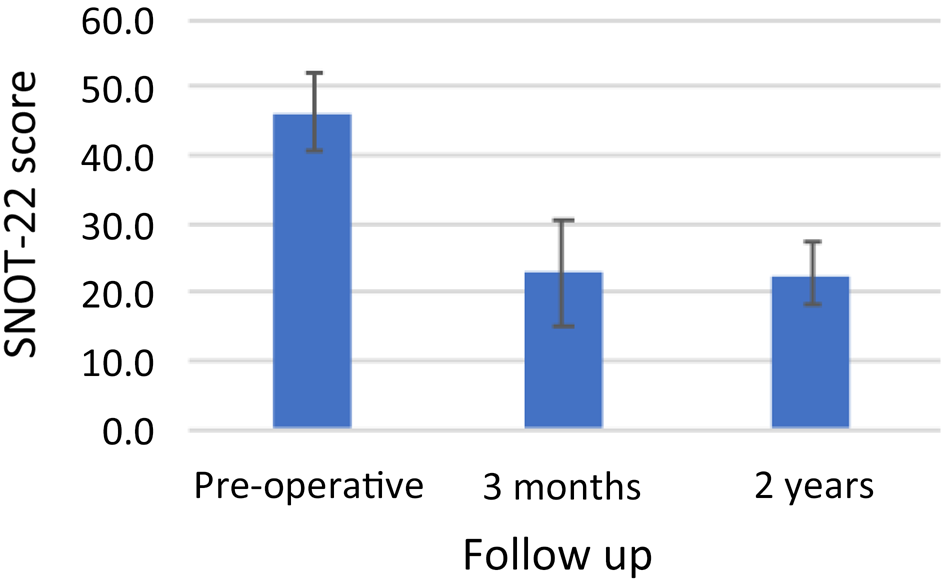
Table 1 and Figure 1 show mean overall SNOT-22 scores from the pre-operative state through to long-term follow up. Error bars represent the standard error. Average SNOT-22 score improved significantly between the pre-operative period (46.2) and three months (22.8) and between the pre-operative period and two years (22.8; p = 0.016 and p = 0.003, respectively). There was no statistically significant difference between three-month and two-year follow up (p = 0.97).
Average total Sino-Nasal Outcome Test (SNOT)-22 score over the two-year follow up. Error bars show standard error.

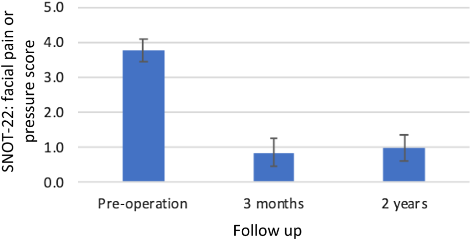
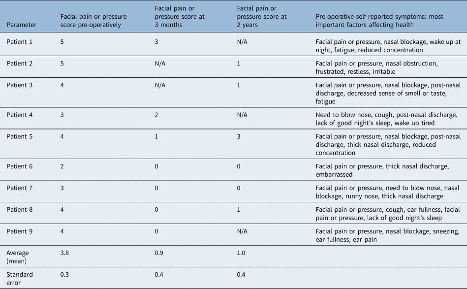
Table 2 shows the scores for one question on ‘facial pain or pressure’, scored from 0 to 5. This was the question closest to the main indication for the surgery, which was frontal pain, pressure or headache. Table 2 also shows the patient self-selected five most important symptoms, which forms part of the SNOT-22 patient-reported outcome measures. There was a significant improvement in facial pain or pressure at three months which lasted for the two years (p > 0.01 for both). There was no significant difference between three months and two years (p = 0.83). Figure 2 shows this long-term improvement which was greater compared with overall symptoms. This symptom was in the five most important items for eight of the nine patients pre-operatively.
Average total Sino-Nasal Outcome Test (SNOT)-22 score for symptoms of pain or pressure over the two-year follow up. Error bars show standard error.

Score for facial pain or pressure and self-reported most important symptoms

Table shows the scores for one question on ‘facial pain or pressure’, scored from 0 to 5. N/A = not applicable
Discussion
In this series, there were no complications, and all patients were discharged on the day of surgery. This fits with the literature, which generally supports the safety of the procedure.Reference Weiss, Church, Kuhn, Levine, Sillers and Vaughan10–Reference Abreu, Balsalobre, Pascoto, Pozzobon, Fuchs and Stamm12 In terms of narrative, one might expect a balloon procedure to be safer. However, vigilance is still required especially in cases of anatomical variance. The distal part of the balloon is inflated out of direct vision, and although in this series there were no complications, it is possible to imagine the potential for a serious complication such as a large skull base defect. As the procedure becomes more widespread, we must continue to monitor for complications and look for methods to reduce risk.
Results of the study show that there was a statistically significant improvement of overall symptoms at three months and at two years compared with the pre-operative period. There was a significant improvement in facial pain or pressure symptoms at both three-month and two-year follow up. Pain or pressure was singled out as it most closely matches the main indication for the procedure, which was persistent frontal pain or pressure or headache. These symptoms were confirmed for each patient in a retrospective review of the case notes. To further the confirmation, all but one patient identified pain or pressure in the self-selected five most important symptoms. These findings support the growing body of evidence within the literature showing the efficacy of the procedure.
The main clinical trial was performed in the USA and was called the Clinical Evaluation to Confirm Safety and Efficacy in the Paranasal Sinuses (‘CLEAR’) trial.13 This involved 115 non-randomised patients, and it showed good safety of the procedure with no major complications and a lasting benefit on SNOT-20 at six months. The revision rate was 9.2 per cent.13
Levine et al. performed a large retrospective study of 1036 patients.Reference Levine, Sertich, Hoisington, Weiss and Pritikin14 Although a large number of patients had a hybrid procedure involving a combination of traditional endoscopic sinus surgery and balloon sinuplasty, the results for efficacy were promising, and this study also supports the safety of the procedure.
Friedman et al. performed a 70-patient study comparing sinuplasty and traditional endoscopic sinus surgery using SNOT-20 as the outcome.Reference Friedman, Schalch, Lin, Mazloom, Neidich and Joseph15 At short-term follow up, the improvements from balloon sinuplasty were comparable if not slightly better.
Koskinen et al. undertook a control study comparing endoscopic sinus surgery and balloon sinuplasty with a six-year follow up via telephone interview, which is the longest follow-up time known for the technique.Reference Koskinen, Myller, Mattila, Penttilä, Silvola and Alastalo16 This study showed balloon sinuplasty to be slightly less effective with reports of slightly more acute exacerbations, thick nasal discharge and revisions in the balloon group over six years. This might be expected in a less invasive procedure.
In general, the literature supports balloon sinuplasty as a safe and effective procedure, and there are multiple other small studies supporting this.Reference Weiss, Church, Kuhn, Levine, Sillers and Vaughan10–Reference Abreu, Balsalobre, Pascoto, Pozzobon, Fuchs and Stamm12
As with many techniques, there is a lack of evidence from high-quality randomised control trials. The use of high-quality multiuser databases in the future may revolutionise the monitoring of technique efficacy, ultimately improving patient care and allocation of resources.
In this study, one patient had a revision procedure. There was no improvement after the revision procedure. This fits with some of the well-known evidence from endoscopic sinus surgery procedures where more revisions tend to lead to diminishing improvements. However, one patient is not enough to draw any definitive conclusions, and as always, pre-operative patient selection is important.
In the national sinonasal audit,Reference Hopkins, Browne, Slack, Lund, Topham and Reeves4 there was a 4.7 per cent revision rate for patients undergoing surgery for chronic rhinosinusitis without polyps in the first 12 months. Additionally, 5 per cent of patients were on the waiting list for a revision. Based on this revision rate, it appears that balloon sinuplasty may have a roughly equivalent or possibly slightly higher revision rate.
In this series, the procedure was successfully completed in all cases. The illuminated guidewire was found to be an effective tool for finding the frontal sinus. However, there are some potential issues reported in the literature with this technique. There was one case where an inter-sinus septal cell had been cannulated and dilated, which caused stenosis of the natural sinus drainage pathway. This could possibly be because the light tip can cause illumination of the frontal sinus when it is not in the correct position. On balance, the light tip method does seem to be an effective and safe method, but the operator must be vigilant about limitations and report future complications.
In all our cases, the frontal sinus was easily reached. From the literature, there is a good success rate. However, occasionally some difficulty was experienced in accessing the frontal sinus.Reference Hopkins, Noon, Bray and Roberts9 Sometimes, difficulty could be anticipated because anatomical variations, such as large agger nasi cells, could be seen on the CT scan. A double-ended probe or seeker could be used to atraumatically find the natural ostium. Hopkins et al. reported that in one case, an agger nasi cell was unroofed using a curette prior to balloon insertion and dilatation of the frontal sinus drainage pathway.Reference Hopkins, Noon, Bray and Roberts9
This study has the limitation that the technique was used for the frontal sinus and offers no addition to the evidence for the efficacy of balloon sinuplasty in other anatomical sites. Patient selection in frontal sinus pain groups can be challenging. Some clinicians advocate that patients are seen by a neurologist to rule out other causes of frontal pain, but this was not standard practice in this series.
Operatively, the frontal sinus has traditionally been a challenging area with the known complication of post-operative scarring. Some patients may choose not to have traditional frontal sinus endoscopic sinus surgery because of the risk of complications. Balloon sinuplasty may offer a faster, safer and effective procedure for a cohort of patients who may not have chosen or been appropriate for the traditional approach. The number of patients suitable for this type of treatment is likely to be relatively small, even in larger centres. This poses a challenge for recruitment to trials. A limitation of this study is the small number of participants. It is hoped that in the future multiple small studies could be combined for further analysis.
Friedman et al. performed an economic analysis of balloon dilatation.Reference Friedman, Schalch, Lin, Mazloom, Neidich and Joseph15 In their study, the procedure was performed under local anaesthetic, and there were significant savings. In the UK, cost savings are likely through increasing capacity for operating lists and reducing overnight stays.
Balloon sinuplasty may have a role in the acute setting, and there has been some evidence of its efficacy in this setting.Reference Hopkins, Noon, Bray and Roberts9 Traditionally, acute frontal sinusitis with complications would often require management using an open procedure. This is usually effective, but there are known complications such as scarring in a cosmetically undesirable area. A short intranasal procedure with intranasal catheter would offer benefits if it could be shown to be effective and safe, but it could be challenging in a highly vascularised and inflamed nasal cavity.
• Evidence for the long-term efficacy and safety of balloon sinuplasty is lacking
• Limited evidence suggests that this new procedure is safe and effective, and this study looks at a group of patients who had frontal sinus procedures with a two-year follow up
• There were no complications and no safety concerns in this small series
• Using the Sino-Nasal Outcome Test-22 patient outcome questionnaire, there was a significant and lasting benefit to patient symptoms
• This new data adds to the evidence supporting the efficacy and safety of balloon sinuplasty
• Novel aspects of this study include a relatively long follow up (two years) and using the technique predominantly for the frontal sinus
The potential to use this technique under local anaesthetic, either in day surgery or in an out-patient setting may offer significant opportunities for economic savings and patient benefits.
Conclusion
This small study supports the growing evidence of balloon sinuplasty being a safe and effective method for treating frontal sinus symptoms. There were no complications, and the technique was successfully completed in all patients. There were particularly effective results when looking at the symptoms of pain or pressure. Faced with a diagnostic dilemma between sinogenic pain and headache, with well selected patients, balloon sinuplasty seems to have long-term benefit. This appears to be an attractive alternative to a neuropathic pain medication or much larger frontal sinus procedures with their associated risk of side effects and complications.
Competing interests
None declared