Introduction
Classical osteoblastoma is a benign bone lesion accounting for less than 1 per cent of all bone tumours, presenting predominantly in the younger age groups. It commonly affects the spine, sacrum and long tubular bones and usually presents with pain and deformity. Osteoblastoma of the larynx is a rare tumour, with only three case reports in the literature.Reference Urbaczyk and Miszczyk1–Reference Agarwala, Graham and Panella3 The differential diagnosis for osteogenic lesions of the larynx includes dystrophic ossification, osteosarcoma and osteoblastoma. Its potentially life-threatening anatomical site renders laryngeal osteoblastoma clinically significant.
We present the unique case of an osteoblastoma arising from the vocal fold in a 76-year-old man presenting with acute airway obstruction.
Case report
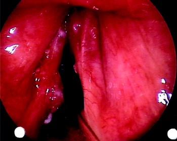
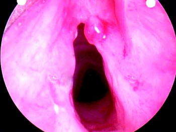
A 76-year-old man had initially presented to the senior author eight months prior to the current presentation, with a long history of voice change clinically resembling muscle tension dysphonia. Laryngoscopy had revealed a papillomatous lesion on the left vocal fold and hypertrophic changes on the right vocal fold (Figure 1). Laser resection of the left vocal fold lesion had been uneventful, and the histopathological apperance had been benign.
Papillomatous vocal fold lesion identified on the left vocal fold a few months before main presentation.

Twenty days prior to his acute airway obstruction presentation, the patient had experienced haemoptysis. On flexible nasoendocopy, a large polyp had been seen in the anterior larynx. Elective admission for microlaryngoscopy and biopsy had been planned.
However, within two weeks of review, whilst awaiting elective surgery, the patient developed rapid onset upper airway obstruction and cardiopulmonary arrest. At a peripheral hospital, he underwent cardiopulmonary resuscitation involving a difficult intubation. He was transferred to our institution for surgical tracheostomy and definitive management.
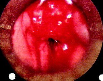
At operation, a bulky tumour arising from the right anterior vocal fold and ventricle was found, extending back to almost occlude the larynx (Figure 2). The tumour was debulked and a sample sent for histopathological analysis.
Intra-operative view of tumour arising from right vocal fold and ventricle and extending back to almost occlude the larynx.

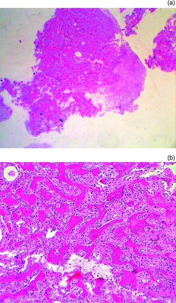
The tumour was received piecemeal for histopathological analysis, and showed fragments of trabeculated bony tissue rimmed by plump, uniform osteoblasts (Figure 3). Ulcerated, oedematous granulation tissue was present on the surface. On the deep aspect, the osteoblastic proliferation abutted against collagenous fibrous tissue. The histological pattern of the osteogenic tissue was typical of an osteoblastoma. The tumour had a uniform, nodular architecture sharply demarcated from surrounding granulation tissue and fibrous tissue. Although there was marked osteoblastic activity, no cytological atypia was present. The zonation and myofibroblastic appearance of myositis ossificans was not seen. The material was received fixed, and no cytogenic or molecular studies were performed.
(a) Low power view showing well demarcated, nodular architecture of lesion (H&E; ×2). (b) Medium power view showing bony trabeculae and osteoblastic rimming (H&E; ×20).

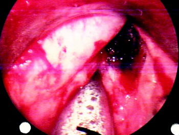
After the formal histological report had been received, the patient underwent complete laser resection of the tumour, without complication (Figure 4). He was decannulated within two weeks of surgery and closely followed up with fortnightly flexible laryngoscopy.
Residual defect following laser excision of tumour.

At 11 weeks of follow up, keratinisation of the left vocal fold and a new right vocal fold lesion were identified (Figure 5). Laser resection of the lesion confirmed pyogenic granuloma. No further abnormality was noted after four further months of follow up. Despite the benign histopathology of the new right vocal fold lesion, its clinically aggressive course warranted close monitoring.
New lesion identified on right vocal fold at 11-week follow-up review.

Discussion
Osteoblastoma, as defined in the World Health Organization classification,Reference Fletcher, Unni and Mertens4 is a tumour of male teenagers and young adults with a predilection for the spine and sacrum but also occurring in the femur, tibia and foot. Cementoblastoma of the jaw, which is found attached to a tooth, is considered to be an osteoblastoma. This classification does not list soft tissue or larynx as a site of osteoblastoma. The published work has very few case reports of osteoblastoma arising from laryngeal cartilages.Reference Urbaczyk and Miszczyk1–Reference Agarwala, Graham and Panella3 Agarwala et al. and Ledeboer et al. reported osteoblastoma of thyroid and cricoid cartilage, respectively, in middle-aged men presenting with the gradual onset of a neck lump, dysphagia and hoarseness.Reference Ledeboer, Kerrebijn, Govaere and den Bakker2, Reference Agarwala, Graham and Panella3 Presumably, these cases arose within ossifying cartilage, whereas our case was extraosseous. Consequently, similar histology does not necessarily imply that the lesion pathogenesis is that of classical osteoblastoma. For discussion purposes, reference is made to classical osteoblastoma, bearing in mind the uncertainty of pathogenesis in the extraosseous larynx.
Classical osteoblastoma histology closely resembles osteoid osteoma, with proliferation of osteoid and woven bone in a loose stroma containing dilated capillaries. Differential diagnoses include osteoid osteoma, aneurysmal bone cyst, well differentiated osteosarcoma, giant cell tumour, ossified haematoma and heterotopic ossification. In the larynx, the principal differential diagnosis of abnormal bone is reactive bone, heterotopic ossification, osteosarcoma or osteoblastoma. Reactive conditions usually show a range of changes such as fibroplasia, neovascularisation, and osteoid formation and zonation, in keeping with their development from an underlying process such as trauma. An important differential diagnosis is osteosarcoma, which requires identification of tumour osteoid as well as cytological atypia, the distinction of which is subjective and can be difficult. Classical osteoblastoma and other osteoblastic tumours with a similar histological appearance are distinguished by clinical and radiological context.
The biological behaviour of benign osteoblastoma is unpredictable. There are reports of aggressive behaviourReference Kenan, Floman, Robin and Laufer5, Reference Ishihara6 and, rarely, of transformation into osteosarcoma with metastatic potential.Reference Merryweather, Middlemiss and Sanerkin7, Reference Hermann, Klein, Springfield and Abdelwahab8 There are variants of osteosarcoma, so-called osteoblastoma-like osteosarcoma, which are histologically very similar and cause diagnostic confusion; in such cases, one distinguishing feature is the presence of permeative margins.Reference Hermann, Klein, Springfield and Abdelwahab8 An entity referred to as ‘aggressive osteoblastoma’, defined by radiographic and histological appearance, falls between benign osteoblastoma and osteosarcoma. Clinically, these tumours have a more aggressive course, and in most cases this is the only diagnostic clue. Radiographically, they tend to extend beyond the cortical margins; histologically, areas of epithelioid osteoblasts, hyperchromatic nuclei, atypia and numerous giant cells of osteoclastic type are present.Reference Merryweather, Middlemiss and Sanerkin7, Reference Canale9 Even though the histological apperance of the index tumour was benign, its clinical course was in keeping with an aggressive osteoblastoma. Another recognised entity is ‘pseudo-malignant osteoblastoma’, a non-aggressive tumour with atypical and bizarre cytology.Reference Kenan, Floman, Robin and Laufer5, Reference Cheung, Wu, Lam and Fu10
The key to successful treatment of classical osteoblastoma is accurate localisation and complete excision of the tumour nidus. With such an approach, long-term follow-up recurrence rates of up to 10–19 per cent are reported.Reference Canale9, Reference Lucas, Unni, Mcleod, O'Connor and Sim11, Reference Kroon and Schurman12 Other treatment options include radiotherapy; however, this is controversial. The Mayo Clinic has documented 34 new cases of sarcoma in previously irradiated bones over 27 years;Reference Sim, Cupps, Dahlin and Ivins14 in 11 patients, sarcoma arose from bones within the radiation field and, in 16, in a pre-existing bone lesion, one of which was an osteoblastic lesion. Nevertheless, there are reports of successful use of primary radiotherapy treatment of osteoblastomas in non-resectable sites (e.g. petrous bone or recurrent osteoblastoma) in both adults and children, with no recurrence or transformation after long-term follow up.Reference Singer and Deutsch15–Reference Berberoglu, Oguz, Aribal and Ataoglu18
• A rare case of obstructing osteoblastoma of the larynx is reported, which presented with rapid onset airway obstruction
• Osteoblastoma must be distinguished, both on histological and clinical grounds, from the more aggressive osteosarcoma or osteoblastoma variants
• Treatment should be surgical excision, which, if available, may be achieved by transoral laser microsurgery
• Due to rapid growth, careful follow up is essential in order to ensure early recognition of recurrence and prevention of potentially life-threatening airway obstruction
Treatment options for laryngeal osteoblastoma include radiation, excision and laser therapy. However, the rarity of this tumour in the larynx precludes establishment of evidence-based management recommendations. The common treatment approaches in the reported cases of laryngeal osteoblastoma have been mainly surgical excisionReference Ledeboer, Kerrebijn, Govaere and den Bakker2, Reference Agarwala, Graham and Panella3 with radiotherapy in one case.Reference Urbaczyk and Miszczyk1 However, we recommend transoral laser tumour excision. Jako and Strong introduced laser surgery in laryngeal microsurgery in the early 1970s; by the 1980s, it was mainly used for treatment of laryngeal papillomatosis.Reference Steiner and Werner19 Today, laser surgery is widely accepted as an alternative to traditional endoscopic resection techniques, open partial laryngectomy and radiotherapy, especially for early glottic cancers.Reference Ambrosch20 Although there are no reports of laser surgery being used to treat osteoblastoma, from our previous successful experience with laser treatment of benign and malignant glottic lesions, one can extrapolate and aim for a similar outcome. The major advantages of laser therapy are organ preservation and rendering possible all treatment options, including laser re-excision, surgical excision and radiotherapy, in patients found to have a local recurrence or metachronous head and neck tumour.
Acknowledgement
The authors thank Dr Alicia Jenkins, Department of Medicine, St Vincent's Hospital, for her editorial assistance.