Implications
The fundamental study could provide a reference for illuminating the regulation mechanism of PI3K-Akt-mTOR signaling in lipid metabolism homeostasis in liver, and give some hint about mechanism of non-pathological hepatic steatosis in goose, and this could be conductive to further research about the breeding of specialized strains of goose for fatty liver.
Introduction
Birds, especially some wild waterfowl, have specific capability of fatty liver production, because they are more likely to show non-pathological hepatic steatosis as a result of energy storage before migration. Fatty liver in waterfowl is due to the excessive triacylglycerol accumulation in liver, which has a close relation with de novo fatty acid synthesis, fatty acid oxidation and very low density lipoprotein (VLDL) assembly and secretion.
Phosphatidylinositol-3 kinases (PI3K) is a lipid kinase which generates the second messenger phosphatidylinositol-3,4,5-trisphosphate (PIP3) (Vara et al., Reference Vara, Casado, de Castro, Cejas, Belda-Iniesta and González-Barón2004). The accumulation of PIP3 recruits Protein kinase B (Akt) to the cell membrane where it is phosphorylated and activated by Phosphoinositide-dependent kinase (PDK) 1 and 2 (Osaki et al., Reference Osaki, Oshimura and Ito2004). Akt activation regulates numerous downstream effectors such as mammalian target of rapamycin (mTOR), which forms a stoichiometric complex with raptor. Raptor has a positive role in nutrient-stimulated signaling to the downstream effector S6 kinase 1 (S6K1) which controls activation of translation and ribosome biosynthesis (Fingar et al., Reference Fingar, Richardson, Tee, Cheatham, Tsou and Blenis2004).
Activation of PI3K-Akt-mTOR pathway is accompanied by the aberrant lipid metabolism (CyCLe, 2011). PI3K-Akt activation could lead to the increase of the protein levels of Sterol regulatory element-binding proteins (SREBP) as well as the gene expression of Fatty acid synthases (FAS) (Krycer et al., Reference Krycer, Sharpe, Luu and Brown2010; Jeon and Osborne, Reference Jeon and Osborne2012). In addition, previous study has proposed a potential link between PI3K-Akt-mTOR pathway and fatty acid oxidation (Soliman, Reference Soliman2011). Inhibition of mTOR pathway is associated with the impaired induction of the transcription factors, including Carnitine palmitoyltransferase 1 (CPT1), Peroxisome proliferator activated receptor (PPAR) and their target genes (Sipula et al., Reference Sipula, Brown and Perdomo2006). VLDL assembly and secretion is also linked with PI3K-Akt pathway in mammal. Inhibition of PI3K-Akt can abolish insulin-induced suppression of VLDL assembly and secretion by regulating the secretion and degradation of Apolipoprotein B (ApoB) and the gene expression of hepatic Microsomal triglyceride transfer protein (MTTP) (Sidiropoulos et al., Reference Sidiropoulos, Meshkani, Avramoglu-Kohen and Adeli2007). Thus, PI3K-Akt-mTOR signaling pathway plays an integral role in regulating several pathways including the de novo lipid biosynthesis, fatty acid oxidation, and VLDL assembly and secretion.
The current study was designed to investigate how the inhibiting PI3K-Akt-mTOR pathway affects the lipids metabolism homeostasis including lipogenesis, fatty acid oxidation and VLDL assembly and secretion in goose hepatocytes. The goose primary heptocytes were isolated and treated with inhibitors of PI3K-Akt-mTOR pathway as LY294002 (PI3K inhibitor), rapamycin (mTOR inhibitor) and NVP-BEZ235 (the dual inhibitor of PI3K and mTOR), then the activity of PI3K-Akt-mTOR signaling, and the expression levels of factors regulating lipid metabolic homeostasis were measured. This study may provide insight into the mechanism of non-pathological hepatic steatosis in goose, and the further research for the mechanism of PI3K-Akt-mTOR signaling in regulating lipid metabolism.
Material and methods
Cell isolation and culture
Primary culture hepatocytes were isolated from three 30-day-old Tianfu meat geese from the Experimental Farm for Waterfowl Breeding in Sichuan Agricultural University. The modification of the ‘two-step procedure’ described by Seglen (Reference Seglen1976) was used. The method differed from that of Seglen was that the liver was removed before the pre-perfusion step. Cell viability was >90% as assessed by the trypan blue dye exclusion test. First, the abdomen of the goose was opened through a midline incision. The liver was taken out after dissecting the surrounding structures. The portal cannula was then placed. The liver was perfused with pre-perfusion solution at 37°C. Subsequently, the collagenase solution (3 mg/ml) was repeatedly circulated through the liver. Ten minutes later, the liver capsule was disrupted. The resulting cell suspension was filtered and washed at 500 r.p.m. for 3 min. Freshly isolated hepatocytes were diluted to 1×10⁶ cells/ml. Culture medium was composed of lower glucose DMEM (containing 4.5 g/l glucose; GIBCO, MA, USA) supplemented with 100 IU/ml mixture of penicillin and streptomycin (Sigma, MO, USA) and 100 ml/l fetal bovine serum (GIBCO). Hepatocytes were then plated in 60-mm culture dishes at 1×10⁶ cells per dish. Cultures were incubated at 40°C in a humidified atmosphere containing 5% CO2, and the media were renewed after 3 h. After 24 h, the medium was changed to serum-free medium under the same additions and culture was continued for 24 h. Cells were then incubated in fresh medium supplemented with LY294002 (Calbiochem, MA, USA), rapamycin (Calbiochem) or NVP-BEZ235 (Selleck, HOU, USA) for 24 h to obtain total RNA and protein (the control cells were cultured with serum-free media). All of the animal experiments were performed according to the guidelines for the Care and Use of Laboratory Animals of Sichuan Agricultural University.
MTT assay
Hepatocytes were plated at a density of 0.5×10⁴ cells/well in a 96-well culture dish. After incubated for 48 h, cells were treated with LY294002 (0, 10, 20, 30, 40, 50 μmol/l), rapamycin (1, 10, 30, 50, 90 nmol/l) and NVP-BEZ235 (0.1, 1, 10, 500, 1000, 2000 nmol/l). The viability was examined at incubation for 6, 12, 24, 36 h according to the process by Natali (Natali et al., Reference Natali, Siculella, Salvati and Gnoni2007). Cell monolayers were incubated for 4 h with 1 mg/ml MTT (Beyotime, Jiangsu, China), which is converted from the yellow tetrazolium compound to the purple formazan derivative by mitochondria of living cells. After removal of the unconverted MTT, the formazan product was dissolved in DMSO and the formazan dye absorbance was measured at 490 nm.
Measurement of protein content in culture cells
The protein contents of acetyl CoA carboxyl’s α (ACCα), FAS, CPT1, ApoB, PI3K, Akt and S6K were determined using respective ELISA kits according to the manufacturer’s instructions (MyBioSource Inc., CA, USA). First, 50 μl of standard or sample was added to the appropriate well of the antibody pre-coated plate, then 10 μl of biological was added to the sample wells (biological was not added to the Standard wells). To each well, 100 μl of enzyme conjugate was added and the plate was incubated for 1 h at room temperature. The plate was washed five times, and after complete removal of excess solution by tapping on absorbent paper, 100 μl of substrates A and B were added to each well. After 15 min of incubation at room temperature, 100 μl of stop solution was added and the absorbance at 450 nm was read using a plate reader. Protein content in the samples were calculated from polynomial second order or exponential standard curves obtained from the standards included in each assay.
RNA preparation and quantitative PCR
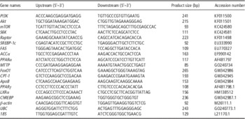
Total RNA was isolated using the Trizol reagent (TakaRa, Dalian, China) and treated with RNase-free DNase I (TakaRa) to remove contaminated genomic DNA. The first strand cDNA was synthesized using the ImProm-II Reverse Transcription System (TakaRa) according to the manufacturer’s instructions. Real-time PCR was performed with SYBR Green PCR Master Mix (TakaRa). All reactions were performed at least in duplicate. Specific primers were designed according to goose gene sequences and are listed in Table 1. The amplification specificity of the primers used has been determined by DNA sequencing in our laboratory. The relative expression ratio of target gene was calculated using the Multicolor Real-Time PCR Detection System CFX software (Bio-Rad, CA, USA) through the calibrator-normalized relative quantification method
 $${\rm 2}^{{{\minus}\Delta \Delta C_{t} }} $$
after normalization to mRNA expression levels of β-actin, UBC and 18S (housekeeping genes). For the target gene expression analysis, the normalized target gene expression level for each sample was compared with the positive control sample. The relative mRNA levels are expressed as the n-fold difference in the normalized target gene expression level between each treated and control sample. The final values were calculated by extracting the cube root of the three relative mRNA levels of each gene relative to β-actin, 18S and UBC.
Primer sequences for real-time PCR

Intracellular lipid accumulation
Hepatocytes were stained with Oil Red O to observe the amount of intracellular lipid accumulation. Cells (1×10⁶ cells/ml) were cultured with LY294002 (0, 10, 40 μmol/l), rapamycin (10, 90 nmol/l) or NVP-BEZ235 (0.5, 2 μmol/l) on six-well culture plates, fixed in 10% formaldehyde, and stained using a method described previously (Mori et al., Reference Mori, Itabe, Higashi, Fujimoto, Shiomi, Yoshizumi, Ouchi and Takano2001). Briefly, the wells were fixed with 10% formaldehyde for 1 h, rinsed with phosphate-buffered saline (PBS) for three times, and then stained with Oil Red O for 30 min. Wells were then treated with 60% propylene glycol (vol/vol) for 1 min to remove free Oil Red O, and then rinsed with PBS for three times. The Oil Red O was extracted with the addition of isopropanol and Oil Red O determined in aliquots from wells following shaking the culture plates 30 min at room temperature. Then cells were examined by phase contrast microscopy at 200× magnification.
In addition, intracellular lipid accumulation was examined using Oil red O extraction. The steps for Oil red O extraction were similar to the described above for Oil red O staining. Cells were washed with PBS for three times and fixed with 10% formaldehyde for 1 h. After washing with PBS three times, the cells were stained with 1% filtered Oil red O for 30 min. Then, Oil red O solution was removed. Intracellular lipid levels were agitatedly extracted with 100% isopropanol solution of 2000 μl for 15 min in Shaker. Finally, a hole with DMSO was used to adjust zero and optical density (OD) value of each hole was monitored by a spectrophotometerat 510 nm.
Measurement of intracellular VLDL concentration
The intracellular VLDL concentration was measured using a chicken VLDL ELISA kit (GBD, MN, USA). As described in the instructions, the microtiter plate had been pre-coated with an antibody specific to VLDL. The enzyme-substrate reaction was terminated by the addition of a sulfuric acid solution and measured spectrophotometrically at a wavelength of 450 nm. The concentration of VLDL in the samples was determined by comparing the OD value of the samples to the standard curve.
Immunoblotting analysis
Cells were lysed on ice for 3 min in RIPA buffer supplemented with 1 mmol/l phenylmethanesulfonyl fluoride and protease inhibitor cocktail (Sigma). After centrifugation at 14 000 r.p.m. for 5 min, the supernatant was harvested as the total cellular protein extract and stored at −80°C. The protein concentration was determined by using Bio-Rad protein assay reagent (Richmond, CA, USA). Equal amounts of protein (100 μg) were separated by 6% SDS-PAGE and transferred onto PVDF membrane in 20 mmol/l TrisHCl (pH 8.0), containing 150 mmol/l glycine and 20% (vol/vol) methanol. Membranes were blocked with sealing fluid and incubated with antibodies to S6K, S6K phosphorylation (P-S6K) (Bioss, Beijing, China) and ACCα (Cell Signaling Technology, MA, USA). A goat anti-rabbit horseradish peroxidase-conjugated IgG at 1 : 1000 (Santa Cruz Biotechnology, CA, USA) was used as the secondary antibody and the signals were detected using an ECL western blot detection kit (Santa Cruz Biotechnology). Then, the membranes were stripped with a Re-Blot Plus solution (Chemicon International, CA, USA). After analysis, the membranes were blotted with an anti-α-tubulin antibody (1 : 1000; Biosynthesis, Beijing, China) to normalize for protein content.
Statistical analysis
All data were expressed as mean±SD. The data were subjected to ANOVA testing and the means were assessed for significance by Tukey’s test. ANOVA and t-tests were performed using the SAS 9.13 statistical software (SAS Institute Inc., NC, USA). The differences were considered to be statistically significant as P<0.05. Every experiment was repeated with three biological samples, and each of samples was run in triplicate.
Results
Cell viability
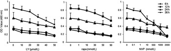
In order to determine the cell viability after inhibiting PI3K-Akt-mTOR pathway, goose hepatocytes were cultured with LY294002 (0, 10, 20, 30, 40, 50 μmol/l), rapamycin ( 0, 1, 10, 30, 50, 90 nmol/l) and NVP-BEZ235 ( 0, 0.1, 1, 10, 500, 1000, 2000 nmol/l). Cell viability was determined after culturing for 6, 12, 24, 36 h by OD measurement at 490 nm as Figure 1. The results demonstrated that the cells have lower viability with the higher concentration of LY294002, rapamycin and NVP-BEZ235, and the prolonged-time incubation increased cell viability.
Cell viability was examined by MTT assay after treatment LY294002, rapamycin or NVP-BEZ235. LY=LY294002; rapa=rapamycin; NVP=NVP-BEZ235.

The activity of PI3K-Akt-mTOR pathway
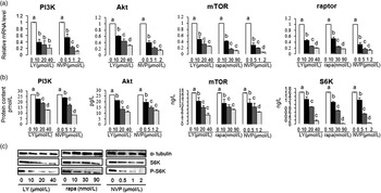
To demonstrate whether the PI3K-Akt-mTOR signaling is inhibited by LY294002, rapamycin and NVP-BEZ235 in goose hepatocytes, the gene expression of the pathway downstream substrates (PI3K, Akt, mTOR, raptor) were analyzed. As illustrated in Figure 2a, incubation with LY294002 or NVP-BEZ235 for 24 h resulted in a decrease of mRNA expression (PI3K, Akt, mTOR, raptor) in hepatocytes. Accordingly, rapamycin reduced the mRNA expression of downstream targets (mTOR and raptor) in a dose-dependent manner.
The effects of LY294002, rapamycin and NVP-BEZ235 on activation of PI3K-Akt-mTOR pathway in hepatocytes. (a) The mRNA expression (PI3K, Akt, mTOR, raptor) by Real-Time PCR, (b) protein expression (PI3K, Akt, mTOR, S6K) by ELISA kits; (c) protein expression of S6K and P-S6K by western blotting. The different letters indicated significant differences (P<0.05). LY=LY294002; rapa=rapamycin; NVP=NVP-BEZ235.

In a parallel experiment, we summarized the effects of LY294002, rapamycin and NVP-BEZ235 on enzyme activities of their downstream activators in goose primary hepatocytes (Figure 2b). The results showed that LY294002 and NVP-BEZ235 both inhibited the enzyme activities of PI3K, Akt, mTOR and S6K, while rapamycin inhibited the enzyme activities of mTOR and S6K.
To further examine whether inhibition of PI3K-Akt-mTOR pathway affects its downstream signaling functions, the protein expression of known downstream target S6K in this pathway was analyzed by western blotting. As shown in Figure 2c, expression levels of S6K and P-S6K were suppressed by LY294002. Similar experiments were conducted with rapamycin and NVP-BEZ235. The data showed that rapamycin and NVP-BEZ235 both could decrease the expression of S6K and P-S6K. These results suggested that the activity of PI3K-Akt-mTOR signaling could be inhibited by LY294002, rapamycin or NVP-BEZ235.
Intracellular lipid accumulation
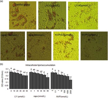
The lipid droplet deposition in hepatocytes was measured using Oil Red O staining method. As shown in Figure 3, after treatment with LY294002 (10, 40 μmol/l), rapamycin (10, 90 nmol/l) or NVP-BEZ235 (0.5, 2 μmol/l), the increase of inhibitors concentration was accompanied by the decrease of lipid droplet deposition in goose primary hepatocytes (Figure 3a). The results detected by the Oil red O extraction showed that lipid accumulation was also reduced with the increasing inhibitors concentration (Figure 3b). Above results indicated that inhibition of PI3K-Akt-mTOR pathway could decrease lipid accumulation in primary cultures of goose hepatocytes.
The effects of LY294002, rapamycin and NVP-BEZ235 on intracellular lipid accumulation in hepatocytes. (a) After Oil red O staining, intracellular lipid accumulation by Phase-contrast microscopy at a magnification of 200×. (b) After Oil red O extraction, intracellular lipid accumulation by monitoring OD value at 510 nm. The different letters indicated significant differences (P<0.05). LY=LY294002; rapa=rapamycin; NVP=NVP-BEZ235.

De novo lipid biosynthesis
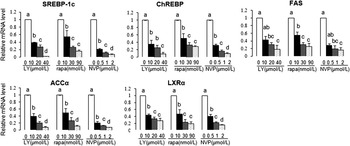
In order to determine whether the inhibition of PI3K-Akt-mTOR signaling leads to the changes of lipid biosynthesis, the relative mRNA abundances of lipogenic genes were determined. As shown in Figure 4, compared with the control group, LY294002, rapamycin or NVP-BEZ235 all decreased the mRNA abundances of Liver X receptor α (LXRα), Sterol regulatory element-binding protein 1C (SREBP-1c), Carbohydrate response element binding protein (ChREBP), ACCα and FAS.
The effects of LY294002, rapamycin and NVP-BEZ235 on mRNA expression of lipogenic genes (SREBP-1c, ChREBP, FAS, ACCα, LXRα) in hepatocytes. The different letters indicated significant differences (P<0.05). LY=LY294002; rapa=rapamycin; NVP=NVP-BEZ235.

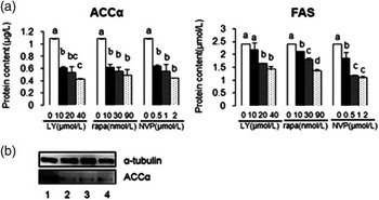
The protein concentrations of FAS and ACCα were analyzed by ELISA analysis after incubation with LY294002, rapamycin or NVP-BEZ235 (Figure 5a). It was shown that the protein contents of FAS and ACCα were inhibited by LY294002, rapamycin or NVP-BEZ235. Accordingly, the result of western blotting analysis showed that LY294002, rapamycin or NVP-BEZ235 all were able to suppress ACCα protein expression (Figure 5b). These data suggested that inhibiting PI3K-Akt-mTOR pathway decreased the mRNA expression and protein expression of genes involved in lipid biosynthesis, leading to reduction of hepatic lipids accumulation.
The effects of LY294002, rapamycin and NVP-BEZ235 on protein contents of ACCα and FAS in hepatocytes. (a) Protein contents of ACCα and FAS using ELISA kits; (b) protein contents of ACCα using western blotting. The different letters indicated significant differences (P<0.05). LY=LY294002; rapa=rapamycin; NVP=NVP-BEZ235; 1: control group, 2: 20 μmol/l LY294002, 3: 30 nmol/l rapamycin, 4 : 1 μmol/l NVP-BEZ235.

Fatty acid oxidation
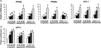
Some previous studies found that PI3K-Akt-mTOR signaling played an important role in fatty acid oxidation. To determine whether the analogous conclusion was observed in goose primary hepatocytes, cells were incubated with LY294002 (10, 20, 40 μmol/l), rapamycin (10, 30, 90 nmol/l) or NVP-BEZ235 (0.5, 1, 2 μmol/l) for 24 h. As shown in Figure 6a, the mRNA expression levels of Peroxisome proliferator activated receptor γ (PPARγ), Peroxisome proliferator activated receptor α (PPARα) and CPT-1 were enhanced along with the increase concentration of LY294002, rapamycin and NVP-BEZ235. In addition, the protein content of CPT-1 after the incubation with the PI3K-Akt-mTOR inhibitors was examined using ELISA analysis (Figure 6b). The results showed that the protein concentration of CPT-1 was elevated by LY294002, rapamycin or NVP-BEZ235 in a dose-dependent manner. The above results suggested that inhibition of PI3K-Akt-mTOR pathway increased the expression of PPARγ, PPARα and CPT-1 involved in fatty acid oxidation.
The effects of LY294002, rapamycin and NVP-BEZ235 on fatty acid oxidation in hepatocytes. (a) mRNA expression of PPARγ, PPARα and CPT-1 by Real-Time PCR; (b) protein content of CPT-1 by ELISA kits. The different letters indicated significant differences (P<0.05). LY: LY294002; rapa=rapamycin; NVP=NVP-BEZ235.

VLDL assembly and secretion
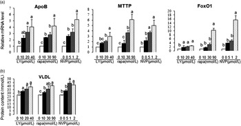
In order to examine the effects of PI3K-Akt-mTOR inhibition on the VLDL assembly and secretion, the transcriptional levels of key factors involved in VLDL assembly and secretion were measured. After the treatment with LY294002, rapamycin or NVP-BEZ235, the mRNA levels of MTTP, Forkhead box O1 (FoxO1) and ApoB showed a dose-dependent increase (Figure 7a). Accordingly, intracellular VLDL concentration was examined by ELISA analysis. As expected, the level of intracellular VLDL was also increased in a dose-dependent manner after treatment with LY294002, rapamycin or NVP-BEZ235 (Figure 7b).
The effects of LY294002, rapamycin and NVP-BEZ235 on VLDL assembly and secretion in hepatocytes. (a) mRNA expression of MTTP, FoxO1, ApoB by Real-Time PCR; (b) intracellular VLDL concentration by ELISA kits. The different letters indicated significant differences (P<0.05). LY=LY294002; rapa=rapamycin; NVP=NVP-BEZ235; VLDL=very low density lipoprotein.

Discussion
Effects of LY294002, rapamycin or NVP-BEZ235 on PI3K-Akt-mTOR signal pathway
PI3Ks is a family of related intracellular signal transducer enzyme that is capable for generating specific inositol lipids. LY294002, a potent inhibitor of PI3Ks, is being used increasingly as a pharmacologic probe to block PI3K-Akt signaling in mammalian cells (Hao et al., Reference Hao, Zhu, Li, Liu, Zhao, Liu, Xing, Feng and Duan2013). Our data showed that the mRNA level of genes involved in PI3K-Akt-mTOR pathway (PI3K, Akt, mTOR and raptor) and the protein content of their downstream substrates (PI3K, Akt, S6K and P-S6K) were inhibited significantly by LY294002. The results were entirely consistent with conclusion that LY294002 inhibited the PI3K-Akt-mTOR pathway signaling by interfering with the function of PI3K activity.
The activity of mTOR kinase was required for PI3K-Akt downstream signaling. Our results have shown that rapamycin decreased downstream targets expression of mTOR kinase (mTOR, S6K and raptor) and the protein expression of S6K and P-S6K. The results were consistent with the previous studies in primary cultures of rat hepatocytes (Wang and Proud, Reference Wang and Proud2006; Brown et al., Reference Brown, Stefanovic-Racic, Sipula and Perdomo2007). The regulation of PI3K-Akt-mTOR signaling network is a complicated process. In order to further verify the inhibition of PI3K-Akt-mTOR pathway, a PI3K/mTOR dual inhibitor NVP-BEZ235 was used to incubate cells for 24 h, and the expression levels of PI3K, Akt, mTOR, raptor and S6K were significantly decreased by NVP-BEZ235. The result suggested that NVP-BEZ235 not only blocked the activities of mTOR and the downstream target S6K, but also abolished the activation of PI3K-Akt pathway signal pathway.
Effects of inhibiting PI3K-Akt-mTOR pathway on de novo lipid synthesis
Lipid biosynthesis is essential for the maintenance of cellular homeostasis. Accumulating evidence supports the idea that PI3K-Akt-mTOR pathway stimulates the lipid synthesis via inducing the expression of SREBP, which activates the transcription of the genes FAS and ACC, the two key enzymes of fatty acid biosynthesis (Laplante and Sabatini, Reference Laplante and Sabatini2010; Hao et al., Reference Hao, Liu, Zhao, Liu, Lv, Chen, Niu and Duan2011). Our previous studies demonstrated that overfeeding-induced elevated hepatic lipogenesis, accompanied with up-regulation of gene expression associating with de novo lipid synthesis, which was the main cause of fatty liver formation in goose (Han et al., Reference Han, Wang, Li, Wang and Zhang2008). Similarly, present results demonstrated that the inhibition of PI3K-Akt-mTOR signal pathway reduced the intracellular lipid accumulation, associated with the decrease of SREBP-1c, FAS and ACCα in goose primary hepatocytes, suggesting PI3K-Akt-mTOR signal pathway plays a key role in regulating goose hepatic lipogenesis.
LXR is a strong activator of the SREBP-1c promoter (Yoshikawa et al., Reference Yoshikawa, Shimano, Amemiya-Kudo, Yahagi, Hasty, Matsuzaka, Okazaki, Tamura, Iizuka and Ohashi2001). Insulin induced the up-regulation of LXR, which led to an increase in mRNA level of LXR target genes; and LXR knockout mice suppressed insulin-mediated increase in lipogenic and cholesterogenic enzymes (Tobin et al., Reference Tobin, Ulven, Schuster, Steineger, Andresen, Gustafsson and Nebb2002). LXRα agonist T0901317 induced triglyceride accumulation through activation of SREBP-1 and its target genes in goose primary hepatocytes (Han et al., Reference Han, Wang, Xu, Li, Ye, Jiang and Zhuo2009). Whether the regulation of lipid synthesis by PI3K-Akt-mTOR pathway is linked with the LXRα is unknown. In our studies, the inhibition of PI3K-Akt-mTOR pathway by LY294002, rapamycin or NVP-BEZ235 resulted in the decrease in mRNA level of LXRα, and the inhibition effect was elevated with the increase of inhibitors concentration. Thus, the decrease of lipid synthesis induced-by inhibiting PI3K-Akt-mTOR pathway maybe via the regulation of LXRα in goose primary hepatocytes.
Liver, the main organ of lipogenesis, is responsible for the conversion of excess dietary carbohydrates into triglycerides by the de novo lipogenesis. The transcription factor ChREBP has emerged as a major mediator of glucose action in the regulation of both glycolysis and lipogenesis. It acts in synergy with SREBP to induce the expression of lipogenic genes such as ACC and FAS as the direct targets of ChREBP (Denechaud et al., Reference Denechaud, Bossard, Lobaccaro, Millatt, Staels, Girard and Postic2008; Strable and Ntambi, Reference Strable and Ntambi2010). Liver-specific inhibition of ChREBP markedly improved hepatic steatosis by specifically decreasing lipogenesis in obese ob/ob mice (Dentin et al., Reference Dentin, Benhamed, Hainault, Fauveau, Foufelle, Dyck, Girard and Postic2006). In this study, the transcription level of ChREBP-1c was significantly decreased by the inhibition of PI3K-Akt-mTOR pathway, which indicated that similar with the transcription factors SREBP-1c, ChREBP also played a very critical role in regulation of lipid synthesis mediated-by PI3K-Akt-mTOR pathway in goose hepatocytes.
Effects of inhibiting PI3K-Akt-mTOR pathway on fatty acid oxidation
Hepatic fatty acid oxidation consists of two distinct pathways, peroxisomal and mitochondrial fatty acid β-oxidation (Reddy and Hashimoto, Reference Reddy and Hashimoto2001). CPT-1 is considered to catalyze the rate-limiting step in mitochondrial fatty acid β-oxidation (Mascaró et al., Reference Mascaró, Acosta, Ortiz, Marrero, Hegardt and Haro1998). CPT-1 is a direct positive PPARα-regulated gene. The PPARα knockout mouse impaired the lipid oxidation capability and showed a massive accumulation of lipids in liver (Monsalve et al., Reference Monsalve, Pyarasani, Delgado-Lopez and Moore-Carrasco2013). Our present studies found that PI3K-Akt-mTOR pathway inhibitors LY294002, rapamycin or NVP-BEZ235 increased the mRNA expression of the transcription factors PPARα and PPARγ and their target genes CPT1 in a dose-dependent manner. PI3K-Akt-mTOR pathway was associated with the regulation of fatty acid oxidation. The activation of insulin signaling triggered a strong induction of the Akt-mTOR cascade, and then followed a decrease of fatty acid oxidation. However, the dual PI3K-mTOR inhibitor, NVP-BEZ235 effectively reverted the decrease of fatty acid oxidation both in vitro and in vivo (Evert et al., Reference Evert, Calvisi, Evert, De Murtas, Gasparetti, Mattu, Destefanis, Ladu, Zimmermann and Delogu2012). Sustained mTOR activity may contribute to the development of steatosis in the hepaRG cell line by impairing lipid homeostasis via decreasing the expression of the transcription factors PPARα and PPARδ (Parent et al., Reference Parent, Kolippakkam, Booth and Beretta2007). The treatment with rapamycin in the skeletal muscle cells increased fatty acid oxidation by increasing activities of CPT I and II both in vivo and in vitro (Sipula et al., Reference Sipula, Brown and Perdomo2006). Our studies indicated the inhibition of PI3K-Akt-mTOR pathway stimulated the fatty acid oxidation by increasing the gene expression of PPARα and PPARγ and their target genes CPT1, which led to the decrease of hepatic lipid accumulation in primary cultures of goose hepatocytes.
Effects of inhibiting PI3K-Akt-mTOR pathway on VLDL assembly and secretion
Lipid secretion and distribution is important for maintaining lipid homeostasis. ApoB and MTTP are essential for the assembly and secretion of triglycerides-rich apoB-contained VLDL lipoprotein. MTTP is necessary for the assembly of the nascent lipoprotein particles. MTTP knockout mice caused a striking reduction in VLDL assembly and secretion in the liver (Liao et al., Reference Liao, Hui, Young and Davis2003). ApoB is a key component whose synthesis rate controls the overall rate of VLDL production (Nguyen et al., Reference Nguyen, Leray, Diez, Serisier, Bloc’h, Siliart and Dumon2008). FoxO1 is a transcription factor that plays a key role in the hepatic VLDL assembly and secretion. FoxO1 stimulated the MTTP expression, elevated the ApoB secretion, and augmented the VLDL production though binding and stimulating MTTP promoter activity (Kamagate and Dong, Reference Kamagate and Dong2008a; Kamagate et al., Reference Kamagate, Qu, Perdomo, Su, Kim, Slusher, Meseck and Dong2008b). Hepatic steatosis occurs in birds when the increase of lipogenesis exceeds the rate of VLDL assembly and secretion. Overfeeding-induced liver steatosis was associated with the elevated de novo lipid synthesis, and the suppression of VLDL secretion into the blood and peripheral tissues (Han et al., Reference Han, Wang, Li, Wang and Zhang2008). The inhibition of PI3K abolished the insulin-induced the inhibition of VLDL secretion by decreasing the intracellular degradation of ApoB, and elevating the ApoB assembly and synthesis by the overactivation of PI3K-Akt-mTOR pathway, and the reduced Akt phosphorylation was accompanied by a significant increase of VLDL secretion in the fructose-fed hamster hepatocytes (Taghibiglou et al., Reference Taghibiglou, Rashid-Kolvear, Van Iderstine, Le-Tien, Fantus, Lewis and Adeli2002). The direct effect of inhibiting PI3K-Akt-mTOR pathway on the VLDL assembly and secretion in avian species have not been reported previously. Our result in goose hepatocytes showed that the inhibition of PI3K-Akt-mTOR pathway decreased the intracellular VLDL concentration, and the transcriptional levels of MTTP, ApoB and FoxO1, which indicated that inhibition of PI3K-Akt-mTOR signaling might stimulate FoxO1 over-activation, which led to the reduction of the hepatic lipid accumulation by inducing the hepatic VLDL assembly and secretion.
In conclusion, our studies demonstrated that inhibition of PI3K-Akt-mTOR pathway reduced hepatic lipid accumulation by regulating several major pathways of hepatic lipid metabolism, including decreasing the lipid synthesis, enhancing the fatty acid oxidation, and increasing the VLDL assembly and secretion in goose hepatocytes.
Acknowledgments
The work was supported by the National Natural Science Funds of China (No. 31101712), and the Research Fund for the Doctoral Program of Higher Education of China (No. 20115103120006).