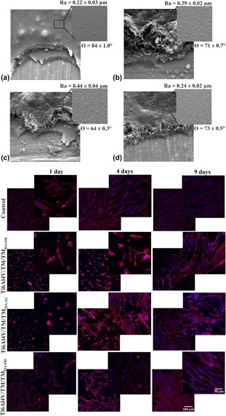
Introduction
With the evolution of regenerative medicine toward the development of new biomaterials that aim at the regeneration of tissues, some researchers have focused their work on the introduction of cells into structures that can serve as support for cell proliferation. The architecture of this biomaterial defines the shape of the new tissues to be formed [Reference Goonoo and Luximon1].
The materials to be used for tissue engineering can be considered as a matrix for the growth of cells, tissues, and organs and thus need to provide both the physical/chemical and biological support for cell adhesion, proliferation, migration, and differentiation [Reference Liao, Chan and Ramakrishna2, Reference Park, Karajanagi, Wolak, Aanestad, Daheron, Kobler, Guerra, Heaton, Langer and Zeitels3]. According to Boudriot et al. [Reference Boudriot, Bernhard, Roland, Andreas and Joachim4], there are two ways to maximize cell growth in tissue engineering: by direct mixing the components in the carrier used as an extracellular matrix, or its coating, and by the addition of cell cultures during its development.
Within the context of cell cultures, the introduction of stem cells has become the extreme importance for tissue engineering due to the possibility of modulating the immune system and avoiding allogeneic rejection [Reference Wang, Thissen and Kingshott5]. By definition, stem cells are immature cells capable of self-regenerating, which, when properly stimulated, are able to migrate to other regions where they give rise to other cell types, being able to replace or regenerate the tissues in a living organism [Reference Preston, Alison, Forbes, Direkze, Poulsom and Wright6]. Mesenchymal stem cells (MSCs) are considered multipotent progenitors, capable of differentiating into three cell lines (osteogenic, adipogenic, and chondrogenic) or more (myogenic, cardiomyogenic, etc.) [Reference Miao, Jin, Zhun, Huang, Qian and Zhang7, Reference Bernardi, Luisi, Fernandes, Dalberto, Valentim, Bogo Chies, Fossati and Pranke8]. Due to this ability, MSCs are widely used in in vitro or in vivo studies [Reference Romanov, Svintsitskaya and Smirnov9, Reference Caplan10].
In regard to the approach that considers directly mixing components, there are some publications in the literature suggesting the use of bioactive particles to favor the osteogenic differentiation of stem cells, facilitating the osseointegration process. Dimitrievska et al. [Reference Dimitrievska, Bureau, Antoniou, Mwale, Petit, Lima and Marple11] obtained higher levels of bone mineralization from the osteogenic differentiation of MSCs on TiO2 surfaces and hydroxyapatite (HA) nanoparticles to Ti6Al4V substrates in a dexamethasone-free culture medium. Sanaei-Rad et al. [Reference Sanaei-Rad, Kashi, Seyedjafari and Soleimani12] observed higher rates for osteogenic differentiation of adipose tissue-derived stem cells on scaffolds of poly(lactic acid-co-glycolic acid (PLGA) polymer when incorporating HA in them). Tae Young et al. [Reference Young, Kang, Venkatesan, Chang, Bhatnagar, Chang, Salameh, Kim and Kim13] obtained similar percentages of the alkaline phosphatase marker and of the proteins (osteocalcin and osteopontin), when promoting the osteogenic differentiation of adipose tissue on HA and HA together with the polymer fucoidan.
One of the ways to incorporate these particles into the biomaterials is through the use of hybrid coatings prepared by the sol–gel method [Reference Owens, Singh, Foroutan, Alqaysi, Han, Mahapatra, Kim and Knowles14]. Among these, the successful development of hybrid coatings with silicon-based precursors (silanes) [Reference Ballarre, Seltzer, Mendoza, Orellano, Mai, García and Ceré15, Reference Rodríguez-Cano, Cintas, Fernández-Calderón, Pacha-Olivenza, Crespo, González-Martín and Babiano16, Reference Zomorodian, Brusciotti, Fernandes, Carmezim, Silva, Fernandes and Montenor17, Reference Liu, Zhan, Yu and Li18] stands out. Chen and Ko (2013) obtained a superior osteogenic differentiation behavior of MSCs derived from bone marrow on the coating of the precursor silane methacryloxypropyltrimethoxysilane (MPTMS) compared to titanium substrates, aiming the application for dental implants. In addition, Martínez-Ibáñez et al. [Reference Martínez-Ibáñez, Juan-Díaz, Lara-Saez, Coso, Franco, Gurruchaga, Suay and Goñi19] have shown that the degradation of these silane coatings leading to the release of silicon may have a positive influence on the osteogenic differentiation of MSCs.
Finally, it is known that for the subsequent implantation of the biomaterial, it is necessary to guarantee its condition of sterilization. The sterilization process is an important issue for the use of biomaterials in the biological environment, since it eliminates microorganisms from the material, thus minimizing the incidence of infections and preventing the transmission of diseases [Reference Park, Navarrete, Baier, Meyer, Tannenbaum, Boyan and Schwartz20, Reference Galante, Ghisleni, Paradiso, Alves, Pinto, Colaço and Serro21]. In literature, authors have demonstrated that the chemical, mechanical, and, consequently, biological properties of biomaterials are susceptible to changes according to the sterilization process used [Reference Park, Navarrete, Baier, Meyer, Tannenbaum, Boyan and Schwartz20, Reference Costa, Lopes, Tipple, Castillo, Hu, Deva and Vickery22]. In the case of metal implants, this fact directly affects the osseointegration process [Reference Shi, Xu, Violin and Lu23]. Recently, Antonini et al. [Reference Antonini, Malfatti, Reilly, Owen and Takimi24] demonstrated that autoclaving sterilization in addition to altering the morphology of nanotexturized Ti6Al4V implants compromised the viability of hESC-MP (human embryonic stem cell-derived mesenchymal progenitor) cells—behavior opposed to that observed for the air plasma process.
The objective of the present work was to evaluate the effect of the sterilization process (steam autoclave, hydrogen peroxide plasma, and ethylene oxide) on the osteogenic differentiation of MSCs on a hybrid coating on a Ti6Al4V substrate. The hybrid coating was obtained from the silane precursors tetraethoxysilane (TEOS) and methyltriethoxysilane (MTES) with HA bioactive particles.
It is worth noting that the incorporation of bioactive particles may impair the barrier effect of hybrid silane coatings, as has been reported in the literature [Reference Owens, Singh, Foroutan, Alqaysi, Han, Mahapatra, Kim and Knowles14, Reference Heise, Wirth, Hohlinger, Hernandez, Ortiz, Wagener, Virtanen and Boccaccini25], because their dispersion may interfere with the uniformity of the formed layer, impairing the resistance behavior to corrosion. Therefore, to associate corrosion protection and bioactive properties with the proposed architecture for the studied coating, in the present work we opted for the application of a bilayer coating composed of TEOS/MTES/HA on TEOS/MTES.
Results and discussion
Characterization of hybrid coating before and after sterilization
It was observed that the sterilization processes modified the morphology of the hybrid coating in relation to the nonsterilized sample, presenting an increase in surface topographical irregularity (Fig. 1). In addition, it was observed that the active coating layer showed expansion and increased porosity after the sterilization process. However, the protective coating layer was morphologically unchanged, and it is important to note that it did not contain bioactive particles and was cured at 450 °C. Likewise, the sterilization processes used did not compromise the cohesive behavior between the protective layer and the bioactive layer of the coatings. This behavior was observed in previous complementary studies, in which it was observed that the sterilization processes studied modified the surface and electrochemical properties of the coating formed by TEOS/MTES [Reference Baldin, Garcia, Henriques, Ely, Birriel, Brandalise and Malfatti26]; and on the dispersion of HA particles therein, the morphology of the sterilized coatings was further altered compared to the nonsterilized [Reference Baldin, Malfatti, Rodói and Brandalise27].
SEM micrographs of the nonsterilized hybrid coating and sterilized by different processes: (a) Ti6Al4V/TM/TMHA, (b) Ti6Al4V/TM/TMHA/OE, (c) Ti6Al4V/TM/TMHA/AC, and (d) Ti6Al4V/TM/TMHA/PH. (R a = mean micrometer roughness and θ = contact angle value obtained in the solution wettability test (DMEM) by the sessile drop method).

The surface roughness observed by SEM/FEG was also consistent with the increase in the average roughness of the sterilized coatings, especially for the samples sterilized by ethylene oxide (Ti6Al4V/TM/TMHA/OE) and by autoclave (Ti6Al4V/TM/TMHA/AC) compared to nonsterilized ones. Sterilization processes increased wettability (which is observed by the decrease in the drop contact angle of Dulbecco’s modified Eagle’s medium (DMEM) with the bioactive coating) of the sterilized bioactive coatings, accentuating their hydrophilic properties, as authors have reported [Reference Wang, Chen, Wang and Gu28]. Similar results have already been published by Wittenburg et al. [Reference Wittenburg, Lauer, Oswald, Labudde and Franz29] and Han et al. [Reference Han, Tsoi, Matinlinna, Zhang and Chen30] for glass and zirconia surfaces sterilized by similar processes.
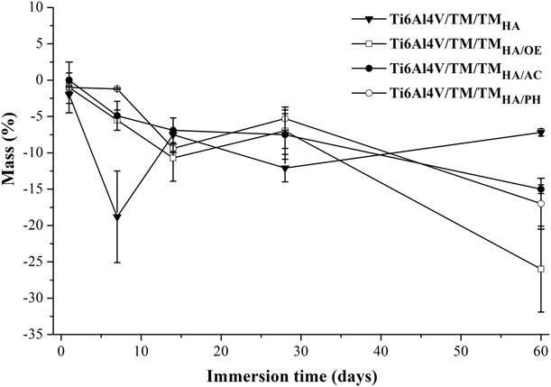
The degradation of the sterilized bioactive coatings in DMEM solution was evidenced by the increase in mass loss as a function of immersion time (Fig. 2). For the nonsterilized and autoclaved coating, the highest degradation was observed in 7 and 28 days. On the other hand, for samples sterilized in ethylene oxide and hydrogen peroxide plasma, this was observed after 14 and 60 days of immersion. The degradation of these samples began to be slower than the other samples. Nevertheless, after 60 days, due to the progressive mass loss, these samples had a higher percentage of mass loss. In a previous study, the evaluation of the electrochemical properties of the coating composed of TEOS/MTES and HA before and after the sterilization processes showed the degradation of the same after 672 h of immersion; such behavior can be observed in Fig. 2 by Baldin et al. [Reference Baldin, Malfatti, Rodói and Brandalise27].
Variation of mass as a function of time of the nonsterilized bioactive coating and sterilized by the different processes, immersed by 1, 7, 14, 28, and 60 days in DMEM solution.

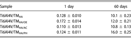
The hydrolytic degradation behavior of the bioactive coatings may be related to their wettability after sterilization. All the sterilized samples showed increased wettability in DMEM solution, and this may have facilitated the breakup of the Si–O–Si bonds by hydrolysis, thus contributing to a greater dissolution of the coating over time, which is evidenced by the increased concentration of silicon in solution as shown in Table I. Romero-Gavilan et al. [Reference Romero-Gavilan, Silva, Cañads, Palla, Izquierdo, Gurruchaga, Goñi and Suay31] evaluated the hydrolytic degradation of silane-based coatings with different percentages of TEOS and concluded that the degradation is favored when water retention occurs in them, allowing a greater release of Si compounds, which, on the other hand, contributed to bone regeneration in the study performed by these authors.
Direct determination of Si (mg/L) by HR-CS GF AAS.

The concentrations of silicon in solution at the end of the immersion test show that the sterilized sample of hydrogen peroxide plasma showed the highest concentration of silicon in solution, followed by ethylene oxide and autoclave. According to Zhai et al. [Reference Zhai, Lu, Wu, Chen, Lin, Naoki, Chen and Chang32], the release of silicon occurs in the form of Si(OH)3O− when in aqueous medium, pH below 9, by the hydrolysis reaction. The silicon migration results corroborate the results obtained for the hydrolytic degradation of coatings (Fig. 2), as Ti6Al4V/TM/TMHA/OE and Ti6Al4V/TM/TMHA/PH samples had the highest percentages of mass loss and the highest concentration of silicon in solution after 60 days of immersion in DMEM solution. Juan-Díaz et al. [Reference Juan-Díaz, Ibánez, Sáez, Izquierdo, Gurruchaga, Goñi and Suay33] analyzed the hydrolytic degradation in water of silane coatings, measuring the release of silicon by the ICP-MS technique and obtained a concentration close to 10 ppm in solution. These results are consistent with those found in the present work. The authors also detected the migration of silicon from the in vivo biodegradation of these coatings and verified that the presence of Si in solution after 1 and 2 weeks positively influenced the regenerations of the cortical and trabecular bones.
Behavior of MSCs on sterilized hybrid coatings
Characterization of MSCs
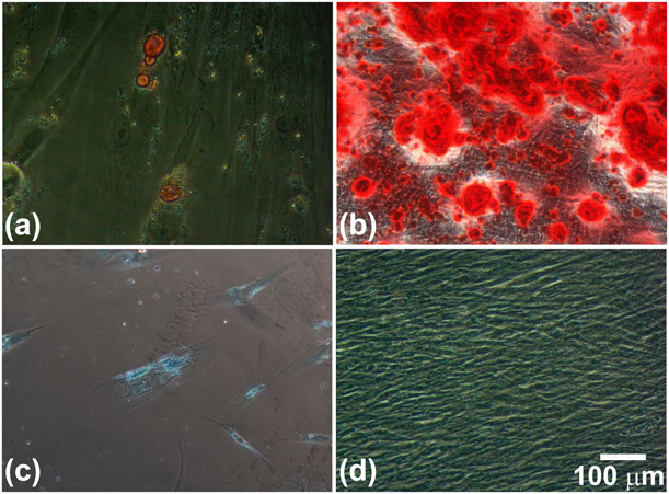
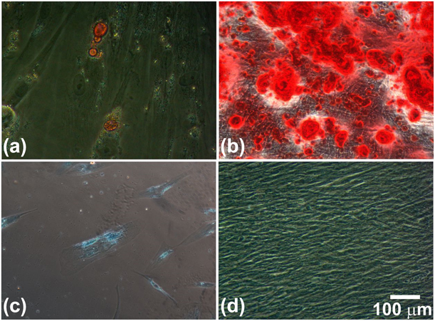
Figure 3 shows the characterization of MSCs after adipogenic, osteogenic, and chondrogenic differentiation. The immunophenotypic analysis of the 3 primary cultures showed highly positive results for the MSC markers, which is a contrary behavior observed for hematopoietic cell markers that showed low expressiveness (Table II).
Cell differentiation images: (a) adipogenic, (b) osteogenic, (c) chondrogenic, and (d) control cells not submitted to differentiation protocols.
Mean of immunophenotypic expression (n = 3) of mesenchymal and nonmesenchymal cell markers.

Cytotoxicity, adhesion, and viability
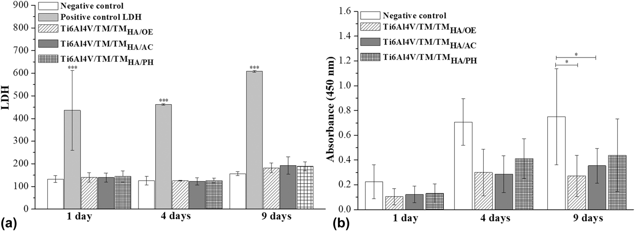
Figure 4(a) shows the cytotoxic potential of the sterilized coatings to the MSCs, when the supernatant solution in which they were immersed for the times of 1, 4, and 9 days was analyzed.
(a) Cytotoxicity of the bioactive coating sterilized by different processes measured by the dosing of LDH in the supernatant after 1, 4, and 9 days of exposure of the MSCs on the coatings and (b) viability of MSCs after 1, 4, and 9 days of exposure on the bioactive coating sterilized by the different processes. (* corresponds to the statistically significant differences using the ANOVA test P ≤ 0.05, and *** corresponds to the statistically significant differences using ANOVA test P ≤ 0.001).

The results obtained for the coatings and the negative control show that there was no significant difference between them. However, when compared to the positive control, considered as the maximum release of the enzyme lactate dehydrogenase (LDH) in the culture medium (cell death control wells), a significant difference was noticed, evidencing the nontoxic behavior of the coatings for trunk mesenchymal cells.
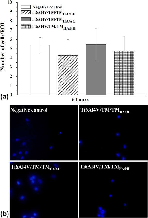
Figure 5(a) shows the results obtained in the MSC adhesion test on sterilized hybrid coatings after 6 h. Considering the amount of cells adhered to the coatings, the results were statistically similar to each other and to the control group. Likewise, by the analysis of the images presented in Fig. 5(b) (cell nuclei stained with DAPI), it was possible to observe similar adhesion behaviors between the samples, in which the adhered cells were shown to be distributed on all surfaces analyzed, regardless of the sterilization process.
(a) Adhesion behavior of the MSCs on the bioactive coating sterilized with the different processes and (b) MSCs stained with DAPI visualized in inverted light microscopy with a magnification of 400×.
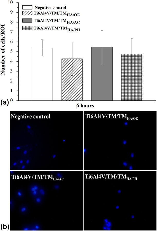
Figure 4(b) shows the cell viability events of the bioactive coatings sterilized by the three different procedures, treated with the WST-8 reagent after 1, 4, and 9 days.
Compared to the control sample, the gold standard of stem cell culture, the coated samples presented lower values of cell viability, except for Ti6Al4V/TM/TMHA/PH that presented a higher viability in relation to the other sterilized samples and did not present any statistical differences from the control sample. For all of the analyzed samples, the cell proliferation was identified on the same ones by the increase in the cell viability as a function of the time, being this more significant for the sterilized samples in autoclave and plasma of peroxide of hydrogen. In addition, despite the small cell viability differences observed between the sterilization methods, the hydrogen peroxide plasma-sterilized sample showed the highest viability after 4 and 9 days.
In the literature, some authors recommend the wettability parameter as a key factor for adhesion and cell viability, and most of these references analyze the use of surfaces with greater wettability to increase the behaviors previously mentioned [Reference Huang, Elklooly, Liu, Zhang, Yang, Shen and Feng34, Reference Hirano, Kozuka, Asano, Kakuchi, Arai and Ohtsu35, Reference Junkar, Kulkarni, Drasler, Rugelj, Mazare, Flasker, Drobne, Humpolicek, Resnik, Schmuki, Mozetic and Iglic36]. However, in this work, the inverse behavior is observed, as the sample Ti6Al4V/TM/TMHA/PH presents a higher contact angle value than the others. A similar result was already obtained in previous studies carried out by Baldin et al. [Reference Baldin, Garcia, Henriques, Ely, Birriel, Brandalise and Malfatti26] related to the biological study of MG-63 cells in protective silane coatings.
In the present study, a direct relationship between the surface roughness of the coatings after sterilization and the cellular viability behavior was not observed, conversely to some studies [Reference Likibi, Jiang and Li37, Reference Qian, Ross, Jia, Xing and Zhao38, Reference Jaidev and Chatterrjee39]. In this same sense, Chen et al. [Reference Chen, Ko, Kuo, Lin, Wu, Yang, Lou and Lin40] evaluated the adhesion, proliferation, and differentiation behaviors of MSCs on substrates of titanium grade 2, submitted to different processes. The authors observed, in the early stages of cell culture, that the influence of the chemical modification of the surfaces by the deposition of silane coatings prevailed over the influence of surface roughness.
The lower behavior regarding cell viability presented by the sterilized sample with ethylene oxide (Ti6Al4V/TM/TMHA/OE) after 9 days may be related to the retention of sterilization products, which, when released as a function of time, promote damages as already reported by some authors (Matuska and Mcfetridge, 2015) [Reference Matuska and Mcfetridge41].
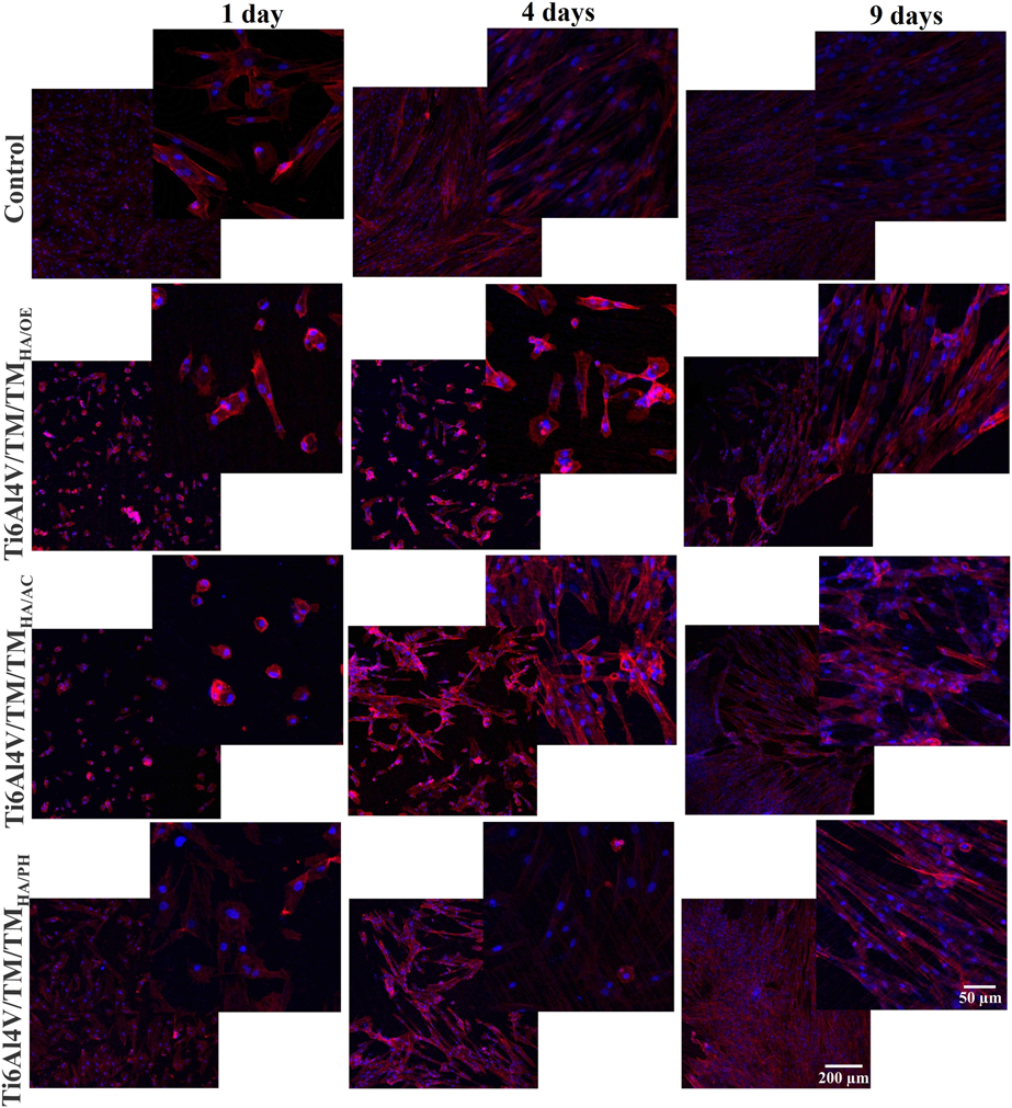
The image obtained in confocal microscopy, of the morphology of the MSCs on the bioactive coatings sterilized by the different processes after 1, 4, and 9 days (Fig. 6), is consistent with the adhesion and cell viability behaviors. After 1 day, a higher distribution of the cellular nuclei (labeled in blue) was observed in the sample sterilized by hydrogen peroxide plasma, compared to samples sterilized by autoclave or by ethylene oxide. In addition, after 9 days, the MSCs extended on the surfaces containing the bioactive coatings independently of the sterilization process. Increased cell density was observed with cytoplasmic extensions elongation, denoting the cytocompatibility of the proposed coating. The predominance of such behavior was more evident for the Ti6Al4V/TM/TMHA/PH sample.
Morphology of the stem cells on the bioactive coating sterilized by the different processes after 1, 4, and 9 days obtained by confocal microscopy.
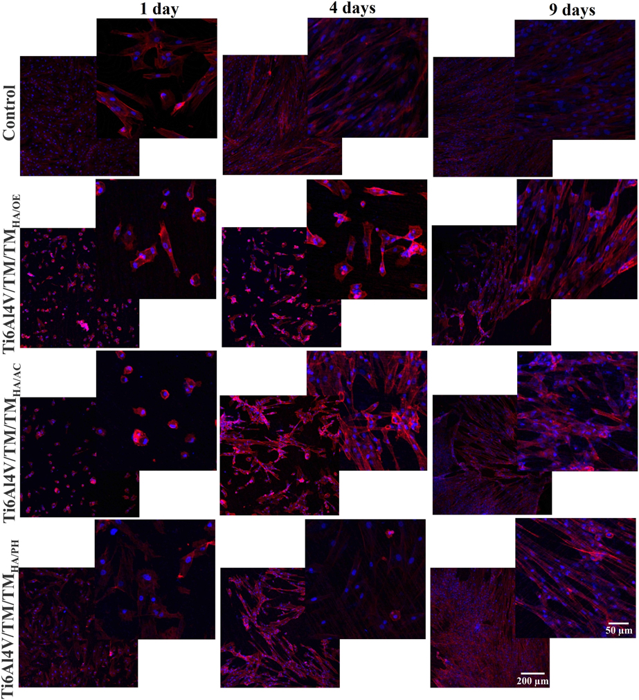
For the Ti6Al4V/TM/TMHA/OE sample, the contraction of the extracellular material was observed, with the cells presenting in a more rounded form until the 4th day of the test. This behavior is in accordance with the previously observed results for cell viability and is an evidence of the effects caused by residual residues from the ethylene oxide sterilization process as previously mentioned.
Osteogenic differentiation
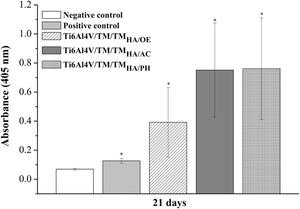
Figure 7 shows the results obtained for bone matrix mineralization through differentiated stem cells after 21 days of assay by using the Alizarin Red S marker. According to Rogina et al. [Reference Rogina, Antunovic, Pribolsan, Mihalic, Vukasovic, Ivkovic, Marijanovic, Ferrer, Ivankovic and Ivankovic42], extracellular mineralization is considered one of the main indicators for osteogenic differentiation, which is determined by the indirect measurement of the presence of calcium salts.
Bone mineralization indirectly quantified by the deposition of calcium resulting from the osteogenic differentiation of the MSCs in contact for 21 days with the bioactive coating sterilized by different processes. (* corresponds to the statistically significant differences using the ANOVA test P ≤ 0.05).

Figure 7 shows that the sterilized samples presented higher levels of calcium deposition when compared to the positive and negative controls, evidencing the promising behavior of osteogenic differentiation provided by the presence of the coating, a similar behavior to that found by Chen and Ko [Reference Chen and KO43]. Among the sterilized samples, in spite of not detecting the statistical difference (P ≤ 0.05), the mineralization was of a higher intensity for samples sterilized by hydrogen peroxide plasma and autoclave than ethylene oxide.
Curran, Chen, and John [Reference Curran, Chen and John44], and Phillips et al. [Reference Phillips, Petrie, Creighton and Garcia45] evaluated the influence of different functional groups present in silane compounds on the osteogenic differentiation behavior of stem cells. In both studies, the authors verified higher rates of the osteocalcin protein in groups of hydrophobic to hydrophilic character, thus favoring osteogenic differentiation.
Kenry et al. [Reference Kenry, Lee, Loh and Lim46] have observed that SiO2 coatings may interfere with stem cell osteogenesis even at surface parameters. Thus, the behavior exhibited by Ti6Al4V/TM/TMHA/PH may be related to the increased hydrolytic degradation with the silicon release observed for this dip in DMEM solution immersion. Juan-Díaz et al. [Reference Juan-Díaz, Ibánez, Sáez, Izquierdo, Gurruchaga, Goñi and Suay33] related the highest levels of mineralization due to the differentiation of stem cells, derived from adipose tissue, with the highest percentages of silicon release in silane coatings. Shie et al. [Reference Shie, Ding and Chang47] evaluated the behavior of osteoblastic cells as a function of the release of silicon from the precursor TEOS with calcium nitrate and observed the existence of a limiting concentration of silicon released in DMEM solution (4 mM), which favored cell viability and consequently promoted a greater mineralization (presence of calcium salts) and being this outdated, the cells tended to apoptosis.
Despite the similar degradation behavior of the Ti6Al4V/TM/TMHA/PH and Ti6Al4V/TM/TMHA/OE samples, similar to that observed for cell viability, the release of residues generated during the ethylene oxide sterilization process may have negatively affected the osteogenic differentiation of this sample in relation to the others.
For autoclave-sterilized coating, the faster degradation in DMEM solution relative to other samples may have promoted an increased exposure of the HA particles to the cell medium. Rogina et al. [Reference Rogina, Antunovic, Pribolsan, Mihalic, Vukasovic, Ivkovic, Marijanovic, Ferrer, Ivankovic and Ivankovic42] evaluated the behavior of MSCs on chitosan scaffolds with different percentages of HA and found that high concentrations of the same (50% addition) may lead to a reduction in mineralization by calcium deposits. Maeno et al. [Reference Maeno, Niki, Matsumoto, Morioka, Yatabe, Funayama, Toyama, Taguchi and Tanaka48] demonstrated the influence of the concentration of calcium ions on the behavior of osteoblastic cells, in which the presence of indices higher than 30 mM can promote the reduction in cellular proliferation as well as the markers indicative of the mineralization event.
Conclusion
The influence of sterilization on the double-layer hybrid coating, on proteic and bioactive properties, and consequently on the osteogenic differentiation behavior of MSCs was evaluated, and the results obtained can be summarized as follows.
The study of hydrolytic degradation of coatings made it possible to predict the effect of sterilization on their stability in DMEM solution. After 60 days of immersion, the sterilizations in ethylene oxide and hydrogen peroxide plasma were the ones that caused the highest percentages of degradation of the coating and, in proportion to this, they presented the highest concentrations of silicon in the solution.
Likewise, the sterilized autoclave coating presented the highest instability of the siloxane network in a short period of time, in contrast to the hydrogen peroxide plasma sterilization, which presented a progressive hydrolytic degradation behavior and, after 60 days, provided the highest release of silicon to the DMEM solution.
Hybrid coatings, independently of the sterilization process, did not present a cytotoxic behavior to the MSCs. In addition, none of the sterilization methods caused changes in the cell adhesion results, as well as in the distribution of the MSCs on the coatings.
Regarding the results of cellular viability, regardless of the sterilization process used, there was an increase in cell proliferation as a function of time. However, after 9 days of analysis, sterilized coatings of hydrogen peroxide plasma and ethylene oxide showed the highest and lowest viability indices, respectively.
The results obtained for the osteogenic differentiation reproduced the results obtained in cellular viability, evidencing once again the positive effect of the sterilization process by hydrogen peroxide plasma. This result may have been favored by the progressive degradation of this coating along with the greater silicon release from the same to the medium. In addition, the lower cell viability index and the contraction of the extracellular material, observed by confocal microscopy for samples sterilized by ethylene oxide, show the negative effects caused by the remaining residues of this process.
Materials and methods
Hybrid coating preparation and sterilization
The bioactive coating was applied over the protective coating, which was elaborated according to the procedure presented in previous works [Reference Baldin, Garcia, Henriques, Ely, Birriel, Brandalise and Malfatti26, Reference Baldin, Malfatti, Rodói and Brandalise27]. The silane precursors were added in a mixture of ethyl alcohol and acetic acid (Neon, 98%), considering the ratio in silane--water ratios (1:2), water--ethyl alcohol (1:1), and water--acetic acid (7:1) [Reference Ballarre, Seltzer, Mendoza, Orellano, Mai, García and Ceré15, Reference Ballarre, Manjubala, Schreiner, Orellano, Fratzl and Ceré49, Reference Ballarre, López, Schreiner, Durán and Ceré50]. For the preparation of the bioactive coating, a sol made up of the silane precursors alkoxysilane Tetraethoxysilane (TEOS) and organoalkoxysilane methyltriethoxysilane. Nitric acid (Neon, 66.2%, Suzano, Brazil) was added to adjust the pH medium (pH ≈ 2). The solution was maintained for 24 h in hydrolysis, and thereafter, the hydroxyapatite particles (Sigma-Aldrich 90%, St. Louis, Missouri) were added in the proportion of 10% (mass) in a solution of TEOS/MTES hydrolyzed precursors for 24 h [Reference Ballarre, Seltzer, Mendoza, Orellano, Mai, García and Ceré15, Reference Omar, Repp, Desimone, Weinkamer, Wagermaier, Cere and Ballarre51].
The homogenization of the solution containing HA particles was performed in a high shear mixer, Model 550 (Sonics, Newtown, Connecticut), with an amplitude of 40% for 6 min. Subsequently, the solution was kept agitated for another 24 h until the deposition of the bioactive coating on a metallic substrate of Ti6Al4V, already coated with a protective coating of same composition, obtained according to the methodology suggested by Baldin et al. [Reference Baldin, Garcia, Henriques, Ely, Birriel, Brandalise and Malfatti26].
The deposition of the bioactive coating was done by the dip coating technique (Marconi, Piracicaba, Brazil), with the application of a single layer of sol, with entry and withdrawal velocity of the solution of 18 cm/min and immersion time of 5 min [Reference Ballarre, López, Schreiner, Durán and Ceré50]. The curing step was then performed in a furnace (Sanchis, Porto Alegre, Brazil) at 60 °C for 1 h, with a heating rate of 10 °C/min.
The hybrid coating composed of a protective and a bioactive layer was sterilized by three different processes: ethylene oxide, steam autoclave, and hydrogen peroxide plasma. The autoclave sterilization of saturated steam and ethylene oxide was carried out at the company Esterilizare in the city of Caxias do Sul/RS/Brazil under the following conditions: autoclave (Baumer, São Paulo, Brazil) with a capacity of 0.56 m3, at 134 °C, and pressure of 0.70 atm for 7 min of exposure; and ethylene oxide at a pressure of 0.65 atm, temperature of 55 °C, and time of 180 min with gas (Chemogas, Grimbergen, Belgium) composed of 90% ethylene oxide and 10% carbon dioxide. The sterilization of the hydrogen peroxide plasma was carried out at the Sterilized Materials Center (CME) of the Pompéia Hospital, located in Caxias do Sul, Brazil, by using the Sterrad NX (Johnson and Johnson Company, Irvine, California) sterilized for 28 min, and the temperature during the sterilization cycle between 45 and 55 °C.
Hybrid coating characterization before and after sterilization
The morphology of the coatings before and after sterilization was observed in top view using the field emission gun scanning electron microscopy (SEM/FEG), brand Tescan-Model Mira3, in the Material Characterization Laboratory at UCS.
The micrometer roughness was analyzed in a contact rugosimeter, Surftest model SJ-301 (Mitutoyo, Kanagawa, Japan), with a cutoff of 0.08 mm.
The contact angle measurements to determine the wettability of the hybrid coating before and after the sterilization processes were performed by the sessile drop method from an apparatus developed by the Corrosion Research Laboratory (LAPEC) of the Federal University of Rio Grande do Sul (Porto Alegre, Brazil), in which a drop of the liquid is deposited on the surface by means of a micropipette. Gout was observed by using a low-magnification lens, and the contact angle was measured by using a goniometer. Contact angle measurements were performed with Sigma-Aldrich® Dulbecco’s modified Eagle’s medium (DMEM) culture medium solution.
The hydrolytic degradation behavior of the hybrid coating before and after the sterilization processes was evaluated by performing the indirect measurement of the mass variation, by the adaptation of ISO 10993-13:2010; in which samples were immersed in a DMEM solution (Dulbecco’s modified Eagle’s medium) supplemented with 10% fetal bovine serum (FBS) and 1% penicillin/streptomycin (P/S) at a temperature of approximately 37 °C and removed at different times (1, 7, 14, 28, and 60 days) [Reference Martínez-Ibáñez, Juan-Díaz, Lara-Saez, Coso, Franco, Gurruchaga, Suay and Goñi19]. After completing each of the times mentioned, the samples were washed with deionized water and dried in an oven at 37 °C for 48 h. Subsequently, the samples had their mass recorded, and the percentage of mass variation was calculated.
The quantification of the silicon released during the degradation of the hybrid coating was performed by the high-resolution continuum source graphite furnace atomic absorption spectrometry technique (HR-CS GF AAS) absorption spectrometry. The atomic absorption spectrometer model contrAA 700 (Analytik Jena, Jena, Germany), from the Federal University of Rio Grande (FURG), was operated with graphite tubes coated with pyrolytic graphite and with an integrated platform. As the radiation source, a xenon arc lamp (Analytik Jena) was used, which allows the emission of radiation within the spectral range from 185 to 900 nm. As for the optical system of the spectrometer, a high-resolution monochromator (Analytik Jena) composed of a prism for preseparation of spectral orders and a diffraction grid of the Echelle type was used.
The monoelementary Si reference solutions used for calibration and for the recovery assay were prepared from stock solutions of 1000 mg/L (Assurance) in HNO3, 5% (v/v) nitric acid and hydrofluoric acid, HF 1% (v/v).
The graphite tubes and platforms were thermally conditioned. The samples and the silicon reference solutions were introduced (5 μL) along with 5 μL of the chemical modifier (Pd 1000 mg/L in 10% HNO3 v/v). The determinations were made directly (without a previous sample decomposition step) using the main Si resonance line at 251.6110 nm, centered on the central pixel (101 ± 1). Si, an external calibration curve of 100–750 μg/L (coefficient of determination of 0.9977), was constructed. For the samples with the highest concentration of Si (60 days), dilutions were made necessary for the adequacy of the readings within the concentration range used for the calibration curve.
Effect of the sterilization process of the bioactive coating on the osteogenic differentiation of MSCs
Obtaining the MSCs
Primary teeth were obtained from the School of Dentistry of UFRGS (Ethics Committee CAAE No. 36403514.6.0000.5347), which were then sent to the Laboratory of Stem Cells of the Faculty of Pharmacy for removal of the pulp from the teeth, extraction of MSCs, and continuity of experiments.
The isolation and manipulation of the pulp tissue of the dental tissue were performed according to Bernardi et al. [Reference Bernardi, Luisi, Fernandes, Dalberto, Valentim, Bogo Chies, Fossati and Pranke8]. Briefly, the pulp of the teeth was maintained in contact with a 0.2% collagenase solution (Gibco, Waltham, Massachusetts) for 30 min and then, after centrifugation at 800 g for 10 min, the resulting cell suspension was seeded into 1 well of a 12-well culture dish in Dulbecco’s modified Eagle’s medium (Sigma-Aldrich) culture medium containing 10% FBS and 1% penicillin/streptomycin (Gibco).
The culture was maintained in a humidified incubator at 5% CO2 and 37 °C for 72 h, when the nonadherent cells were removed by medium exchange. Subsequent exchanges were performed every 3–4 days. After subconfluence, the primary culture was washed once with Hank’s saline solution (HBSS) free of Ca+2 and Mg+2. A 0.25% solution of trypsin in HBSS was used, and the cells were incubated for 10 min at 37 °C. The cells were resuspended in culture medium and plated onto new cell culture bottles. Subsequent passages were performed in a similar way. The passages (subcultures) were carried out when the crop reached a confluence of at least 90%.
Characterization of MSCs
The characterization of MSCs was performed by the morphological analysis of cell cultures, by cell differentiation and by immunophenotyping.
The morphology and confluence of MSCs were routinely observed in the Olympus Inverted Phase Contrast Microscope. Immunophenotypic characterization was performed with the MSCs in the fifth passage of the cell culture. The antibodies used were those specific to identifying MSC markers, such as CD73, CD90, CD105, as well as for confirming the absence of hematopoietic stem cell markers, such as CD34, CD45, and HLA-DR. The presence of the molecules was assessed using 2 μL of antihuman monoclonal antibody (Pharmingen, Becton Dickinson, San Jose, CA) for each of the molecules. 7AA (7-Amino Actinomycin D) (Molecular Probes, Eugene, Oregon) was used to evaluate only living cells. Subsequently, the cells were analyzed on a FACSAria III flow cytometer (BD), equipped with an argon laser at 488 nm. The equipment was adjusted for the conditions of analysis of cell size and complexity, and the fluorescence adjustment was performed with the aid of PE (phycoerythrin–phycoerythrin), FITC (fluorescein isothiocyanate fluorescein), and APC (allophycocyanin–allophycocyanin). Data acquisition was performed using around 10,000 events of each sample, and data analysis was performed by using the software FACSDiva version 6.0 (BD Biosciences, São José, Califórnia).
The cell differentiation step was performed by following the protocols described by Meirelles et al. [Reference Meirelles, Chagastelles and Nardi52]. In vitro cell differentiation in mesenchymal cell types, necessary to confirm the identity of the population obtained, was tested from the fifth passage onward.
Osteogenic differentiation was performed by culturing for 21 days in medium containing 10% FBS, dexamethasone (10−8 M), ascorbic acid 2-phosphate (5 μg/mL), and β-glycerophosphate (10 mM). The deposition of the mineralized matrix was observed by staining with Alizarin Red S at pH 4.2. For adipogenic differentiation of MSCs, cell culture was performed for 3–4 weeks in IMDM medium containing 20% human plasma, dexamethasone (10−7 M), insulin (2.5 μg/mL), indomethacin (5 μM), rosiglitazone (5 μM), and sodium heparin (10 units/mL). Adipocytes were discerned by observation under phase contrast microscopy and/or by staining with the Oil Red O dye. For the chondrogenic differentiation, the adherent cells were cultured in differentiation medium for 21 days. The medium comprised of DMEM and was supplemented with 15 mM Hepes, 6.25 μg/mL insulin, 10 ng/mL TGF-γ1, and 50 nM ascorbic acid-2 phosphate solution (AsAP). Chondrogenesis was demonstrated by staining with Alcian Blue.
After the characterization of the MSCs, the cytotoxicity, viability, morphology, and osteogenic differentiation of the same were evaluated when sowed on sterilized hybrid coatings.
Cytotoxicity
The cytotoxicity of the hybrid coating with respect to the MSCs was evaluated by the colorimetric method (Labtest), through the quantification of the enzyme LDH. This enzyme is part of the metabolism of glucose and can be found in the cells of the main tissues. Due to the rupture of the cell membrane, the enzyme LDH, located in the cytoplasm of the cells, is released into the culture medium and quantification is performed. The dosage of this enzyme in the culture medium increases with cell death. Subsequently, the samples containing bioactive coatings were arranged in 24-well culture plates immersed in DMEM medium supplemented with 10% FBS and 0.1% P/S, from which aliquots of the supernatant were removed for analysis after 1, 4, and 9 days of culture. The controls were: negative, cells cultured directly on the plastic surface of the culture plates, as a control of nontoxicity, and positive, in which the cells were cultured directly into the well and, on the day of dosing, Triton X-100 (Sigma-Aldrich) was added in the concentration of 1% (v/v) to verify the release LDH, by disrupting the cell membrane. Absorbance reading was performed on Multiskan FC Thermo Scientific.
Cell adhesion, viability, and morphology
Adhesion of the MSCs was analyzed by the arrangement of the coated samples in 24-well plates immersed in DMEM culture medium supplemented with 10% FBS and 0.1% P/S. Approximately 3 × 10⁴ cells were seeded in those. For comparison, the negative control was performed on cells cultured directly on the plastic surfaces of the culture plates. After 6 h of seeding, the cells were stained with 4,6-diamidino-2-phenylindole (DAPI), a cell nucleus marker. After numerous washes to remove excess dye, photographs of the surfaces of the samples were taken, with a magnification of 400× (97.2 × 10−3 cm2), at 9 random points with the help of an inverted Olympus microscope. The adhesion result was calculated by the average of the cells obtained at each point.
Cell viability was analyzed using the WST-8 reagent (Sigma-Aldrich). In 24-well plates, 3 × 10⁴ cells/well were seeded in DMEM culture medium supplemented with 10% FBS and 0.1% P/S and after 1, 4, and 7 days of culture, the WST-8 reagent was added, remaining in contact with the cells for 1 h (according to the manufacturer’s instructions). The solution was then read at 450 nm on the same plate reader quoted above. As a negative control, cells were cultured directly into the culture plate well.
The morphology of the MSCs was analyzed by confocal microscopy, by using an Olympus FV1000 microscope (Tokyo, Japan). For this analysis, the cells were seeded onto the samples containing the hybrid coatings in DMEM medium supplemented with 10% FBS and 0.1% P/S. After 1, 4, and 9 days after sowing, paraformaldehyde was used for 1 h at room temperature to set the same. Then, a solution of PBS/Triton (0.1%) (Invitrogen, Carlsbad, Califórnia) was left in contact with the cells for 30 min, followed by the addition of a rhodamine/phalloidin solution (200 μg/mL) in PBS/Triton for 40 min at room temperature and sheltered from light. After the latter solution was removed, the DAPI dye was added for the labeling of the cell nuclei for 1 min at room temperature and protected from light. Successive washes were performed, and the samples with labeled cells were kept refrigerated and protected from light until the microscopy analysis was performed. As a negative control, cells were once again seeded directly onto the culture wells.
Osteogenic differentiation (mineralization)
After obtaining and characterizing the MSCs, they were dispersed on the proposed protective and bioactive hybrid coating and then the osteogenic differentiation of the same was evaluated and measured qualitatively by the bone mineralization.
To perform the assay, samples coated and sterilized by the different procedures were distributed into wells of 24-well cell culture plates and then silicone o-rings were placed around them, ensuring that the seeded cells remained on the biomaterials and that the area of analysis was the same for all groups.
Then, 3 × 10⁴ cells were seeded onto the biomaterials, and the differentiation ability was verified after 21 days of experiment. For the analysis, the triplicates were fixed with 4% paraformaldehyde for 30 min. All groups were then stained with Alizarin Red for 20 min. After washing with Milli-Q water, 500 µL of 10% acetic acid (V/V) was added to the wells containing the cells and/or biomaterials until all stained calcium crystals were completely dissolved. Of these, 50 µL was transferred to a 96-well plate and 100 µL of an ammonium hydroxide solution was added. The plate reader reading was performed immediately, at 405 nm.
The results were expressed as mean ± standard error of the triplicates performed, and the statistical significance of the results was evaluated using t-test and one-way analysis of variance (ANOVA) with the multiple mean comparison test (Tukey) to evaluate the statistical differences in the case of normal distribution. Significance was accepted for P less than 0.05 using the Statistical Package for the Social Sciences (SPSS, version 19.0) for Windows.
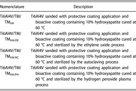
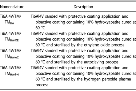
Table III shows the names and descriptions used for the samples during this study.
Nomenclature used for the samples during the development of this study.
Acknowledgments
The present work was developed with the support of the Brazilian government through the National Council of Scientific and Technological Development CNPq (CNPq/PVE 401211/2014-2 and CNPq 308773/2014-4) and the Coordination of Improvement of Higher Education Personnel (Capes). The authors would also like to thank the company Esterilizare RS/Brazil and Hospital Pompéia RS/Brazil, for their support in carrying out the processes and sterilization.