INTRODUCTION
Cryptosporidium parvum is recognized as a major cause of diarrhoea in humans. Although C. parvum infection is more common in the developing world, water-borne outbreaks are an emerging threat in industrialized countries (Goldstein et al. Reference Goldstein, Juranek, Ravenholt, Hightower, Martin, Mesnik, Griffiths, Bryant, Reich and Herwaldt1996). Cryptosporidial infection is typically mild to moderate in immunocompetent individuals but immunocompromised hosts and children can experience protracted diarrhoea causing dehydration, malabsorption and weight loss (Manabe et al. Reference Manabe, Clark, Moore, Lumadue, Dahlman, Belitsos and Chaisson1998; Newman et al. Reference Newman, Sears, Moore, Nataro, Wuhib, Agnew and Guerrant1999). Cryptosporidiosis can be fatal depending on the virulence of the strain and the patients' co-morbidities (Molbak et al. Reference Molbak, Hojlyng, Gottschau, Sa, Ingholt and da Silva1993; Manabe et al. Reference Manabe, Clark, Moore, Lumadue, Dahlman, Belitsos and Chaisson1998; Okhuysen et al. Reference Okhuysen, Chappell, Crabb, Sterling and duPont1999). Currently, no effective treatment is available.
Cryptosporidium research typically requires large numbers of organisms but the parasite cannot be grown reliably either axenically or in tissue culture (Arrowood and Sterling, Reference Arrowood and Sterling1987; Upton et al. Reference Upton, Tilley and Brillhart1994; Arrowood, Reference Arrowood2002; Hijjawi, Reference Hijjawi, Thompson, Armson and Morgan-Ryan2003), nor can it be cryopreserved (Fayer et al. Reference Fayer, Nerad, Rall, Lindsay and Blagburn1991). Although Hijjawi and coworkers reported the complete development cycle of C. parvum in cell-free culture (Hijjawi et al. Reference Hijjawi, Meloni, Ng'anzo, Ryan, Olson, Cox, Monis and Thompson2004), these findings could not be reproduced in our hands (unpublished data) or in the hands of others (Girouard et al. Reference Girouard, Gallant, Akiyoshi, Nunnari and Tzipori2006). Due to these difficulties, C. parvum is currently maintained by serial passage through large animals with subsequent bulk purification from feces (Arrowood and Sterling, Reference Arrowood and Sterling1987). These procedures are costly, time consuming and require facilities suitable for large animals. Supply options for smaller laboratories are thus limited and oocysts that most investigators rely upon can spend prolonged periods of time stored at 4°C in potassium dichromate solution. Such oocysts are not completely stable and changes in viability, density and surface markers have been documented (Medema et al. Reference Medema, Schets, Teunis and Havelaar1998; Young and Komisar, Reference Young and Komisar2005).
Several small animal models have been developed for in vivo studies of C. parvum infection but only 2 groups have explicitly used these models to propagate and amplify C. parvum. Using fecal pellets, Petry and coworkers were able to generate average yields of 108 oocysts per mouse but their dexamethasone-treatment model was labour intensive and led to animal death in several cases (Petry et al. Reference Petry, Robinson and McDonald1995). Compared to our results, Meloni and Thompson (Reference Meloni and Thompson1996) recovered, on average, 10-fold fewer purified oocyst from murine intestines while requiring 100-fold higher infectious doses.
In this study, we demonstrate that high amplification of C. parvum oocysts can be achieved in less than 2 weeks using IFNγR-KO mice. We further demonstrate that these oocysts can be efficiently harvested using a simple 2-step procedure and remain fully viable for both in vitro and in vivo experiments.
MATERIALS AND METHODS
Parasites
Oocysts originating from an Iowa bovine Cryptosporidium parvum isolate were amplified once through C57BL/6 IFNγR-KO mice, purified, and used for subsequent in vivo infection. A stock solution was maintained in 2·5% potassium dichromate (K2Cr2O7, Sigma, P-2588) and stored at 4°C until used.
Infection of mice
This study was approved by the Animal Care Committee of the Research Institute of the McGill University Health Centre. A total of 60 male and female C57BL/6 IFNγR-KO mice between 6 and 8 weeks old were used (Jackson Laboratories, Bar Harbor, ME, USA: mean body weight 25·5±4·8 g). Mice were housed in a conventional facility. At the time of infection (day 0), stock oocysts were washed 5 times in phosphate-buffered saline (PBS: pH 7·2). Solutions at a final oocyst concentration of 2×10⁴ oocysts/ml were prepared and mice were infected by gavage with ~2000 oocysts. Mice were sacrificed between 11 and 14 days post-infection (p.i.) or when pre-determined weight loss or behavioural changes were observed. Only 1 mouse needed to be sacrificed prior to study termination (day 9 p.i.).
Oocyst purification from feces
On day 6 p.i., the bedding was removed and cage bottoms were lined with sterile gauze. Ten fresh fecal pellets were collected daily from each cage of 5 mice starting 7 days p.i. until study termination (day 11–14 p.i.) to determine the kinetics of fecal oocyst excretion. The pellets were suspended in 2 ml of cold 2·5% (w/v) K2Cr2O7 and vortexed thoroughly. Suspensions were stored at 4°C until used. Upon processing, fecal suspensions were mixed with 10 ml of cold distilled water (dH2O) and centrifuged (2000 g for 10 min). Supernatants were discarded and 2 sequential NaCl flotations were performed as previously described with small modifications (Meloni and Thompson, Reference Meloni and Thompson1996). Briefly, resuspended pellets were mixed with 10 ml of a cold saturated NaCl solution and overlaid carefully with 2·5 ml of cold dH2O. Tubes were centrifuged (1600 g for 10 min) and the interphase (1·5 ml) was removed, washed with 10 ml of cold dH2O and centrifuged (2000 g for 10 min). These procedures were repeated once and the pelleted oocysts were resuspended in 2 ml of 2·5% K2Cr2O7 and stored at 4°C.
Oocyst purification from intestinal tissues
Study termination was defined as day 11 (n=10), 12 (n=30) or 14 (n=20) p.i. At termination, mice were sacrificed and the entire gut (duodenum to rectum) was removed, chopped into 0·5–1 cm pieces and placed into 150 ml tubes (Protocol™, Fisher Scientific, Nepean, ON, Canada) containing 10 ml of cold dH2O with 0·02% Tween20 (Fisher Biotech, Fair Lawn, NJ, USA) and 0·05 g Sputasol (stock: 0·1 g DTT, 0·78 g NaCl, 0·02 g KCl, 0·112 g Na2HPO4, 0·02 g KHPO4) per mouse (Meloni and Thompson, Reference Meloni and Thompson1996). Tissues were homogenized for 2 min at 8000 rpm (Polytron PT1300D homogenizer, Kinematica AG, Switzerland) and placed on a rotary mixer (Fisher Scientific) at low speed for 90 min. An ether extraction was performed as previously described (Arrowood, Reference Arrowood2002). Briefly, homogenates were centrifuged at 2000 g for 10 min, supernatants were discarded and oocyst-containing pellets were resuspended in 8 ml of cold dH2O with 0·02% Tween20 and 2 ml of ether (Fisher Scientific BP 337-100). Tubes were vortexed and spun as above. The oocyst-containing pellet was washed in 35 ml of cold dH2O and centrifuged again. A salt flotation procedure was performed as above using 20 ml of a cold saturated NaCl solution overlaid with 5 ml of cold dH2O. After removal of the visible interphase, the materials remaining in the tube were vortexed and overlaid again with another 5 ml of cold dH2O for a second NaCl flotation. The 2 final interphases were combined, washed twice in 35 ml of dH2O as above, resuspended in 4 ml of cold K2Cr2O7 and stored at 4°C.
Oocyst amplification rate
Amplification of the inoculated oocysts was calculated by dividing the total number of oocysts recovered from feces (total of days 7–14) or intestinal tissues (days 11, 12 or 14) by the mean inoculum (2000 oocysts): termed the Oocyst Amplification Factor (OAF). These newly-generated oocysts are hereafter referred to as NRCP1.
Viability of recovered oocysts
Excystation
Oocyst excystation was triggered by initial incubation on ice and subsequent incubation at 37°C in DMEM medium (Wisent, St.Bruno, QC, Canada) with 0·75% (w/v) sodium taurocholate (Sigma, T-4009). Aliquots were removed every 10 min for up to 60 min to monitor excystation by microscopy (% excystation=empty oocyst shells/intact oocysts: 200 oocysts counted).
In vitro infection model
In vitro infectivity of the recovered oocysts was assessed using 3 human intestinal cell lines (HCT-8, CaCo-2, I-407: ATCC # CCL-244, HTB-37, CCL-6, respectively, Rockville, MD) as previously described with slight modifications (Arrowood, Reference Arrowood2002). The cell monolayers were inoculated with a 50:50 mixture of excysted oocysts and sporozoites, and infection of the cells was proven by immunofluorescence using monoclonal antibodies directed against the C. parvum surface protein C3C3 (Centers for Disease Control and Prevention, Atlanta, GA).
In vivo infection
The in vivo infectivity of newly recovered oocysts was tested by exposing 10 naïve IFNγR-KO mice to 2000 oocysts by gavage as described above. Fecal pellets and intestinal tissues of these animals were harvested and processed as described above.
Statistical analysis
Statistical analysis was performed using Windows Excel for basic statistic calculations (average, mean, standard deviation). SPSS version 13.0 was used for Student's t-test, correlation analysis and linear trend test.
RESULTS
Oocyst recovery from fecal pellets
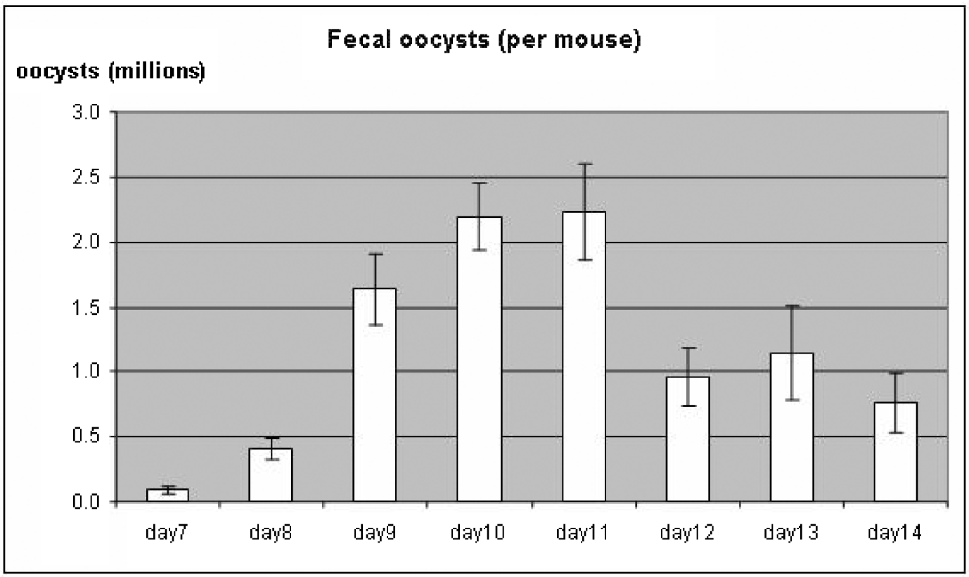
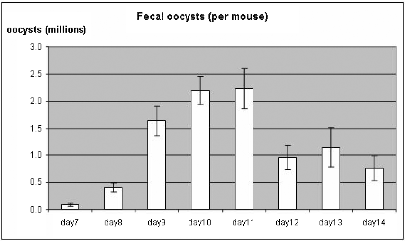
The crude fecal suspensions revealed larger numbers of oocysts than expected. The first NaCl flotation yielded a distinct whitish band of oocysts at the interphase but there was obvious fecal contamination by microscopy. After the second flotation, the interphase was visually free from contamination and no bacterial growth could be detected after 24 h storage in 2·5% K2Cr2O7 solution. Oocyst shedding typically began between days 7 and 9 p.i. and peak oocyst excretion was observed on days 10 and 11 p.i. after which oocyst recoveries declined rapidly (Fig. 1). The mean number of oocysts recovered from fecal samples over the 6–8 day collection period was 2·6×106 oocysts per mouse (range 8×10⁵ to 5·8×106). A total of 1·6×108 oocysts was recovered from all 60 mice with a calculated OAF of approximately 1300. Since only 2 fecal pellets were collected each day per animal, this is certainly an underestimate of the true amount of oocysts recoverable.
Mean oocyst recovery from daily fecal collection (2 pellets per mouse) after first NaCl flotation. Numbers are shown per mouse as the mean±s.e.m. at day 7 post-infection.
Oocyst recovery from intestinal tissues
Oocyst numbers in the intestinal tissue homogenates were visually striking. Following ether extraction, a single NaCl flotation of the obtained material yielded a sharp band of pure oocysts without obvious fecal contamination. Repeat NaCl flotation of the material from the original ether extraction tube also yielded a clean band of oocysts at the interphase. Yields from the first and second flotations accounted for approximately 80% and 20% of the total number of oocysts recovered, respectively. On average 3·8×107 oocysts per mouse were recovered from intestinal tissues (range 5·7×106 – 1·0×108). The total number of oocysts recovered from all 60 mice was 2·3×109 with a calculated OAF of approximately 19 000.
Viability and infectivity of recovered oocysts
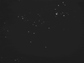
The efficiency and kinetics of excystation of the oocysts recovered from mice were equal if not superior to oocysts purified from bovine feces. Both Iowa bovine oocysts and IFNγR-KO passaged oocysts (NRCP1) readily infected all the human intestinal cell lines tested with similar efficiencies and kinetics (data not shown). The cell lines infected with these oocysts were identical in appearance both by light microscopy and immunofluorescence to the cells infected with Iowa bovine oocysts (Fig. 2). In our in vivo infection model, the NRCP1 oocysts were infectious for naïve IFNγR-KO mice.
Infected HCT-8 cells labelled with C3C3 antibody that detects all Cryptosporidium life stages. Oocysts are recognized by their round shape and size (>3 μm). Secondary antibody FITC-labelled.

A linear trend test revealed that mice euthanized on day 11 (peak of oocyst excretion) yielded significantly more intestinal oocysts than animals sacrificed on day 12 followed by those on day 14 (P<0·001, figure not shown).
DISCUSSION
Our results demonstrate that large numbers of C. parvum oocysts can be purified from both fecal pellets and the intestinal tissues of infected IFNγR-KO mice. Oocyst amplification rates far higher than those reported in other models were achieved: at least 1300-fold (fecal pellets) and ~19 000-fold (intestinal tissues). A number of factors are likely to have contributed to the high amplification rates achieved in this model.
Most importantly, IFNγ is an essential cytokine in both animals and humans to defend against cryptosporidial infection (Chen et al. Reference Chen, Harp and Harmsen1993; Griffiths et al. Reference Griffiths, Theodos, Paris and Tzipori1998; Mead and You, Reference Mead and You1998; Campbell et al. Reference Campbell, Stewart and Mead2002) and mice lacking IFNγ are highly susceptible to cryptosporidial infection (Chen et al. Reference Chen, Harp and Harmsen1993; Theodos et al. Reference Theodos, Sullivan, Griffiths and Tzipori1997; Mead and You, Reference Mead and You1998). In our hands, innocula as low as 10–50 oocysts result in 100% infection rates of IFNγR-KO mice (Ndao et al. submitted for publication). To our knowledge, no studies investigating oocyst amplification in IFNγ-deficient mouse models have been reported. In the dexamethasone-treated C57BL/6 adult mouse model, Petry et al. (Reference Petry, Robinson and McDonald1995) reported a narrow ‘window’ for effective amplification, with a significant proportion of the mice either failing to become infected or dying as a result of exogenous immunosuppression. The NaCl flotation approach has previously been shown to be efficient (Kuczynska and Shelton, Reference Kuczynska and Shelton1999) and is likely to have contributed to our high oocyst recovery rates. In our hands, even repeated NaCl flotations had little impact on the morphological characteristics and viability of isolated oocysts, effects commonly seen with other purification methods (Robertson et al. Reference Robertson, Campbell and Smith1992; Bukhari and Smith, Reference Bukhari and Smith1995; Young and Komisar, Reference Young and Komisar2005).
The total number of oocysts recovered from the IFNγR-KO mice was very high overall but daily excretion in fecal pellets differed widely between animals and in the same animal on different days, a common finding also reported by other investigators (Stehr-Green et al., Reference Stehr-Green, McCaig, Remsen, Rains, Fox and Juranek1987; Ungar, Reference Ungar1990; Petry et al. Reference Petry, Robinson and McDonald1995; Fayer et al. Reference Fayer, Gasbarre, Pasquali, Canals, Almeria and Zarlenga1998). Theoretical factors that may have contributed to the range in oocyst recoveries include stool consistency; oocysts are more stable in humid environments (Weber et al. Reference Weber, Bryan, Bishop, Wahlquist, Sullivan and Juranek1991; Robertson et al. Reference Robertson, Campbell and Smith1992), and variable adherence to fecal debris (Young and Komisar, Reference Young and Komisar2005). Different purification methods have been reported to select for viable or non-viable as well as for empty or intact oocysts (Current, Reference Current, Dubey, Speer and Fayer1990; Bukhari and Smith, Reference Bukhari and Smith1995). Young and Komisar (Reference Young and Komisar2005) have suggested that most purification methods select for a specific oocyst, resulting in a fixed level of oocyst viability. Such differences may have contributed to imperfect definition of the NaCl flotation interphase in our study and inconsistent recovery rates (Medema et al. Reference Medema, Schets, Teunis and Havelaar1998).
In contrast to the variable recovery rates from fecal pellets, the number of oocysts obtained from intestinal tissues was more stable. Higher concentrations of oocysts in the NaCl flotation interphase allowed for easier visualization and extraction and may have contributed to this finding.
Recovery rates from fecal and intestinal tissues were clearly related to the stage of the infection. The day 11 peak in fecal oocyst excretion corresponded closely with the onset of clinical signs of infection and weight loss while correlating with higher oocyst numbers recovered from intestinal tissue.
The amplification model used in the current study was remarkably simple and efficient. Compared to the dexamethasone-treated C57BL/6 mice, the IFNγR-KO model is simpler (i.e. no drug treatment, fewer premature deaths) and resulted in comparable or higher yields. Following infection of the mice on day 0, no further intervention was required until day 7, when the collection of feces began. Although intestinal homogenates cannot be preserved for any significant period of time, fecal pellet homogenates in K2Cr2O7 could theoretically be stored for up to 20 days for batch processing without significant decline in oocyst survival (Robertson et al. Reference Robertson, Campbell and Smith1992). Further simplification of our model by eliminating the labour-intensive collection of fecal pellets would result in a 7% reduction of recovered oocysts while reducing the processing time by an estimated 75%.
Introduction of this model should simplify the workings of small- and medium-sized laboratories that currently depend on the purchase or acquisition of oocysts from larger institutions or commercial sources. This model permits the recovery of large numbers of fresh oocysts within as few as 11 days while avoiding the inevitable changes in oocyst viability that occur with time in storage (Coulliette et al. Reference Coulliette, Huffman, Slifko and Rose2006). Finally, this model is trivial to scale-up or scale-down depending upon the immediate needs of the laboratory. The IFNγR-KO mouse model is therefore simple, rapid, inexpensive and effective, presenting a feasible alternative for the amplification and maintenance of C. parvum in experimental laboratories.
The study was supported by the Sandler Family Supporting Foundation. The study was approved by the Research Ethics Committee of the McGill University Health Centre at the Montreal General Hospital.