Introduction
The treatment of complicated tracheal stenosis is often problematic. Resection and reconstruction is currently the primary treatment for critical airway obstruction, and produces good long-term results. However, extensive resection and reconstruction may be inappropriate for some patients. Insertion of an airway stent to maintain airway patency is a relatively easy and safe procedure with low morbidity and mortality.Reference Grillo and Donahue1–Reference Cavaliere, Venuta, Foccoli, Toninelli and La Face5
The Montgomery T tube is reportedly effective and has a low incidence of complications. The device has two variations: the standard T tube, which is effective in most tracheal stenosis patients; and the long T tube (with extra-long lower limb), which is suited to patients with intrathoracic tracheal stenosis.Reference Montgomery6–Reference Liu, Lee, Huang, Cheng, Hsu and Huang8
While the standard Montgomery T tube is familiar to many practitioners, use of the long T tube is rarely reported. Consequently, the long-term results of long T tube procedures for airway stenosis remain unclear. This report describes outcomes following long T tube stenting over a four-year period.
Materials and methods
Between 2004 and 2006, 11 patients with airway stenosis who were unsuited for surgical excision underwent long T tube stenting at Chang Gung Memorial Hospital. The mean age of the patients was 47.5 years (range 17–82 years), and the mean follow up was 21.5 months (range 4.1–33 months). The patients' underlying diseases were intracerebral haemorrhage (n = 3), kyphoscoliosis (n = 1), coronary artery disease (n = 1) and buccal cancer (n = 1). Nine (82 per cent) patients had severe respiratory distress and four (44 per cent) required mechanical ventilation. Another two patients with supra-glottic and glottic stenosis, after corrosive injury and laryngeal trauma, were admitted with tracheostomy tubes.
The diagnosis of airway stenosis was based on clinical manifestations, computed tomography scanning and endoscopic findings. Since its introduction in the thoracic department of our facility in October 2001, rigid bronchoscopy has been used routinely to correct airway obstruction. In this study, all bronchotherapeutic procedures to improve respiratory function in cases of airway stenosis were performed by a single surgeon (YHL). Procedures were conducted under general anaesthesia. Obstruction of the larynx and upper and lower trachea was graded as follows: grade one, <50 per cent; grade two, 51–70 per cent; grade three, 71–95 per cent; and grade four, 95–99 per cent. The stricture segment was dilated by a rigid scope maintained on the axis of the tracheal tube, for easier airway evaluation and avoidance of tracheal perforation.
Debulking of luminal obstruction (by laser, electrocautery, microdebrider or removal of granulation tissue after anterior airway split) was considered in patients with significant airway stenosis after dilation. These procedures were used to create the patent airway for T tube placement, depending on the patient's operative findings. The T tube was inserted under direct visualisation as previously described.Reference Liu, Wu, Hsieh, Ko, Liu and Lin9 Simple modification of the T tube insertion technique (i.e. shortening the limb of the T tube or inverting the T tube) was used to facilitate appropriate placement. Rigid bronchoscopy was used to confirm the optimal T tube placement, and patients were transferred to the intensive care unit with an endotracheal tube inserted via the horizontal limb of the T tube, for ventilator weaning.
Post-operative follow up included physical examination, radiography and bronchoscopy. All patients were followed up appropriately. Morbidity related to previously reported complications of stent placement, including granulation formation, sputum impaction and tube migration. For each complication, management was discussed by the radiologist, thoracic surgeons and otolaryngologist until consensus was reached. Thus, optimal modalities (i.e. laser ablation, microdebrider and stent revision) were introduced to improve respiratory status and facilitate decannulation.
Results
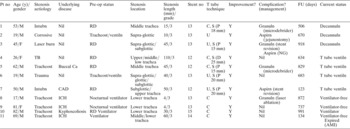
Airway stenosis was confirmed intra-operatively in all patients. All patients had grade three stenosis, indicating significant airway stenosis (Table I).
Demographics and treatment results

Pt no = patient number; y = years; pre-op = pre-operative; FU = follow up; M = male; F = female; intubn = intubation; granuln = granulation; aspirn = aspiration; TB = tuberculosis; tracheost = tracheostomy; Ca = cancer; CAD = coronary artery disease; ICH = intracerebral haemorrhage; RD = respiratory distress; C = conventional; S = tube end shortened by cutter; U = upside-down; P = proximal end; D = distal end; Y = yes; NG = nasogastric tube feeding; decannuln = decannulation; ventiln = ventilation; AMI = acute myocardial infarction
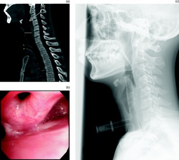
Conventional placement of an extra-long T tube was performed in seven of the 11 (64 per cent) patients. Of the remaining four patients, an inverted T tube was used for stent placement (Figure 1). In six patients, shortening of the horizontal arm of the tube was required in order to place the tube in the correct position. In one patient, revision (i.e. shortening of the stent) was performed two weeks after stent placement.
Pre-operative (a) sagittal computed tomography (CT) scan and (b) bronchoscopy showing an airway obstruction (arrow) involving the supra-glottic region. (c) Post-operative sagittal CT image after Montgomery T tube placement, establishing a patent airway.

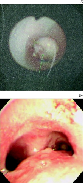
Granulations occurred in four patients. In one patient, granulations over the upper end of the T tube caused severe stenosis in the supra-glottic region. The patient was successfully treated by placing the stent through and above the supra-glottic region. Two patients suffered granulation tissue formation over the posterior glottic and sub-glottic region due to stent irritation. They were successfully treated with microdebrider resection of granulation lesions, via suspension laryngoscopy. One patient revealed unusually exuberant granulation formation at the tip of the lower end of the T tube. He was treated successfully by laser ablation of granulation tissue and shortening of the lower T tube limb, in order to alleviate mucosal irritation from the tube (Figure 2).
(a) Bronchoscopy showing airway obstruction at distal end of Montgomery T tube involving the lower tracheal region, caused by granulation tissue due to T tube irritation of mucosa. (b) Post-operative bronchoscopy after laser ablation and stent shortening had established a patent airway.

Aspiration occurred after stent placement in three of the 11 patients. One patient with a severe supra-glottic and oesophageal stricture following corrosive injury, required feeding and nutrient supplement via a jejunostomy tube. Therefore, aspiration was not problematic. The feeding tube and T tube were removed, and oral feeding was resumed one year after surgery. In two patients with persistent aspiration, supra-glottic placement of the proximal limb of the T tube was followed by nasogastric feeding. In one of these patients, the stent was successfully removed one year after surgery. Recovery of swallowing and phonatory function was uneventful, and the patient remained free of respiratory symptoms six months after completing treatment. In another patient who underwent the revision procedure and shortening of the proximal limb of the T tube, aspiration complications were alleviated two months after surgery.
No peri-operative morbidity or mortality occurred. Three patients were weaned off the ventilator one day after surgery and required no further ventilatory support after an average follow up of 19.4 months (range 4.5–29 months). The average follow up of the remaining patients was 22.3 months (range 4.1–33 months). These patients had a 27 per cent rate of decannulation. The average time interval for decannulation was 14.6 months (range nine to 21.5 months) from the day of T tube insertion.
Discussion
At bronchoscopy, the airway was recannulated and the stent placed within the stenotic segment, thus maintaining the airway calibre. However, the airway stent itself caused some complications. Previous reports have shown that metallic airway stents may induce granulation, exacerabate airway obstruction and present difficulties during removal.Reference Wood, Liu, Vallieres, Karmy-Jones and Mulligan2, Reference Dumon10, Reference Gaissert, Grillo, Wright, Donahue, Wain and Mathisen11 Although a silicone intratracheal stent may have been suitable for some patients in the present study, the device had been unavailable in Taiwan until early 2007. The Montgomery T tube was therefore recommended and employed in all patients to obviate complications related to stent migration and stent removal.
Rigid bronchoscopy is routinely used for stent placement in our department. This approach allows T tube insertion through the original tracheal opening, with no further neck incision, and adequacy of stenting and optimal stent position can be confirmed.Reference Liu, Wu, Hsieh, Ko, Liu and Lin9
In order to maximise the clinical efficacy and therapeutic potential of the T tube, the limb of the T tube was shortened to accommodate the tracheal stenosis and to prevent vocal fold irritation proximally and carinal irritation distally. Another modification involved inverting the T tube during stent placement (an extra-long lower limb was used for subglottic and supra-glottic laryngeal lesions). This ensured accurate T tube placement for all laryngeal and tracheal lesions.
In patients who require ventilation after a stroke and who develop tracheal stenosis, studies have shown that a long tracheostomy bypassing the stenotic segment can stabilise the airway.Reference Levine, Sasaki and Kirchner12 However, in the present study, the T tube was adopted to improve respiratory function, and three of four patients were weaned from their ventilator.Reference Wu, Liu, Hsieh and Ko13 The present results indicate that the T tube may be useful for managing ventilated patients with tracheal stenosis. Additionally, a T tube also allows endotracheal tube insertion via the horizontal limb for ventilation in patients who have failed weaning.
• Treating complicated tracheal stenosis is often problematic. Resection and reconstruction is currently the primary treatment for critical airway obstruction and produces good long-term results
• This study evaluated the efficacy and safety of the extra-long Montgomery T tube for managing major airway obstruction in tertiary care patients in Taiwan
• Eleven patients with major airway stenosis treated with extra-long Montgomery T tubes between April 2004 and December 2006 were retrospectively reviewed
• The use of an extra-long T tube is an effective and safe method for treating major airway obstruction in the supra-glottic to lower tracheal region
Stenosis involving the laryngeal region is a serious condition. Although some surgeons have reported successful resection and reconstruction in patients with supra-glottic stenosis, we believe that supra-glottic airway resection requires a substantially more difficult surgical technique involving mobilisation of the stenotic segment and anastomosis of the healthy airway. Surgical resection is associated with anastomotic leaks and deep neck infection, and may require vigorous cervical debridement and result in possible voice impairment.Reference Couraud, Jougon and Velly14 In the present study, the T tube was introduced into the airway to maintain airway patency, after splitting the stenosis at the supra-glottic and glottic levels and removing granulation tissue in the subglottic region in order to reduce surgical morbidity and improve treatment outcome. Both patients (patient No 2 and 3) were decannulated with acceptable voice quality and experienced no respiratory complications over a period of nine months.
A major complication of T tube insertion was granulation tissue formation, causing airway stenosis and respiratory distress. In this series, such granulation tissue was debulked by laser ablation and microdebridement. Vocal granulations were more effectively treated with the microdebrider, which has advantages in terms of normal tissue preservation and avoidance of thermal injury.Reference Hsieh, Liu, Yueh and Ko15–Reference Zalzal and Collins17
Conclusion
In cases of complex airway stenosis, the use of an extra-long Montgomery T tube was effective and stabilised respiratory status in 100 per cent of patients. However, treatment outcome depends on operator experience, knowledge of airway anatomy, effective management of complications and cooperation between the physicians treating the airway stenosis. The procedure should be considered as a therapeutic option in patients with stenoses at the supra-glottic, glottic, subglottic and tracheal levels. This treatment also allows decannulation and weaning from ventilation.