


Introduction
Cinematic rendering is a photorealistic three-dimensional visualization technique that uses Monte Carlo–based global illumination, volumetric scattering, and physically based light–tissue interaction models to transform CT datasets into lifelike anatomic reconstructions. Reference Eid, De Cecco and Nance1–Reference Dappa, Higashigaito and Fornaro3 Unlike conventional volume rendering, which relies on simplified local illumination and uniform shading, cinematic rendering incorporates soft shadowing, subsurface scattering, ambient occlusion, and material-specific reflectance intended to more closely approximate real-world optical behaviour. Reference Calhoun, Kuszyk, Heath, Carley and Fishman4 These features can improve visualization of complex geometries such as vascular bifurcations and vessel–chamber interfaces.
Cinematic rendering has been described across a range of cardiovascular and thoracoabdominal applications, including postoperative vascular complications, infiltrative cardiac and renal lymphoma, and complex aortic repairs imaged with photon-counting CT, Reference Rowe, Johnson and Fishman5–Reference Forouzannia and Umair9 as well as CHD and thoracic vascular anomalies. Reference Röschl, Purbojo and Rüffer10–Reference Layden, Brassil and Jha12 It has also been used in medical education to support learners’ spatial understanding of anatomy. Reference Binder, Scholz and Ellmann13,Reference Elshafei, Binder and Baecker14
These features are particularly relevant to the evaluation of anomalous coronary artery origins, where subtle differences in vessel takeoff, course relative to the great vessels, and intramural depth can be difficult to appreciate on standard 2D images. Overlapping structures, partial-volume effects, and loss of depth cues may obscure critical segments, complicating characterization of an anomalous coronary artery’s trajectory or assessment of postoperative changes. Accurate characterization of these findings is clinically important because coronary origin and course directly influence risk stratification and management decisions. Reference Kim, Seo and Do15–Reference Brothers, Frommelt and Jaquiss18 This pictorial essay illustrates the application of cinematic rendering to coronary CT angiography studies involving anomalous coronary origins—including interarterial courses and post-surgical assessment following unroofing—and describes, through representative cases, how cinematic rendering’s restoration of volumetric context and soft-tissue differentiation aided our visual assessment of these complex anatomic variants.
Technical background and comparison with conventional volume rendering
Cinematic rendering is adapted from computer-graphics rendering methods developed for film and animation. Rather than relying on the simplified local lighting used in conventional volume rendering, cinematic rendering simulates how light actually behaves as it passes through tissue—reflecting, scattering, and softening—using Monte Carlo path tracing, a method that traces large numbers of simulated light rays through the CT volume to produce natural-appearing shadows, ambient occlusion, and subsurface scattering. Reference Rowe, Johnson and Fishman19–Reference Soyer21
To generate a cinematic rendering image, high-resolution isotropic CT data are normalized, denoised, and segmented into material categories (myocardium, fat, vascular wall, contrast-opacified blood, bone, air), each of which is then assigned optical properties governing how light is absorbed, scattered, or reflected. Myocardium, for example, is modelled with substantial subsurface scattering to reproduce its soft, layered appearance, while contrast-filled blood is modelled with strong forward scattering to increase perceived luminal brightness and improve delineation of the vessel wall. A Monte Carlo global illumination engine then projects millions of simulated photons through the volume, iteratively calculating reflection, refraction, scattering, and absorption to produce the final image; because each photon may undergo numerous interactions, this process is computationally demanding and typically requires graphics-processing-unit acceleration. Reference Zhang, Eagleson and Peters22 Once rendered, the volume can be explored interactively, with adjustable transparency, camera position, lighting, and clipping planes—capabilities that have been described across CHD, postoperative vascular imaging, and thoracic pathology. Reference Rowe, Chu, Zimmerman and Fishman23,Reference Recht, Weisberg and Fishman24
Conventional volume rendering and maximum-intensity projection remain the mainstay of cardiovascular CT visualization, but each has recognized limitations. Volume rendering’s local illumination and uniform shading can produce a smoothed appearance with reduced depth perception and limited soft-tissue separation; maximum-intensity projection offers strong vessel conspicuity but collapses volumetric information into a single plane, which can create false impressions of vessel continuity or overlap in coronary imaging. Reference Calhoun, Kuszyk, Heath, Carley and Fishman4 Cinematic rendering’s global illumination model is intended to produce softer, more natural shadow transitions that may improve visual separation between myocardium, fat, and contrast-enhanced blood; this has been described across cardiovascular applications including postoperative vascular imaging, CHD, and aortic reconstruction. Reference Rowe, Johnson and Fishman5,Reference Zimmerman, Rowe and Fishman6,Reference Röschl, Purbojo and Rüffer10,Reference Layden, Brassil and Jha12 In the coronary anomalies presented in this essay, where subtle differences in vessel takeoff, interarterial course, and intramural depth can be difficult to appreciate on 2D images, we found cinematic rendering’s depth cues helpful in visually tracing vessel course relative to the great vessels—an anatomic distinction relevant to risk stratification. Reference Kim, Seo and Do15–Reference Brothers, Frommelt and Jaquiss18 As above, this is a qualitative, case-based observation rather than a finding from a controlled comparison.
Clinical applications of cinematic rendering in anomalous coronary imaging
Cinematic rendering has been applied across a range of cardiovascular imaging scenarios, where its depth cues and soft-tissue contrast have been described as helpful in clarifying findings that are ambiguous on conventional reconstructions. Early work focused on postoperative vascular imaging—visualizing graft configurations, pseudoaneurysms, and other postsurgical changes following aortic aneurysm interventions, hybrid arch reconstructions, and venous graft complications. Reference Zimmerman, Rowe and Fishman6,Reference Umair, Fishman and Zimmerman8 Cinematic rendering has also been applied to acute and chronic aortic pathology, congenital vascular malformations, and anomalous pulmonary venous drainage. Reference Forouzannia and Umair9,Reference Röschl, Purbojo and Rüffer10,Reference Recht, Weisberg and Fishman24–Reference Lengyel, Lumsden and Chinnadurai26
Anomalous aortic origin of a coronary artery encompasses a spectrum of variants with differing clinical significance; the course of the anomalous vessel—interarterial, retroaortic, prepulmonic, or subpulmonic—directly influences risk stratification, with interarterial courses associated with the highest risk of adverse cardiac events. Reference Angelini16,Reference Brothers, Frommelt and Jaquiss18 In the cases below, cinematic rendering’s restoration of volumetric context and soft-tissue differentiation supported continuous visual tracing of the anomalous vessel from its origin through its course relative to the great vessels.
Normal coronary anatomy
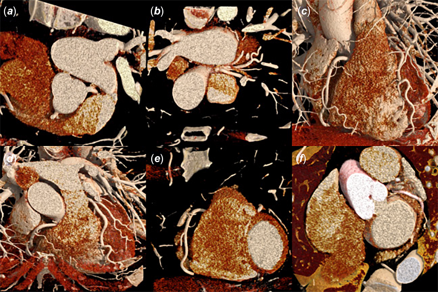
Normal coronary anatomy was reviewed to establish baseline references for comparison (Figure 1). Structures demonstrated include: right coronary artery origin from the right coronary cusp; left main coronary artery origin from the left coronary cusp; left main coronary artery bifurcation or trifurcation into left anterior descending artery, left circumflex artery, and, when present, ramus intermedius; left anterior descending artery within the anterior interventricular groove; right coronary artery in the right atrioventricular groove; and left circumflex artery in the left atrioventricular groove. Reference Kim, Seo and Do15
Normal coronary origins and courses demonstrated with standard CT angiography. (a) Normal coronary origins with RCA arising from the right coronary cusp and LM from the left coronary cusp. (b) LM trifurcation into LAD, ramus intermedius, and circumflex, with early branching of LAD and circumflex also noted. (c) Normal course of RCA and LAD. (d) Normal course of LAD in the anterior interventricular groove, with partially visualized proximal course of RCA. (e) Normal course of RCA in the right AV groove. (f) Normal course of LCx in the left AV groove. These images provide anatomical reference for interpreting aberrant coronary trajectories.

Imaging demonstrations of anomalous coronary variants
Anomalous right coronary artery from left coronary cusp with interarterial course
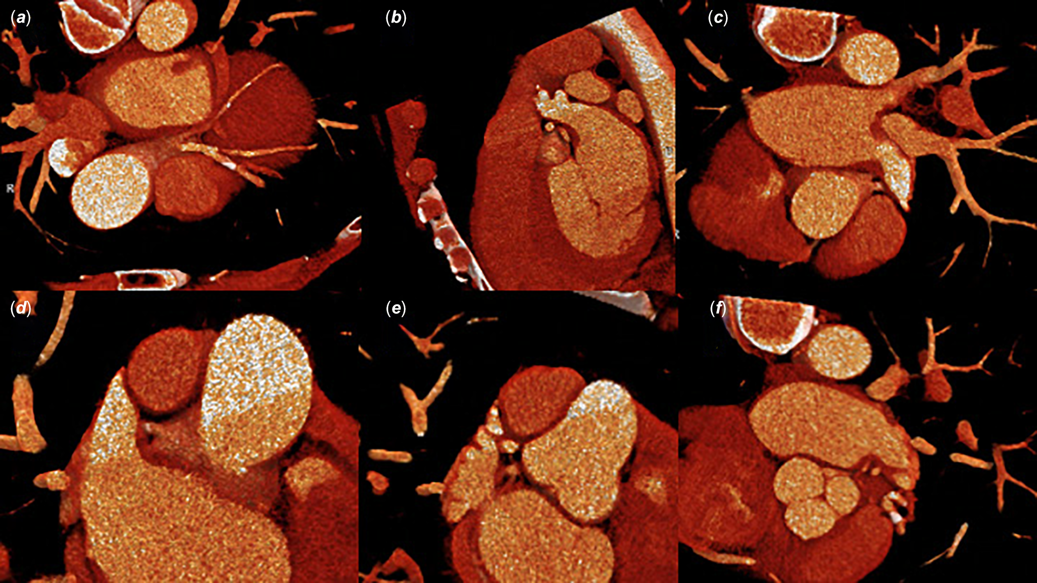
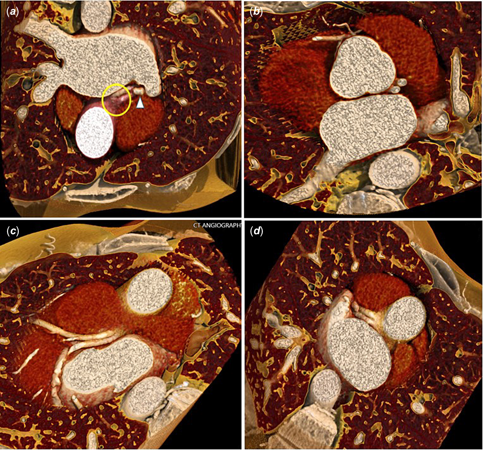
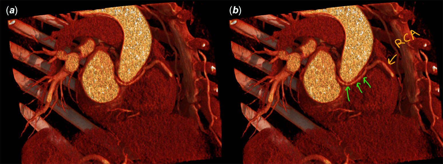
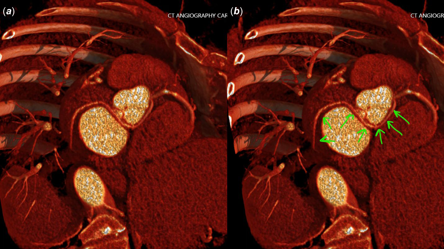
2D CT angiography demonstrated an atypical right coronary artery origin from the left coronary cusp with interarterial transit between the ascending aorta and pulmonary artery (Figures 2,3). Cinematic rendering images below allowed continuous 3D tracking of the vessel with volumetric separation of surrounding anatomy, consistent with an anomalous interarterial course. Reference Kim, Seo and Do15–Reference Brothers, Frommelt and Jaquiss18
Panels (a–f): 3D CR images for characterization of an anomalous RCA trajectory. (a–c) CR allowed continuous 3D tracking of the RCA, providing depth, soft-tissue contrast, and vessel-surface detail consistent with an interarterial anomalous course. The 3D relationship of the RCA to the great arteries, with associated mild narrowing, is visualized as the vessel emerges through the space between the aorta and pulmonary artery.

(a) CR imaging demonstrates the anomalous origin and proximal course of the RCA. (b) The subsequent course of the proximal- to mid-RCA. (c) CR demonstrates narrowing of the RCA in the interarterial course.

Post-surgical assessment following unroofing of anomalous right coronary artery
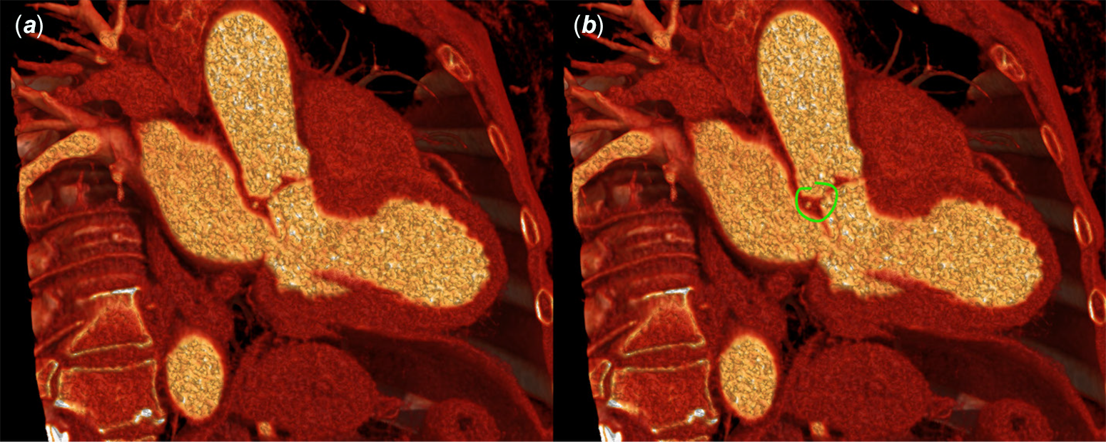
Cinematic rendering imaging of an anomalous right coronary artery arising from the left coronary cusp demonstrates an interarterial course with surgical unroofing of the intramural segment already performed at the time of imaging. Cinematic rendering from the preoperative coronary CT angiography examination was not available due to limited scan resolution; cinematic rendering performed on the postoperative examination allowed detailed visualization of postoperative vessel morphology (Figure 4). Reference Brothers, Frommelt and Jaquiss18
Panels (a–d): Sequential cross-sections of the RCA in anomalous origin demonstrating postoperative anatomy following unroofing. The ostium shows a slit-like configuration with acute angulation, resulting in moderate-to-severe residual ostial narrowing (image a). The intramural segment decreased in depth from 6 mm preoperatively (assessed on 2D coronary CT angiography; CR was not performed preoperatively) to 2 mm post-unroofing (images a and b), a mild interval improvement; however, both the acute angulation and interarterial course continue to contribute to narrowing. Mild residual narrowing is also noted in the proximal interarterial segment (image c), with only minimal improvement compared with the prior examination. The proximal RCA segment after emerging from the interarterial course is shown in (image d).

Absent left main coronary artery: separate origins of left anterior descending artery and left circumflex artery
In contrast to normal coronary anatomy, where the left main coronary artery gives rise to both the left anterior descending artery and left circumflex artery, this example illustrates separate anomalous origins of the left anterior descending artery and left circumflex artery arising directly from the left coronary cusp (Figure 5). While this variant is generally considered benign, accurate identification is important for procedural planning and to avoid misinterpretation as left main disease or anomalous origin from the wrong sinus. Reference Kim, Seo and Do15–Reference Schmitt, Froehner and Brunn17
Separate origins of LAD and LCx directly from the left coronary cusp. Images (a–d) demonstrate different imaging planes showing the separate origins of LAD and LCx from the left coronary cusp, with no true LM present. Panels C and D show external surface renderings demonstrating the separate LAD and LCx origins.

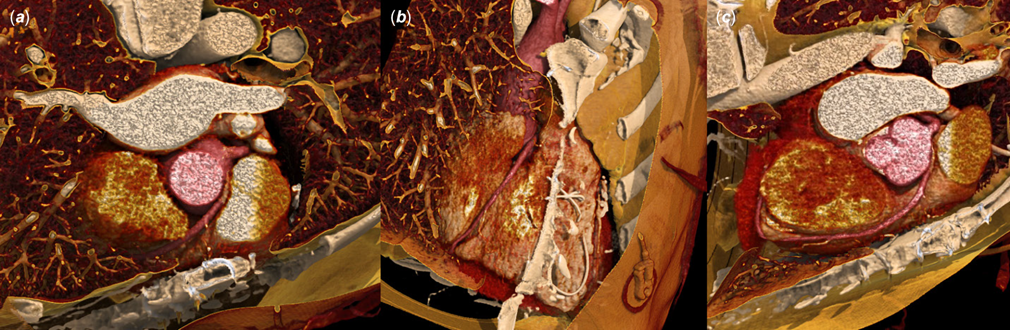
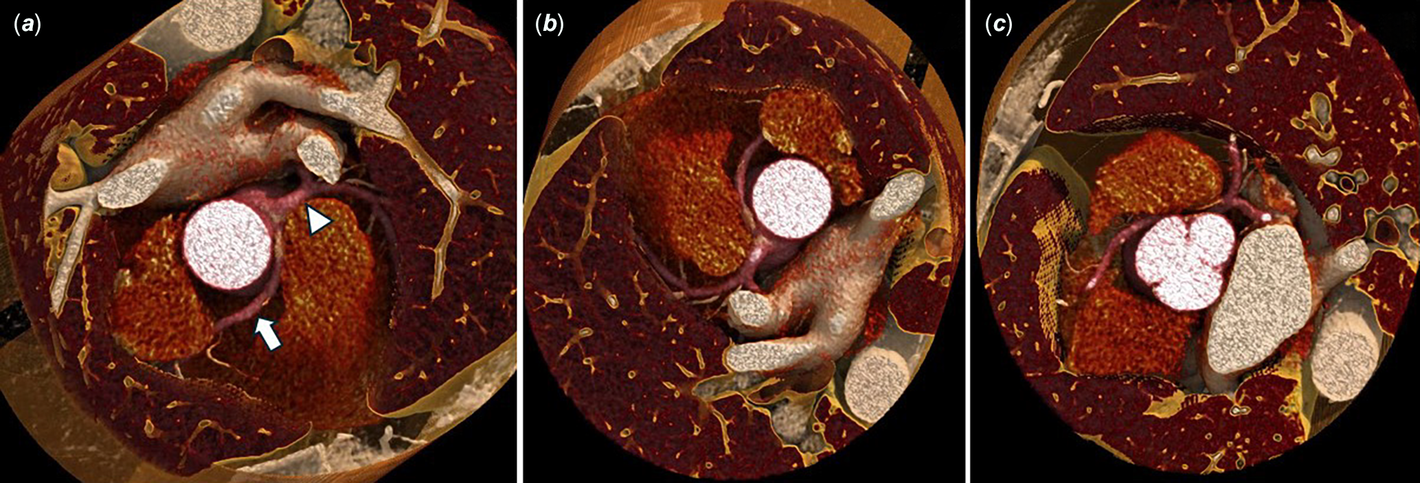
Cinematic rendering provides external surface views delineating the separate ostia of the left anterior descending artery and left circumflex artery arising independently from the left coronary cusp, consistent with absence of a true left main coronary artery (Figures 6 and 7).
Anomalous origin of the RCA from the left coronary cusp. (a) CR demonstrates the anomalous RCA (arrow) arising from the left coronary cusp in close proximity to the LM (arrowhead). (b) Alternative viewing angle showing the spatial relationship between the anomalous RCA origin and the LM at the level of the left coronary cusp. (c) External surface rendering further delineating the anomalous RCA takeoff adjacent to the LM origin.

Figure 6. Long description
Panel A: A cross-sectional view of the heart showing the anomalous right coronary artery (RCA) originating from the left coronary cusp. The RCA is marked with an arrow, and the left main (LM) coronary artery is marked with an arrowhead. Panel B: An alternative viewing angle of the heart highlighting the spatial relationship between the anomalous RCA origin and the LM at the level of the left coronary cusp. Panel C: An external surface rendering of the heart further delineating the anomalous RCA takeoff adjacent to the LM origin.
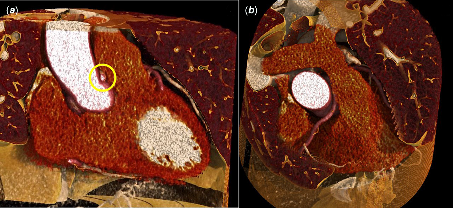
Interarterial course of the anomalous RCA. (a) CR demonstrates the RCA (encircled) coursing between the ascending aorta and main pulmonary artery in an interarterial trajectory. (b) Alternative projection showing the interarterial course of the anomalous RCA as it passes through the space between the great vessels. This high-risk anatomic configuration is associated with increased risk of adverse cardiac events due to potential dynamic compression of the vessel between the aorta and pulmonary artery during exercise or states of increased cardiac output.

Anomalous origin of left circumflex artery from right coronary artery ostium
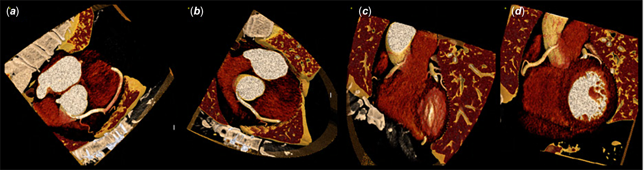
In contrast to normal coronary anatomy, where the left circumflex artery arises from the left main coronary artery off the left coronary sinus and courses within the left atrioventricular groove, this case demonstrates anomalous origin from the right coronary ostium with a retroaortic course (Figures 8–10).
CR angiography demonstrating an anomalous origin of the LCx arising from the RCA ostium. The LCx follows a retroaortic course, traversing the fat plane between the posterior aortic root and the left atrium before reaching the left AV groove. This represents a benign anatomic variant without interarterial compression.

The proximal course of the anomalous LCx is well visualized in this figure, arising from the RCA ostium.

Comprehensive view of the anomalous LCx showing both the proximal course from the RCA ostium and the mid-to-distal vessel as it follows the retroaortic path to the left AV groove.

Discussion
Across the cases presented, we observed that cinematic rendering provided additional visual context beyond what was available on standard coronary CT angiography reconstructions in these specific examples, largely by reintroducing depth cues and soft-shadow behaviour that helped differentiate structures that appeared indistinct on planar images. These observations are consistent with prior descriptive reports of cinematic rendering’s use in visualizing vascular surfaces, congenital variants, and postoperative changes compared with conventional volume rendering, Reference Eid, De Cecco and Nance1–Reference Dappa, Higashigaito and Fornaro3,Reference Rowe, Johnson and Fishman5,Reference Zimmerman, Rowe and Fishman6,Reference Rowe, Johnson and Fishman19 though—as in that prior literature—our observations here are qualitative and not derived from a blinded or quantitative comparison.
We deliberately retained surrounding anatomic structures (the aortic root, pulmonary artery, and adjacent cardiac chambers) in most figures rather than aggressively clipping them. Because the central question in each case is the anomalous vessel’s course relative to the great vessels, we felt that removing this context would undermine the very spatial relationship the figures are meant to illustrate, and would render the images less representative of true anatomy. We have, however, revised the relevant figure legends below to more explicitly orient the reader to the structures of interest before describing the finding.
In the cases presented, cinematic rendering supported continuous visual tracking of the anomalous right coronary artery through its interarterial course, visualization of postoperative anatomy following unroofing—including residual ostial narrowing and reduction of the intramural segment—and depiction of the separate left anterior descending artery and left circumflex artery origins from the left coronary cusp, a variant that can be difficult to characterize definitively on standard reconstructions.
Taken together, these case-based observations are consistent with a growing descriptive literature suggesting cinematic rendering may serve as a useful adjunct to conventional coronary CT angiography interpretation in anomalous coronary origins, though we emphasize that this remains to be evaluated systematically. Reference Soyer21,Reference Fishman27
Limitations and future directions
Several limitations temper broader clinical adoption of cinematic rendering. There is no standardized rendering protocol across vendors: different platforms use proprietary light-transport models, scattering kernels, and material presets, and individual readers may select different colour schemes, windowing presets, and lighting models, complicating standardization. Because cinematic rendering’s lighting, camera angle, transparency, and surface-enhancement settings are all user-adjustable, two readers may generate different renderings from the same dataset—a concern in coronary imaging, where millimetre-scale distinctions determine whether a segment is intramural or interarterial, and one that underscores the need for standardized coronary presets and reproducible workflows.
As discussed above, our observations in this essay are based on a small number of illustrative cases and our own clinical impressions; we have not conducted a blinded reader study or quantitative comparison against conventional volume rendering or maximum-intensity projection, and we did not perform additional volume sculpting beyond what is described above, in order to keep the renderings faithful to true anatomic relationships. Both represent important next steps for establishing cinematic rendering’s incremental diagnostic value in this setting.
Cinematic rendering’s Monte Carlo–based global illumination is also computationally intensive, and real-time interaction typically requires modern graphics-processing-unit-accelerated systems, which may limit access in lower-resource settings. Reference Zhang, Eagleson and Peters22 Looking ahead, integration of cinematic rendering with immersive volume rendering and augmented-reality platforms has shown feasibility for CHD and surgical planning; Reference Böttcher, van Assen and Fari11,Reference Rowe, Schneider and Krueger28 automated segmentation and artificial-intelligence-assisted material mapping may reduce operator dependence and improve reproducibility; and photon-counting CT, with its higher spatial resolution and improved noise characteristics, may pair well with cinematic rendering for visualization of small coronary segments and anomalous origins. Reference Forouzannia and Umair9
Conclusion
In this series, cinematic rendering provided additional visual context for several coronary CT angiography findings involving anomalous coronary origins in the cases presented. The added depth cues and soft-tissue separation aided our tracing of anomalous vessel courses, assessment of their relationship to the great vessels, and evaluation of postoperative anatomy following surgical correction. These case-based observations suggest cinematic rendering may be a useful complement to routine coronary CT angiography review in select cases of anomalous coronary origin, pending more systematic evaluation of its incremental diagnostic value.
Financial support
This research received no specific grant from any funding agency, commercial, or not-for-profit sectors.
Competing interests
None.
Ethical standard
This study was conducted in accordance with the ethical standards of the relevant institutional and national committees on human experimentation and with the Helsinki Declaration of 1975, as revised in 2008. Institutional review board approval and informed consent were not required for this fully anonymized imaging series.
AI use
An AI writing assistant was used to support grammar and language editing during manuscript revision; all scientific content, analysis, and conclusions are the authors’ own.

 Open access

