Introduction
Depression has been identified as a robust risk factor for the development of coronary heart disease (CHD), with an adjusted relative risk of 1.9 (Kuper et al. Reference Kuper, Nicholson, Kivimaki, Aitsi-Selmi, Cavalleri, Deanfield, Heuschmann, Jouven, Malyutina, Mayosi, Sans, Thomsen, Witteman, Hingorani, Lawlor and Hemingway2009). However, some studies have found that only the somatic (e.g. feeling tired or having little energy) but not the cognitive (e.g. feeling down, depressed or hopeless) dimension of depression predicted poor outcome in patients with cardiovascular disease (Ormel & de Jonge, Reference Ormel and de Jonge2011). These findings have stirred controversy regarding the cause–effect relationship between subtypes of depression and cardiovascular disease. Thus, although it had been generally assumed that depression drives cardiovascular disease, it has been more recently hypothesized that somatic symptoms of depression are mainly caused by atherosclerotic disease and related systemic inflammation, whereas cognitive symptoms of depression are thought to be due to the meaning and psychosocial impact of coronary events and unrelated stressors (e.g. personality, history of depression) (Ormel & de Jonge, Reference Ormel and de Jonge2011).
In this regard, some, but not all (e.g. Kuper et al. Reference Kuper, Nicholson, Kivimaki, Aitsi-Selmi, Cavalleri, Deanfield, Heuschmann, Jouven, Malyutina, Mayosi, Sans, Thomsen, Witteman, Hingorani, Lawlor and Hemingway2009; Ormel & de Jonge, Reference Ormel and de Jonge2011), epidemiological studies have found an up to twofold risk for atherosclerosis in depressed versus never depressed cardiovascular patients. In a recent cross-sectional study of 1261 persons from the community, Bus et al. (Reference Bus, Marijnissen, Holewijn, Franke, Purandare, de Graaf, den Heijer, Buitelaar and Voshaar2011) found that the intima–media thickness (IMT) of the carotid artery, a reliable measure of atherosclerosis (Elovainio et al. Reference Elovainio, Keltikangas-Järvinen, Kivimäki, Pulkki, Puttonen, Heponiemi, Juonala, Viikari and Raitakari2005; Whipple et al. Reference Whipple, Lewis, Sutton-Tyrrell, Matthews, Barinas-Mitchell, Powell and Everson-Rose2009; Pizzi et al. Reference Pizzi, Manzoli, Mancini, Bedetti, Fontana and Costa2010), was associated only with the somatic symptom dimension of depression. The authors concluded that subclinical vascular disease may have inflated the depressive symptoms. They further postulated that the vascular origin of depression may explain why treatment of depression in cardiac patients has hardly affected vascular outcome (Bus et al. Reference Bus, Marijnissen, Holewijn, Franke, Purandare, de Graaf, den Heijer, Buitelaar and Voshaar2011). However, these conclusions relied on current depression, and it must be assumed that only lasting depression can contribute to the development of atherosclerosis (Ormel & de Jonge, Reference Ormel and de Jonge2011).
The current study sought to examine the relationship between depression and its symptom dimensions and atherosclerosis, using data from the Gutenberg Health Study (GHS), a major representative, longitudinal community study on cardiovascular risk. We hypothesized that the somatic symptom dimension of depression but not the cognitive symptom dimension would be associated with signs of atherosclerosis as measured by the IMT or carotid plaques. Unlike the previous study by Bus et al. (Reference Bus, Marijnissen, Holewijn, Franke, Purandare, de Graaf, den Heijer, Buitelaar and Voshaar2011), we included medical history of depression in our analyses.
Method
Study population
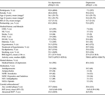
We investigated cross-sectional data from the first 5000 participants enrolled in the GHS from April 2007 to October 2008. The GHS is a population-based, prospective, observational single-center cohort study in the Rhein-Main Region in western mid-Germany. The GHS has been approved by the local ethics committee and by the local and federal data safety commissioners. The primary aim of the study was to evaluate and improve cardiovascular risk stratification. The sample was drawn randomly from the local registry in the city of Mainz and the district of Mainz-Bingen. The sample was stratified 1:1 for gender and residence and in equal strata for decades of age. Inclusion criteria were age 35–74 years and written informed consent. Persons with insufficient knowledge of the German language or physical or mental inability to participate were excluded. The response rate was 60.3%, defined as the recruitment efficacy proportion; that is the number of persons with participation in or an appointment for the baseline examination divided by the sum of the number of persons with participation in or an appointment for the baseline examination plus those who refused and those who were not contactable. Table 1 gives an overview of the study participants with (n = 357) and without (n = 4580) depression.
Sample stratified by caseness of current depression: demographics, medical history, medication and distress (n = 4937)

PHQ-9, Nine-item Patient Health Questionnaire; SES, socio-economic status; CHD, coronary heart disease; MI, myocardial infarction; PAD, peripheral artery disease; FH, family history; BMI, body mass index; s.d., standard deviation; IQR, interquartile range; IMT, intima–media thickness; CI, confidence interval.
a Missing values n = 186.
Materials and assessment
The 5-h baseline examination in the study center comprised the evaluation of prevalent classical cardiovascular risk factors and clinical variables, a computer-assisted personal interview, laboratory examinations from a venous blood sample, and blood pressure and anthropometric measurements. In general, certified medical technical assistants performed all examinations according to standard operating procedures.
Predictor variables
Depression was measured by the nine-item Patient Health Questionnaire (PHQ-9), which quantifies the frequency of being bothered by each of the nine diagnostic criteria of major depression over the past 2 weeks. Responses are summed to create a score between 0 and 27 points. A PHQ-9 sum score of ⩾10 was used for the definition of caseness for depression, yielding a sensitivity of 81% and a specificity of 82% for any depressive disorder (Lowe et al. Reference Lowe, Grafe, Zipfel, Witte, Loerch and Herzog2004). The somatic and cognitive dimensions of depression were defined according to prior studies (Hoen et al. Reference Hoen, Whooley, Martens, Na, van Melle and de Jonge2010; Martens et al. Reference Martens, Hoen, Mittelhaeuser, de Jonge and Denollet2010). Four PHQ-9 items related to problems with sleep, fatigability, appetite and psychomotor agitation/retardation were classified as somatic depressive symptoms, and five items related to lack of interest, depressed mood, negative feelings about self, concentration problems and suicidal ideation were classified as cognitive depressive symptoms. During the computer-assisted personal interview, participants were asked by a physician whether they had ever received a definite diagnosis of any depressive disorder (medical history of lifetime diagnosis of any depressive disorder, medical history of depression).
We defined three sets of predictor variables: current depression as defined by PHQ-9 ⩾10, cognitive and somatic depressive symptoms of depression, and a medical history of depression.
Outcome variables
IMT was assessed using an iE33 ultrasound system (Philips Medical Systems, The Netherlands) with an 11- to 3-MHz linear-array transducer. Evaluation was performed using an automatic computerized system (QLAB; Philips Medical Systems), and triggering was performed according to the Q wave on electrocardiography to enable measurement in complete relaxation of the ventricle. Mean IMT was recorded 1 cm before the carotid bulb over a length of 1 cm at the far wall of both common carotid arteries (CCAs). Only parts of the CCAs without plaques were included in the IMT analysis. Plaques were defined as protrusions into the lumen of 1.5 mm, and screening for plaques was performed for common, internal and external carotid arteries according to previous investigations. To determine normal IMT values, Sinning et al. (Reference Sinning, Wild, Echevarria, Wilde, Schnabel, Lubos, Herkenhoff, Bickel, Klimpe, Gori, Münzel, Blankenberg and Espinola-Klein2011) excluded patients with dyslipidemia, body mass index (BMI) ⩾30 kg/m2, arterial hypertension, diabetes mellitus, smoking, family history of myocardial infarction (FH-MI), a history of coronary artery disease or heart failure, and the remaining 1025 subjects (21.3%) were defined as healthy participants without cardiovascular risk factors. Based on normograms according to age group and gender, an IMT above the 95th percentile was defined as ‘abnormal’ IMT.
The presence of CHD, peripheral artery disease (PAD), stroke and MI were assessed by the following questions: ‘Were you diagnosed with a stenosis of your coronary vessels (a circulatory disorder of the legs or peripheral artery disease, stroke, myocardial infarction)?’
Covariates
Cardiovascular risk factors were defined as follows: smoking was dichotomized into non-smokers (never smoker and ex-smoker) and current smokers [occasional smoker (<1 cigarette/day) and smoker (>1 cigarette/day)]. Obesity was defined as a BMI of ⩾30 kg/m2. Diabetes was defined in individuals with a definite diagnosis of diabetes by a physician or a blood glucose level of ⩾126 mg/dl in the baseline examination after an overnight fast of ⩾8 h or a blood glucose level of ⩾200 mg/dl after a fasting period of <8 h. Dyslipidemia was defined as a definite diagnosis of dyslipidemia by a physician or a low density lipoprotein (LDL)/high density lipoprotein (HDL) ratio of >3.5. Hypertension was diagnosed if antihypertensive drugs were taken, or a mean systolic blood pressure of ⩾140 mmHg (diastolic blood pressure ⩾90 mmHg) in the second and third standardized measurement after 8 and 11 min of rest. A positive FH-MI was defined as at least one MI in a first-degree female relative aged < 65 years or in a first-degree male relative aged <60 years. Socio-economic status (SES) was defined according to Lampert & Kroll (Reference Lampert, Kroll, Richter and Hurrelmann2009), with scores of SES ranging from 3 to 27, where 3 indicates the lowest and 27 the highest SES.
Physical activity was assessed using the Short Questionnaire to Assess Health-enhancing physical activity (SQUASH; Wendel-Vos et al. Reference Wendel-Vos, Schuit, Saris and Kromhout2003), capturing commuting, leisure time, household, work and school activities. The questionnaire does not measure energy expenditure, but indicates the habitual activity level. According to Ainsworth et al. (Reference Ainsworth, Haskell, Leon, Jacobs, Montoye, Sallis and Paffenbarger1993), sleeping, lying, sitting and standing were classified as inactivity. For regression analysis the physical activity score was presented in quartiles (Q1 lowest, Q4 highest quartile).
Statistical analysis
Variables are reported as numbers (percentage), mean (and 95% reference confidence interval) or median [and interquartile range (25th/75th)], as appropriate. The relationship between depression and IMT was analyzed by linear regression analyses. As sex and age may influence the association of depression with IMT and plaques, the interaction terms ‘predictor × sex’ and ‘predictor × age’ were calculated. As there were no significant interaction effects, no separate analyses were performed for sex or age. However, analyses were performed separately for participants with and without a history of established cardiovascular disease (CHD, PAD, MI or stroke).
As predictors, we separately tested medical history of depression, current depression (PHQ-9 ⩾10) and the two depressive symptom dimensions (somatic and cognitive). In a stepwise fashion, adjustments were made (1) for age and sex and (2) additionally for SES medical history (FH-MI, diabetes, hypertension, dyslipidemia) and lifestyle risk factors [smoking (ever), BMI and physical activity] and psychotropic medication.
Furthermore, the associations of depression with abnormal IMT and the presence of atherosclerotic plaques were determined by logistic regression analyses, again using medical history of depression, current depression (PHQ-9 ⩾10) and the two subscales as predictors and adjusting stepwise for the aforementioned variables. All p values correspond to two-tailed tests. Because of the large number of tests applied in this study, p values should be interpreted with caution and in connection with effect estimates. Statistical analyses were performed using SAS for Windows 9.2 TS Level 1M0 (SAS Institute Inc., USA).
Results
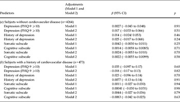
In a first step, we determined the impact of depression on IMT, for participants without (n = 4264) and with (n = 473) a history of cardiovascular disease (CHD, PAD, MI or stroke). Table 2 a shows three sets of predictions: set 1 for categorical depression, set 2 for medical history of depression, and set 3 for the somatic and the cognitive subscales separately. In a stepwise fashion, adjustments were made (1) for sex and age, and (2) for SES, for medical history (FH-MI, diabetes), cardiovascular risk factors [hypertension, dyslipidemia, smoking (ever), BMI and physical activity] and psychotropic medication.
Association of depression with intima–media thickness (IMT)

PHQ-9, Nine-item Patient Health Questionnaire; CI, confidence interval.
Cardiovascular disease includes history of coronary heart disease (CHD), peripheral artery disease (PAD), myocardial infarction (MI) or stroke.
Linear regression with the dependent variable IMT in mm; the predictors depression (PHQ-9 ⩾10) and history of depression were included separately, the predictors somatic and cognitive symptoms of depression were included simultaneously.
Model 1: Age, gender, socio-economic status (SES), interaction age × depression, interaction gender × depression.
Model 2: Model 1 and family history of MI (FH-MI), diabetes, hypertension, dyslipidemia, smoking (ever), body mass index (BMI), activity score, antidepressants, antipsychotics, anxiolytics, antiepileptics, and opioids.
As Table 2 a shows, in participants without evidence for cardiovascular disease, neither the presence of depression nor a history of depression nor the scores of the two subscales were predictive of IMT, regardless of the statistical adjustments made. In regression analyses, we also found no association between history of depression and measures of atherosclerosis (IMT, plaques), regardless of the presence of cardiovascular disease.
Table 2 b shows corresponding analyses for subjects with a history of cardiovascular disease. In participants with a history of cardiovascular disease, neither the presence of depression nor the scores of the two subscales were predictive of IMT, regardless of the adjustments made.
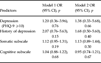
In the next step (see Table 3), we associated current depression and medical history of depression with an increased (‘abnormal’) IMT (>95% percentile according to Sinning et al. Reference Sinning, Wild, Echevarria, Wilde, Schnabel, Lubos, Herkenhoff, Bickel, Klimpe, Gori, Münzel, Blankenberg and Espinola-Klein2011). Again, no associations were found between the presence of depression or the subscales of depression and an increased (‘abnormal’) IMT.
Association of depression with ‘abnormal’ intima–media thickness (IMT), n = 618 (12.8%)

PHQ-9, Nine-item Patient Health Questionnaire; IMT, intima–media thickness; OR, odds ratio; CI, confidence interval.
Logistic regression analysis with the dependent variable ‘abnormal’ IMT (i.e. IMT > 95th percentile of the nomogram as defined by Sinning et al. Reference Sinning, Wild, Echevarria, Wilde, Schnabel, Lubos, Herkenhoff, Bickel, Klimpe, Gori, Münzel, Blankenberg and Espinola-Klein2011); the predictors depression (PHQ-9 ⩾10) and history of depression were included separately, the predictors somatic and cognitive symptoms of depression were included simultaneously.
Model 1: Age, gender, interaction age × depression, interaction gender × depression.
Model 2: Model 1 and family history of myocardial infarction (FH-MI), diabetes, hypertension, dyslipidemia, smoking (ever), body mass index (BMI), activity score, antidepressants, antipsychotics, anxiolytics, antiepileptics, opioids.
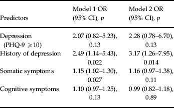
We further tested the association of depression with the presence of carotid plaques. As Table 4 shows, in the crude model (only adjusted for age and sex), there were only associations between history of depression and somatic symptoms of depression and the presence of plaques. In the fully adjusted model, however, the associations disappeared for current somatic symptoms of depression. Only medical history of depression was maintained as a predictor. There were no interactions for age × depression or gender × depression.
Association of depression with carotid plaques, n = 1817 (37.8%)

PHQ-9, Nine-item Patient Health Questionnaire; OR, odds ratio; CI, confidence interval.
Logistic regression analysis with the dependent carotid plaque yes versus no, the predictors depression (PHQ-9 ⩾10) and history of depression were included separately; the predictors somatic and cognitive symptoms of depression were included simultaneously.
Model 1: age, gender, interaction age × depression, interaction gender × depression
Model 2: Model 1 and family history of myocardial infarction (FH-MI), diabetes, hypertension, dyslipidemia, smoking (ever), body mass index (BMI), activity score, antidepressants, antipsychotics, anxiolytics, antiepileptics, opioids.
Discussion
Previous studies had found that only the somatic dimension of depression was strongly related to CHD and to cardiovascular risk factors (Bus et al. Reference Bus, Marijnissen, Holewijn, Franke, Purandare, de Graaf, den Heijer, Buitelaar and Voshaar2011). We therefore hypothesized that somatic depression would also be associated with increased IMT. Contrary to our hypothesis, we did not find any associations between the somatic symptom dimension of depression and IMT, regardless of gender or history of cardiovascular disease. This finding was maintained after stepwise adjustments were made for potential confounders (SES, medical history, cardiovascular risk factors and psychotropic medication). We also found no association between the presence of depression and the cognitive symptom dimension of depression and IMT. Neither were measures of depression associated with ‘abnormal’ IMT (above the 95% percentile). The presence of carotid plaques was associated with somatic symptoms of depression and a medical history of depression in the age- and sex-adjusted model. After fully adjusting for potential confounders, a strong relationship between medical history of depression and the presence of carotid plaques was maintained, with an odds ratio (OR) of 3.17. The association of a previous diagnosis of depression with carotid plaques may reflect long-term consequences of previous depressive episodes. Carotid plaques need time to develop. Therefore, it is more likely that a previous diagnosis of depression as compared to current depressive symptoms is associated with atherosclerotic plaques. We assume that the long-term atherogenic effect of depression results largely from an unhealthy lifestyle (Ye et al. Reference Ye, Muntner, Shimbo, Judd, Richman, Davidson and Safford2013). Unlike the study in a psychiatric sample by Smith et al. (Reference Smith, Blumenthal, Babyak, Doraiswamy, Hinderliter, Hoffman, Waugh and Sherwood2009), we did not find a relationship between late age of onset of depression and atherosclerotic disease.
Thus, our findings from a large community cohort contradict the vascular depression hypothesis, which conceives of depression as the consequence rather than the cause of atherosclerosis. The finding that, after controlling for medical and behavioral risk factors, only recurrent depression was associated with plaques is consistent with the hypothesis that long-standing depression contributes to atherosclerotic disease.
Limitations
Unlike previous studies, we included not only current depression but also history of depression. Because of the cross-sectional design of our study, however, causal inferences about the direction of the associations are precluded. In addition, medical history of depression was only associated with the presence of plaques and not with measures of IMT. We cannot rule out that age was a factor, as the sample in the Dutch study was an average of 10 years older than our community sample, and therefore their median IMT was higher (0.88 mm) than ours (0.65 mm). Nevertheless, the rate of CHD was comparable (7% in both samples). We also cannot rule out methodological differences, namely their use of the Beck Depression Inventory (BDI) versus the PHQ in our study. Compared to previous studies, our study is the largest, based on a representative community sample, differentiating the two symptom dimensions of depression, somatic and cognitive symptoms.
We cannot rule out the possibility that some of the associations may have been confounded by current medication use not yet considered in our analysis, and that the main findings may reflect differences in the severity of co-occurring somatic diseases that have not yet been identified in our study.
The inconsistency of findings in the field may indicate that the relationship between depression and cardiovascular disease is even more complex than previously assumed. There is evidence that symptoms of depression in cardiac patients differ from symptoms in psychiatric patients, with a preponderance of somatic symptoms in cardiac patients, particularly in men (Poole et al. Reference Poole, Dickens and Steptoe2011). Cardiac patients and community subjects may be unaware that they suffer from depression, and they may instead report fewer stigmatizing somatic than cognitive symptoms. Typical symptoms of somatic depression (e.g. sleep disorder, fatigue) are often seen in severe depression; thus somatic depression may indicate more severe depression. In analyzing the relationship between depression and indicators of cardiovascular disease, it has become common practice to adjust for medical history and lifestyle factors such as FH-MI, diabetes, hypertension, dyslipidemia, smoking, obesity or physical inactivity. These lifestyle factors, however, are strongly associated with depression, thus attenuating the relationship. Based on our findings, the pessimism of Bus et al. (Reference Bus, Marijnissen, Holewijn, Franke, Purandare, de Graaf, den Heijer, Buitelaar and Voshaar2011) regarding the ineffectiveness of psychocardiological interventions does not seem to be justified. It may, however, be necessary to resort to a dual approach, targeting both depression and lifestyle variables.
Declaration of Interest
None.