Introduction
Schistosomiasis is a parasitic disease of considerable medical and veterinary importance throughout tropical and subtropical regions, caused by dioecious digeneans of the genus Schistosoma. Schistosomes infect an estimated 180–200 million people worldwide, the majority of whom are in sub-Saharan Africa, and cause chronic diseases that can lead to severe liver, intestinal and bladder pathology and even death (Vos et al., Reference Vos, Abajobir, Abate and Murray2017). Schistosomiasis is also a disease affecting domestic livestock such as cattle, sheep and goats throughout Africa, the Middle East, Asia and some countries bordering the Mediterranean (De Bont and Vercruysse, Reference De Bont and Vercruysse1997, Reference De Bont and Vercruysse1998); however, the true impact of veterinary schistosomiasis is largely unknown.
Schistosomes have a two-host life cycle with an asexual stage occurring in a specific intermediate freshwater snail host and a sexual stage within the definitive mammalian host from which eggs are voided in the urine or feces, depending on the schistosome species involved. The sexual stage of these dioecious parasites enables interactions between male and female worms within their definitive hosts. Most Schistosoma species are host specific and geographically separated, which helps to maintain species barriers; however, given the opportunity, during the sexual stage within the mammalian host, heterospecific crosses between species can occur and have been demonstrated in controlled laboratory experiments. This can lead to either parthenogenesis or hybridization depending on the phylogenetic distance of the species involved. Hybridization, with the production of generations of viable hybrid offspring, will result from heterospecific crosses between closely related species. Hybrids often exhibit several enhanced phenotypic characteristics (hybrid vigour) such as higher fecundity, faster maturation time, higher infectivity, increased pathology and the ability to infect both intermediate snail hosts of the parental species, thereby widening their intermediate host spectrum (Taylor, Reference Taylor1970; Southgate et al., Reference Southgate, Tchuem Tchuenté, Sène, De Clercq, Théron, Jourdane, Webster, Rollinson, Gryseels and Vercruysse2001; Webster, Reference Webster2003).
Advancements in molecular tools, and in particular advances in DNA sequencing, have allowed greater exploration of inter-species interactions particularly between Schistosoma haematobium, which causes human urogenital schistosomiasis, with several of its sister species (Leger and Webster, Reference Léger and Webster2017). One such interaction, discovered through multi locus (mitochondrial cox1 and nuclear ITS) genetic analysis of human miracidia, is with the livestock parasite S. bovis. Schistosoma haematobium/S. bovis hybrids have been identified from samples from school-aged children in Senegal (Huyse et al., Reference Huyse, Webster, Geldof, Stothard, Diaw, Polman and Rollinson2009; Webster et al., Reference Webster, Emery, Webster, Gouvras, Garba, Diaw, Seye, Tchuente, Simoonga, Mwanga, Lange, Kariuki, Mohammed, Stothard and Rollinson2012), Mali (Soentjens et al., Reference Soentjens, Cnops, Huyse, Yansouni, De Vos, Bottieau, Clerinx and Van Esbroeck2016), Niger (Brémond et al., Reference Brémond, Sellin, Sellin, Nameoua, Labbo, Theron and Combes1993) and Benin (Moné et al., Reference Moné, Holtfreter, Allienne, Mintsa-Nguéma, Ibikounlé, Boissier, Berry, Mitta, Richter and Mouahid2015), and more recently in the Mediterranean Island of Corsica (Boissier et al., Reference Boissier, Grech-Angelini, Webster, Allienne, Huyse, Mas-Coma, Toulza, Barré-Cardi, Rollinson, Kincaid-Smith, Oleaga, Galinier, Foata, Rognon, Berry, Mouahid, Henneron, Moné, Noel and Mitta2016). The biological, epidemiological or pathological consequences of this hybridization are as yet unknown but certainly warrant further investigation due to the potential risk of zoonotic transmission, reservoirs of infection, increased transmission and pathology related to hybrid vigour.
The Senegal River Basin (SRB) represents a dynamic transmission hotspot for human and animal schistosomiasis with recognized hybrid zones (Meurs et al., Reference Meurs, Mbow, Vereecken, Menten, Mboup and Polman2012; Webster et al., Reference Webster, Diaw, Seye, Webster and Rollinson2013; Boon et al., Reference Boon, Van Den Broeck, Faye, Volckart, Mboup, Polman and Huyse2018). Since 1985 the SRB has undergone marked anthropogenic ecological changes, which in turn have had a major impact on the transmission of water-borne parasitic diseases such as schistosomiasis (Southgate et al., Reference Southgate, Tchuem Tchuenté, Sène, De Clercq, Théron, Jourdane, Webster, Rollinson, Gryseels and Vercruysse2001; Picquet et al., Reference Picquet, Ernould, Vercruysse, Southgate, Mbaye, Sambou, Niang and Rollinson1996). The town of Richard-Toll (Lower SRB Valley) was exclusively a focus for human intestinal schistosomiasis (S. mansoni) with previous epidemiological studies reporting the lack of urogenital schistosomiasis in this area (Talla et al., Reference Talla, Kongs, Verlé, Belot, Sarr and Coll1990, Reference Talla, Kongs and Verlé1992; Stelma et al., Reference Stelma, Talla, Polman, Sturrock and Gryseels1993). From the early 2000s, technicians reported cases of suspected urogenital schistosomiasis in adults from the local Richard-Toll hospital. Through the molecular characterization of schistosome miracidia isolated from adults visiting the hospital in 2012, this study aimed to gain insights in the epidemiology of urogenital schistosomiasis in this non-target (not included in the mass preventative chemotherapy interventions) group of individuals in the Lower SRB Valley.
Methods
Study area
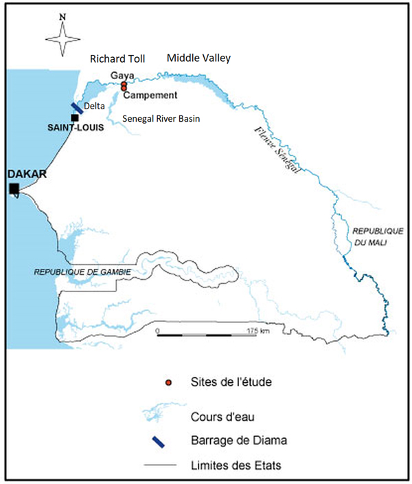
The study area included two neighbourhoods (Campement and Gaya) of Richard-Toll in the Lower Valley of the SRB. Richard-Toll (16°27N, 15°42W) is located 106 km from Saint-Louis on the main road No. 2 in the Department of Dagana, on the South bank of the Senegal River. It is boarded to the North by the Senegal River, to the South by the rural Community of Mbane, to the East by the Gaya's rural Community and to the West by the rural Community of Ronkh (Fig. 1).
Map showing the location of the study sites Gaya and Campement located in the town of Richard-Toll.

Miracidia sampling
In 2012, schistosome miracidia were isolated from 11 infected adult patients that visited the Richard-Toll hospital and who were living in Gaya (n = 5) and Campement (n = 6). Individual urine samples from each patient were filtered through a Nytrel® filter to capture the schistosome eggs and the filter was then put in a petri dish containing freshwater and exposed to artificial light to induce miracidial hatching. Individual miracidia were captured under a dissecting microscope in 3–5 µL of water using a pipette and stored on indicating Whatman FTA cards to capture and preserve the DNA (Webster et al., Reference Webster, Rabone, Pennance, Emery, Allan, Gouvras, Knopp, Garba, Hamidou, Mohammed, Ame, Rollinson and Webster2015).
DNA preparation from FTA cards
FTA discs containing the DNA from the individual miracidium were punched out of the card using a 2 mm Harris Micro Punch and the discs were placed in separate eppendorf tubes. DNA from each individual miracidium was alkaline eluted from the disc as described in Webster et al. (Reference Webster, Rabone, Pennance, Emery, Allan, Gouvras, Knopp, Garba, Hamidou, Mohammed, Ame, Rollinson and Webster2015). Negative controls, FTA discs with no sample, were included.
Identification of schistosome species
Each miracidium was genetically characterized by multi locus analysis of the mitochondrial cox1 region and the nuclear ITS rDNA region (Webster et al., Reference Webster, Emery, Webster, Gouvras, Garba, Diaw, Seye, Tchuente, Simoonga, Mwanga, Lange, Kariuki, Mohammed, Stothard and Rollinson2012).
Diagnostic multiplex PCR (rapid diagnostic PCR) for identification of mtDNA
The mitochondrial cytochrome oxidase subunit 1 (COX1) fragment was amplified from each miracidium using a developed diagnostic PCR that can be used to distinguish rapidly between S. bovis and S. haematobium mtDNA (Webster et al., Reference Webster, Rollinson, Stothard and Huyse2010).
Positive controls with S. haematobium and S. bovis genomic DNA from the Schistosomiasis Collection at the Natural History Museum were used to compare the profiles (Emery et al., Reference Emery, Allan, Rabone and Rollinson2012). Negative controls, with water, were used to check for contamination.
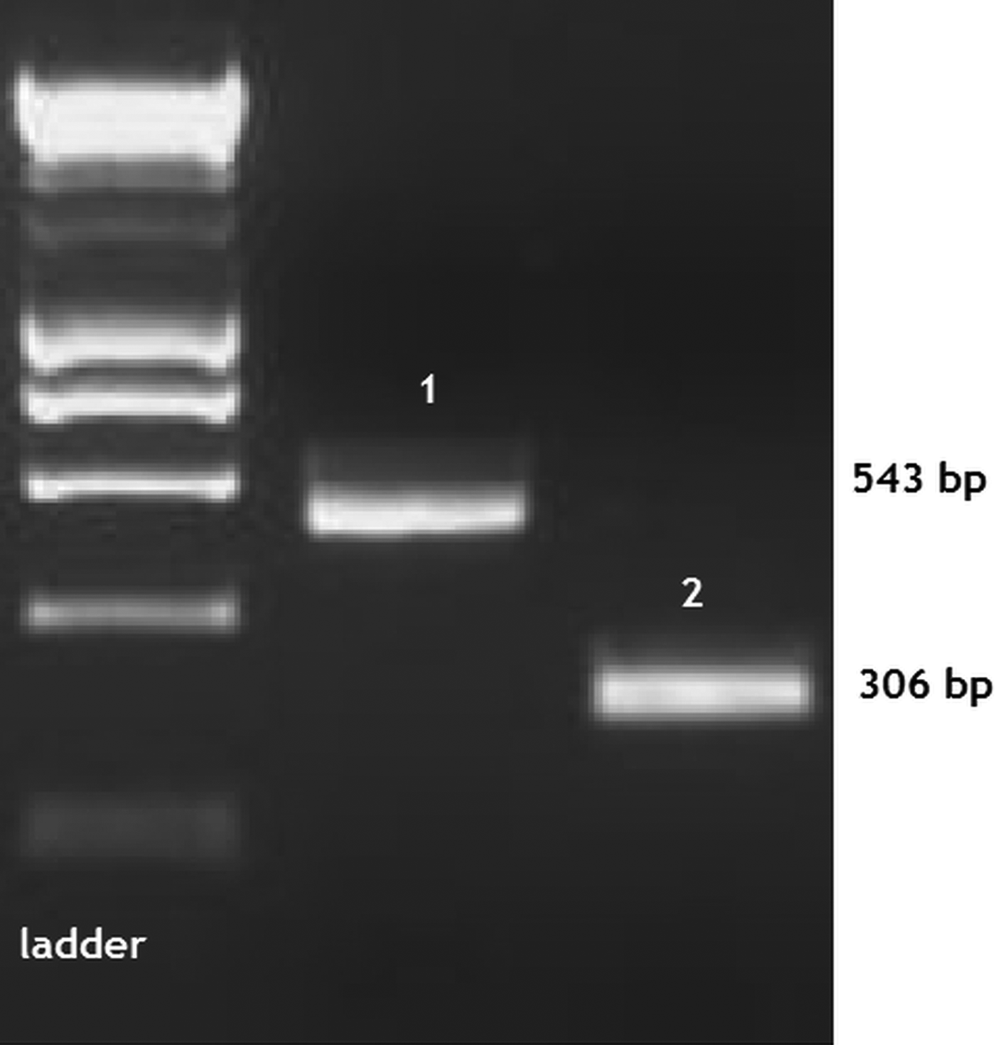
Four microlitres of each PCR product were analysed by electrophoresis on a 2% GelRed agarose gel. Hyperladder IV was used to size the amplicon bands, which were visualized using a UV gel doc system. Species identification of the mtDNA was made according to Webster et al. (Reference Webster, Emery, Webster, Gouvras, Garba, Diaw, Seye, Tchuente, Simoonga, Mwanga, Lange, Kariuki, Mohammed, Stothard and Rollinson2012) and the bands were assigned to either a S. haematobium or S. bovis mtDNA profile (Fig. 2).
Rapid diagnostic multiplex cox1 gel image showing Schistosoma haematobium (1) and S. bovis (2) profiles of different sized PCR amplicon bands. Miracidia were from patients from Richard-Toll (Campement and Gaya). The bands were compared using HyperLadder IV.

Amplification of the nuclear rDNA ITS 1 and 2 for species identification
The complete ITS (1 + 2) nuclear ribosomal DNA region was amplified as described in Webster et al. (Reference Webster, Emery, Webster, Gouvras, Garba, Diaw, Seye, Tchuente, Simoonga, Mwanga, Lange, Kariuki, Mohammed, Stothard and Rollinson2012). The PCR products were visualized on a 0.8% GelRed agarose gel as described above.
Purification and sequencing
All cox1 PCR products that presented a non-S. haematobium profile were sequenced along with a small selection of the S. haematobium cox1 products. All the ITS 1–2 PCR products were sequenced to give a nuclear and mitochondrial genetic profile for each miracidium (Webster et al., Reference Webster, Emery, Webster, Gouvras, Garba, Diaw, Seye, Tchuente, Simoonga, Mwanga, Lange, Kariuki, Mohammed, Stothard and Rollinson2012). The PCR products were first purified using the QIAquick® PCR Purification Kit according to the manufacturer's protocol.
Purified PCR products were sequenced in both directions using a dilution of the PCR primers and the sequences were analysed using an Applied Biosystems, 377 automated sequencer.
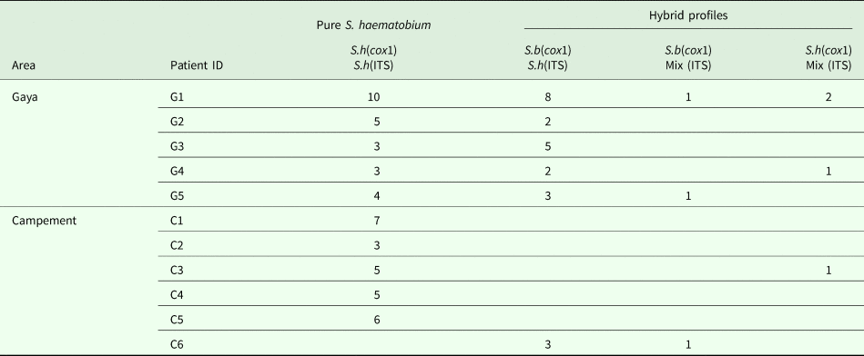
The sequences were viewed and manually edited using Sequencher V4.5 software (Gene Codes Corp., Ann Arbor, Michigan, USA). The identity of the mt cox1 sequences were confirmed using the Basic Local Alignment Search Tool (BLAST) and by comparison to the reference sequences (Webster et al., Reference Webster, Emery, Webster, Gouvras, Garba, Diaw, Seye, Tchuente, Simoonga, Mwanga, Lange, Kariuki, Mohammed, Stothard and Rollinson2012). At the polymorphic positions of the ITS 1 + 2 between S. haematobium and S. bovis, any occurrence of double chromatogram peaks were recorded to identify mixed ITS sequences. The mt and nuclear DNA genetic profiles for each individual miracidium were recorded (Table 1).
Showing the number of individual miracidia that presented the different genetic profiles from samples from five adult patients from Gaya (G1–5) and six adult patients Campement (C1–6).

S.h, Schistosoma haematobium; S.b, S. bovis.
Results
Molecular profiles were obtained from 81 miracidia from 11 patients (six from Campement and five from Gaya). Sequences matched those found in Huyse et al. (Reference Huyse, Webster, Geldof, Stothard, Diaw, Polman and Rollinson2009) Genbank Accession Numbers: FJ58850-62.
Rapid diagnostic cox1 PCR
The rapid diagnostic cox1 PCR from each miracidium showed one of two bands (543 or 306 bp), which are specific to S. haematobium and S. bovis, respectively (Fig. 2). Sixty-eight per cent and 32% of the miracidia tested gave a S. haematobium or S. bovis cox1, respectively.
Nuclear ITS 1 + 2 rDNA
The majority of the miracidia presented a pure S. haematobium ITS sequence but seven miracidia presented a mixed S. bovis/S. haematobium sequence (double chromatograms at the polymorphic sites between S. bovis and S. haematobium).
Pure S. haematobium vs S. haematobium/S. bovis hybrids
Thirty-seven per cent of the miracidia presented a S. bovis/S. haematobium hybrid genetic profile and 63% presented a pure S. haematobium genetic profile. Interestingly many more hybrids were found from patients from Gaya compared with Campement with all five patients from Gaya producing hybrid miracidia and only two out of the six patients from Campement producing hybrids. Of the hybrid miracidia, 83% were from patients from Gaya whereas only 17% were from patients from Campement.
Discussion
The data have confirmed the occurrence of S. haematobium/S. bovis hybrids in adults in the town of Richard-Toll (Lower Valley of the SRB). The hybrid profiles are typical of those found from miracidia analysed from children from neighbouring areas (Webster et al., Reference Webster, Emery, Webster, Gouvras, Garba, Diaw, Seye, Tchuente, Simoonga, Mwanga, Lange, Kariuki, Mohammed, Stothard and Rollinson2012; Boon et al., Reference Boon, Van Den Broeck, Faye, Volckart, Mboup, Polman and Huyse2018). Richard-Toll is an area with a high demographic growth rate and hosts the biggest Senegalese food-processing industry, which attracts thousands of seasonal workers during the period of the harvests. It is not known if the infected patients from this study acquired their infections from neighbouring areas or if there is active transmission in Richard-Toll in the Gaya and Campement regions.
Natural interspecific hybridization of S. haematobium/S. bovis has been observed in the Delta of the Senegal River (Huyse et al., Reference Huyse, Webster, Geldof, Stothard, Diaw, Polman and Rollinson2009; Webster et al., Reference Webster, Diaw, Seye, Webster and Rollinson2013) and also in the Middle Valley (Boon et al., Reference Boon, Van Den Broeck, Faye, Volckart, Mboup, Polman and Huyse2018). Taylor (Reference Taylor1970) and Webster et al. (Reference Webster, Diaw, Seye, Webster and Rollinson2013) also demonstrated, in the laboratory, the feasibility of hybridization between S. haematobium and S. bovis. Several hypotheses were put forward by Huyse et al. (Reference Huyse, Webster, Geldof, Stothard, Diaw, Polman and Rollinson2009) and Webster et al. (Reference Webster, Diaw, Seye, Webster and Rollinson2013) to explain the mechanism enabling introgressive hybridization and to predict any potential consequence. In these particular areas of Richard-Toll, it is necessary to undertake more studies related to the epidemiology of human and livestock schistosomiasis transmission and also to identify active local transmission areas that harbour the Bulinus intermediate snail hosts.
In conclusion, this study provides conclusive evidence for the occurrence of urogenital schistosomiasis in Richard-Toll, the presence of S. haematobium/S. bovis hybrids in this area and also hybrid infections in adults. Since the construction of the Diama Dam at the mouth of the Senegal River and the Manantali Dam in Mali, the transmission of both urogenital and intestinal schistosomiasis has been extremely dynamic along the SRB with foci appearing and disappearing at a rapid rate. There are four schistosome species (S. mansoni and S. haematobium infecting humans and, S. bovis and S. curassoni infecting animals) circulating within these foci complicating the epidemiology and also possibly leading to hybridization or competition between species. These dynamics need to be monitored closely so that the best forms of control can be implemented and more importantly new foci are not missed from treatment programmes. Our data were obtained from infected adults that were visiting a hospital clinic and these patients would not actively be incorporated into a national treatment programme, which is not only detrimental to the health of these patients but also to the potential success of control in these types of highly dynamic schistosomiasis foci.
Acknowledgements
Thanks to the Programme d'Appui à l'Enseignement Supérieur de l'Union Economique Monétaire Ouest Africain (PAES/UEMOA) and the University Gaston Berger for financing the fieldwork and Mariama Sene-Wade's travel and accommodation in London. Thanks also to the Natural History Museum Sequencing Facility for generating the sequence data. Thanks to the Natural History Museum for hosting Mariama Sene-Wade during her research visit in 2013. Thanks also to the patients for their participation in the study.
Financial support
The authors would also like to acknowledge the Programme d'Appui à l'Enseignement Supérieur de l'Union Economique Monétaire Ouest Africain (PAES/UEMOA) and the University Gaston Berger and the Natural History Museum's Departmental Investment Fund for the financial support facilitating the molecular work.
Conflict of interest
None.
Ethical statement
All adult patients had come to the clinic at Richard-Toll for urogenital schistosomiasis diagnosis. All infected patients gave oral consent for the characterization of their infections. All patients received treatment, from the clinic, with Praziquantel. All data were analysed anonymously.