Introduction
Superior canal dehiscence syndrome describes audiovestibular symptoms caused by the (partial) absence of the bony covering of the superior semicircular canal. It is a relatively new diagnosis, first described in 1998 by Minor et al.Reference Minor, Solomon, Zinreich and Zee 1 Superior semicircular canal dehiscence has a radiological incidence of 1 per cent in the general population, but the anatomical incidence and the incidence of the associated syndrome appears to be lower.Reference Crovetto, Whyte, Rodriguez, Lecumberri, Martinez and Eléxpuru 2
Superior canal dehiscence syndrome patients develop symptoms related to hearing and balance, including autophony, aural fullness, sound- or pressure-induced vertigo or oscillopsia, chronic disequilibrium, conductive hyperacusis, tinnitus, and hearing loss.Reference Minor, Solomon, Zinreich and Zee 1 , Reference Minor 3 Examination findings include vertical torsional eye movements when tragal pressure or supra-threshold sound stimulation is applied. Audiometry typically shows an air–bone gap in the lower frequencies and supranormal bone conduction thresholds.Reference Minor 3 , Reference McCall, McKenna, Merchant, Curtin and Lee 4 The diagnosis is supported by a dehiscence of the superior semicircular canal shown on high-resolution computed tomography (CT) imaging of the temporal bone.Reference Crovetto, Whyte, Rodriguez, Lecumberri, Martinez and Eléxpuru 2 Decreased vestibular-evoked myogenic potential thresholds and increased vestibular-evoked myogenic potential amplitudes also provide supportive evidence for the diagnosis.
The dehiscence is most commonly located in the apical region of the superior semicircular canal, at the arcuate eminence.Reference Crovetto, Whyte, Rodriguez, Lecumberri, Martinez and Eléxpuru 2 , Reference Lookabaugh, Kelly, Carter, Niesten, McKenna and Curtin 5 Rarely, superior canal dehiscence syndrome is caused by a dehiscence of the medial wall of the superior semicircular canal, bringing the superior semicircular canal into contact with the superior petrosal sinus.
Here, we review the literature and report three further cases of superior canal dehiscence syndrome caused by erosion of the superior semicircular canal by the superior petrosal sinus.
Case reports
Case one
A 51-year-old man presented with balance problems provoked by physical strain. The problems had commenced a year before presentation, with a short episode of transient vertigo provoked by head movement. He described left-sided hearing loss since the age of nine years, when he sustained a head injury. He also experienced pulsatile tinnitus and abnormal perception of internal sounds. A Valsalva manoeuvre induced nystagmus in the vertical plane. Pure tone audiometry demonstrated a sensorineural loss with non-serviceable hearing on the left side and normal hearing on the right side. The CT scans revealed a small defect in the bony part of the left superior semicircular canal, adjacent to the superior petrosal sinus. In light of saccule and/or inferior vestibular nerve hypofunction on the left-hand side, vestibular-evoked myogenic potentials could not be used to further support the diagnosis.
Case two
A 40-year-old woman presented with a 4-year history of episodic vertigo, pulsatile tinnitus, aural fullness in the left ear, and autophony with difficulty hearing in background noise. The episodes of vertigo lasted seconds and were provoked by temperature and pressure changes, speaking, laughter, and head movement. She had a heightened awareness of internal sounds, including her own eye movements, her footsteps and the turning of her neck. Her audiogram demonstrated no significant hearing asymmetry, although left-sided supranormal bone-conducted hearing was measured. Vestibular-evoked myogenic potentials testing showed a significantly lower threshold on the left side in comparison to the right. Computed tomography and magnetic resonance (MR) imaging showed a short segment of dehiscence of the left superior semicircular canal, in close relation with the superior petrosal sinus.
Case three
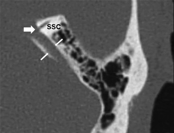
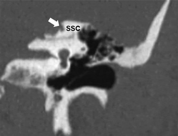
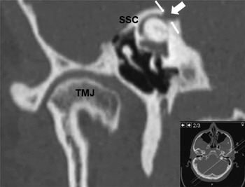
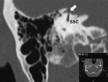
A 54-year-old man visited our clinic with complaints of left-sided hearing loss, aural fullness, hyperacusis and ongoing tinnitus that was sometimes pulsatile in nature. He did not have any complaints of balance disturbance or vertigo. The symptoms had been present for 30 years. His past medical history mentioned a minor head trauma as a child. Audiometry showed a mixed-type hearing loss on the left side. The CT report did not mention any abnormalities; however, after review, a dehiscence of the superior semicircular canal was observed medial to the arcuate eminence, in contact with the superior petrosal sinus (Figures 1–4). This was confirmed by a MR venogram. No other causes of the mixed hearing loss were found.
High-resolution, axial computed tomography image of the left petrous bone in case three. The broad arrow indicates the dehiscence in the medial part of the superior semicircular canal (SSC), which is in contact with the superior petrosal sinus (indicated by the narrow arrows).

High-resolution, coronal computed tomography image of the left petrous bone in case three. The medial location of the superior canal dehiscence is shown (broad arrow). SSC = superior semicircular canal

Pöschl projection high-resolution computed tomography image of the left petrous bone in case three. The dehiscence (indicated by the broad arrow) of the superior semicircular canal (SSC) is seen as a ‘cookie bite’ (narrow arrows) out of its bony cover, formed by the groove containing the superior petrosal sinus. TMJ = temporomandibular joint

Stenvers high-resolution computed tomography image of the left petrous bone in case three. The dehiscence of the medial part of the superior semicircular canal (SSC, indicated by the broad arrow) is clearly visible.

Diagnosis
In all three cases, the combination of specific audiovestibular symptoms, combined with the radiological finding of a dehiscent bony cover of the superior semicircular canal, are strongly suggestive of the diagnosis of superior canal dehiscence syndrome.
Discussion
Superior canal dehiscence is most frequently found at the apex of the superior semicircular canal, but can also be found at the posteromedial border, in close relation to the superior petrosal sinus.
The exact aetiology of semicircular canal dehiscence is yet to be determined. Takahashi et al. have hypothesised that semicircular canal dehiscence may arise during fetal development. Adhesion of the membranous labyrinth to the dura mater may develop if the superior semicircular canal has protruded directly to the middle cranial fossa. If such an adhesion develops before complete ossification of the bony labyrinth, it may prevent the complete bony covering of the superior semicircular canal apex. This hypothesis does not explain posteromedial dehiscences adjacent to the superior petrosal sinus. It also does not explain the observation that the onset of symptoms in most superior canal dehiscence syndrome patients takes place in adult life.
Suggested risk factors for the development of superior canal dehiscence syndrome symptoms in semicircular canal dehiscence patients include infection, neoplasm, and (minor) head trauma or barotrauma.Reference Carey, Minor and Nager 6 , Reference Takahashi, Tsunoda, Shirakura and Kitamura 7
In Carey's study of semicircular canal dehiscence, including dehiscences adjacent to the superior petrosal sinus, the structure of the bone surrounding the site of the semicircular canal dehiscence is lamellar, indicating no abrupt bony change. This finding perhaps points towards an error in development, rather than post-traumatic erosion of the bone.Reference Carey, Minor and Nager 6
Superior canal dehiscence syndrome caused by posteromedial dehiscence of the superior canal, resulting in contact between the superior semicircular canal membrane and the superior petrosal sinus wall, seems to be quite rare. We searched the relevant literature published until January 2017, using PubMed and Embase databases, with the search terms ‘canal dehiscence’ and ‘superior petrosal sinus’. We identified 4 articles with a total of 27 cases of posteromedial semicircular canal dehiscence adjacent to the superior petrosal sinus causing superior canal dehiscence syndrome. These articles describe clinical symptoms and signs associated with superior canal dehiscence syndrome, and positively identify a superior canal dehiscence in contact with the superior petrosal sinus on high-resolution CT.Reference McCall, McKenna, Merchant, Curtin and Lee 4 , Reference Lookabaugh, Kelly, Carter, Niesten, McKenna and Curtin 5 , Reference Koo, Hong, Kim and Kim 8 , Reference Puwanarajah, Pretorius and Bottrill 9
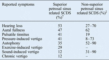
It is estimated that semicircular canal dehiscence with posteromedial localisation accounts for 4–9 per cent of ears with semicircular canal dehiscence.Reference Crovetto, Whyte, Rodriguez, Lecumberri, Martinez and Eléxpuru 2 , Reference McCall, McKenna, Merchant, Curtin and Lee 4 , Reference Lookabaugh, Kelly, Carter, Niesten, McKenna and Curtin 5 In 14 of the 27 reported cases, the related symptoms are adequately described.Reference McCall, McKenna, Merchant, Curtin and Lee 4 , Reference Koo, Hong, Kim and Kim 8 , Reference Puwanarajah, Pretorius and Bottrill 9 The symptoms reported by these 14 patients did not differ considerably from the symptoms reported by the 3 patients in this study. When we combined these patients with ours (n = 17), we found that the most common symptoms of superior canal dehiscence syndrome associated with posteromedial semicircular canal dehiscence in communication with the superior petrosal sinus wall were: hearing loss (53 per cent), aural fullness (47 per cent), pulsatile tinnitus (41 per cent) and pressure-induced vertigo (41 per cent) (Table I).Reference Minor 3 , Reference McCall, McKenna, Merchant, Curtin and Lee 4 , Reference Koo, Hong, Kim and Kim 8 – Reference Zhou, Gopen and Poe 10 Sound-induced vertigo appeared to be the least common symptom, appearing in only 12 per cent of the patients; however, 29 per cent of the superior canal dehiscence syndrome patients experienced exercise-induced vertigo, which may be triggered by the increased body and blood temperature, causing caloric stimulation of the superior semicircular canal through its close relation with the superior petrosal sinus.
Superior canal dehiscence syndrome symptoms according to dehiscence localisation*

*Superior canal dehiscence syndrome caused by a medial dehiscence in contact with the superior petrosal sinus versus other dehiscence localisations. † n = 17;Reference McCall, McKenna, Merchant, Curtin and Lee 4 , Reference Koo, Hong, Kim and Kim 8 , Reference Puwanarajah, Pretorius and Bottrill 9 ‡ n = unknown.Reference Minor 3 , Reference Zhou, Gopen and Poe 10 SCDS = superior canal dehiscence syndrome
Compared to patients with a posteromedial semicircular canal dehiscence associated with the superior petrosal sinus, patients with ‘normal’ apical superior canal dehiscence syndrome experience tinnitus less often (in 19 per cent), especially pulsatile tinnitus.Reference Zhou, Gopen and Poe 10 , Reference Liu, Bi, Li, Li, Dong and Zhao 11 Patients with apical semicircular canal dehiscence also experience exercise-induced vertigo less frequently, but suffer from sound-induced vertigo more frequently (occurring in 31–90 per cent).Reference Minor 3 , Reference Zhou, Gopen and Poe 10 However, a distinct superior canal dehiscence syndrome symptom or combination of symptoms reliably differentiating between the two types of semicircular canal dehiscence cannot be identified.
The diagnosis of superior canal dehiscence syndrome should be based on a combination of clinical criteria and high-resolution CT findings. However, an apparent dehiscence on a CT scan does not necessarily result in a diagnosis of symptomatic superior canal dehiscence syndrome.Reference Crovetto, Whyte, Rodriguez, Lecumberri, Martinez and Eléxpuru 2 , Reference Carey, Minor and Nager 6 Conversely, the diagnosis of superior canal dehiscence syndrome can still be made when a bony cover over the superior semicircular canal is debatable, but the history, audiometry and vestibular-evoked myogenic potentials testing clearly suggest superior canal dehiscence syndrome.
In order to diagnose semicircular canal dehiscence on high-resolution CT of the temporal bone, 0.5–0.6 mm collimated axial images with multiplanar reconstructions should be used.Reference Crovetto, Whyte, Rodriguez, Lecumberri, Martinez and Eléxpuru 2 On axial images, the bony groove of the superior petrosal sinus can be recognised in communication with the superior semicircular canal (Figure 1).Reference McCall, McKenna, Merchant, Curtin and Lee 4 On coronal high-resolution CT reconstructions, the medial location of the semicircular canal dehiscence can be recognised (Figure 2). However, given the uncommon medial location of the dehiscence, the diagnosis can be easily missed, especially on normal axial and coronal high-resolution CT reconstructions. The characteristic sign indicative of semicircular canal dehiscence caused by the superior petrosal sinus is the presence of a ‘cookie bite’ out of the bone covering the medial part of the superior semicircular canal on Pöschl projection CT images (Figure 3). Furthermore, the resolution of CT is such that it is not always possible to differentiate a very thin bony covering from a true dehiscence because of the ‘partial volume effect’.Reference Crovetto, Whyte, Rodriguez, Lecumberri, Martinez and Eléxpuru 2 In order to ascertain the location of the superior petrosal sinus, CT scans with contrast or a MR venogram of the brain can be used.
If avoidance of provocative stimuli is not enough to control symptoms, patients can opt for surgical management. This can be performed via either a middle fossa or transmastoid approach. As of yet, there are no known reports that directly compare the two surgical approaches for superior canal dehiscence syndrome caused by a posteromedial location of the dehiscence, but there are theoretical arguments for plugging via the transmastoid approach. Given the medial location of the dehiscence, it might be necessary to retract the temporal lobe in order to visualise the dehiscence when performing the middle fossa approach. Furthermore, mobilising the superior petrosal sinus off the site of dehiscence could cause profuse bleeding and make the procedure more difficult.Reference McCall, McKenna, Merchant, Curtin and Lee 4 The transmastoid approach with plugging of the superior canal has a reported success rate of 94 per cent, with preservation of hearing.Reference Beyea, Agrawal and Parnes 12 However, the location of the dehiscence in the superior semicircular canal was not specified,Reference Beyea, Agrawal and Parnes 12 and the success rate of this procedure has not yet been described separately for posteromedial semicircular canal dehiscence.
-
• Superior canal dehiscence syndrome is most commonly caused by dehiscence at the superior semicircular canal arcuate eminence
-
• Thirty cases of superior canal dehiscence syndrome caused by posteromedial dehiscence, in contact with superior petrosal sinus, have now been reported
-
• Pulsatile tinnitus, hearing loss, aural fullness and pressure-induced vertigo are the most common symptoms
-
• Dehiscence at the superior petrosal sinus is recognised by a ‘cookie bite’ out of the superior semicircular canal medial wall
-
• This typical finding can be identified on Pöschl projection high-resolution computed tomography imaging
Conclusion
Pulsatile tinnitus, hearing loss, aural fullness and pressure-induced vertigo are the most common symptoms in superior petrosal sinus related superior canal dehiscence syndrome. Although no pathognomonic set of symptoms of SPS-related SCDS can be identified, pulsatile tinnitus and exercise induced vertigo are more frequent, while sound induced vertigo and autophony are less frequent findings compared to SCDS in which the dehiscence has a more common apical location. Superior petrosal sinus related semicircular canal dehiscence can be recognised on high-resolution CT axial images by the bony groove of the superior petrosal sinus in close contact with the medial aspect of the superior semicircular canal. However, given the uncommon localisation of the dehiscence, the diagnosis can be easily missed. Additional Pöschl high-resolution CT reconstructions may facilitate its identification through the typical ‘cookie bite’ defect.