Introduction
Rapid, divergent evolution of male genitalia that result in even closely related species having distinct genitalic morphologies is one of the most general patterns in animal evolution (Eberhard, Reference Eberhard1985; Shapiro & Porter, Reference Shapiro and Porter1989; Hosken & Stockley, Reference Hosken and Stockley2004). Numerous hypotheses have been proposed to explain this trend (summary, Eberhard, Reference Eberhard1985; also Jocqué, Reference Jocque1998; Simmons and Siva-Jothy, Reference Simmons, Siva-Jothy, Birkhead and Møller1998; Simmons, Reference Simmons2001; Singh & Kulanthinal, Reference Singh and Kulathinal2005). Those most commonly cited in recent studies are mechanical lock-and-key (Shapiro & Porter, Reference Shapiro and Porter1989), sexual selection by cryptic female choice (Eberhard, Reference Eberhard1985) and sexually antagonistic coevolution (Chapman et al., Reference Chapman, Arnqvist, Bangham and Rowe2003). The lock-and-key hypothesis predicts species-specific female structures that match those of the males, and that can prevent coupling with males of other closely related species. The antagonistic coevolution hypothesis also predicts a physical match, except in cases in which species-specific behaviour rather than morphology of the female has selected for the species-specific traits of the male (Eberhard, Reference Eberhard2004a,Reference Eberhardb). The cryptic female choice hypothesis, in contrast, predicts frequent lack of complementary structures or behaviour in the female. It also predicts frequent cases in which males perform movements with their genitalia during copulation that are apparently designed to stimulate the female. Tsetse flies (Glossina spp.) have highly modified, species-specific male genitalia (Buxton, Reference Buxton1955; Potts, Reference Potts and Mulligan1970) and are, thus, appropriate subjects to test these ideas.
A second reason for studying the reproduction of tsetse flies is that they are vectors of human and animal trypanosomiasis in substantial parts of sub-Saharan Africa, where they have important medical and economic impacts (Gooding & Krafsur, Reference Gooding and Krafsur2005; Feldman et al., Reference Feldmann, Dyck, Mattioli, Jannin, Dyck, Hendrichs and Robinson2005). A detailed understanding of the sexual behaviour of these flies and, in particular, of the events that occur during copulation may aid efforts to control wild populations using the sterile insect technique (SIT) as part of area-wide integrated pest management programs (Wall & Langley, Reference Wall and Langley1993). The use of sterile males to reduce the density of wild populations depends on successful copulations of the released sterile males with wild females that result in sperm transfer, and unsuccessful copulations can also reduce the productivity of the colonies used to produce males for sterilization and release.
Events that occur during copulation in Glossina affect not only insemination of the female, but also her tendency to ovulate and to remate. Mechanical stimuli sensed during copulation are responsible in G. morsitans Westwood for triggering ovulation by the female (Saunders & Dodd, Reference Saunders and Dodd1972). This study ruled out stimuli from the sperm, the spermatophore, products of the accessory glands, testes and ejaculatory ducts of the male and humoral factors from the spermathecae of inseminated females as being possible triggers. By interrupting copulation and then pairing the female with one or more additional males, it was found that the effects of these stimuli ‘add up’ over the course of a copulation. Artificial mechanical stimulation of the uterus with a glass bead also increased the rate of ovulation, but not as much as natural copulation (Chaudhury & Dhadialla, Reference Chaudhury and Dhadialla1976).
Rapid induction of female resistance to further mating is a second consequence of both chemical and physical stimuli associated with copulation in G. morsitans (Gillot & Langley, Reference Gillot and Langley1981). Male accessory gland substance(s), as well as mechanical distension of the uterus (where the spermatophore is deposited) and/or stimulation of the tip of the female abdomen (which is clasped by the male during copulation), increased female refractoriness.
A third set of processes that may be affected by events during copulation includes intromission and sperm transfer. Little active female co-operation seems to be needed by the male to accomplish the early stages of copulation successfully, as 40 of 64 males paired in tubes copulated with recently freeze-killed females and performed the terminal ‘jerking’ behaviour that is associated with ejaculation (Jaenson, Reference Jaenson1979a,Reference Jaensonb). Not all copulations (=genitalic couplings) result in insemination, however (Vanderplank, Reference Vanderplank1948; Jaenson, Reference Jaenson1979a,Reference Jaensonb; Vreysen & van derVloedt, Reference Vreysen and van der Vloedt1990), and internal female responses during copulation may be important, as only 10 (25%) of these males transferred sperm and spermatophore material to the female's uterus (Jaenson, Reference Jaenson1979a). ‘Normal’ copulations by Glossina in captivity can also show substantial rates of failure; copulations in captivity of five species (palpalis, fuscipes, tachinoides, morsitans and swynnertoni) showed failure rates ranging from 19 to 33% (Vanderplank, Reference Vanderplank1948). Insemination can apparently be affected by the female. There was a significant correlation between the degree of filling of the spermathecae and female receptivity in females of G. pallidipes of different ages (Davies-Cole & Chaudhury, Reference Davies-Cole and Chaudhury1990). Under coercive circumstances, in which the female could not bend the tip of her abdomen ventrally out of reach of the genitalic clasp of the cerci of a male that had mounted her in the confines of a glass tube, females (apparently of pallidipes, morsitans and swynnertoni) often mated but then separated after only abbreviated genitalic coupling; ‘the act lasts only a few minutes and it is doubtful if any sperm is passed’ (Vanderplank, Reference Vanderplank1947; see also Jaenson, Reference Jaenson1979a). Short copulations of this sort usually result in no sperm transfer, although a few sperm were found in occasional females (Vanderplank, Reference Vanderplank1948). The powerful male genitalic clamps on the female (Vanderplank, Reference Vanderplank1948; this study) make it clear that females cannot directly force separation, so premature separations are probably ultimately due to male decisions, perhaps due to female resistance to intromission or deeper penetration.
In summary, the events during copulation are likely to be especially important for understanding reproduction in tsetse flies; ‘… the time spent in copulation prior to accessory secretion or sperm transfer is therefore an important component of the mating experience of female tsetse flies’ (Tobe & Langley, Reference Tobe and Langley1978). This study of Glossina pallidipes Austen 1903 provides the most detailed description of copulatory behaviour ever made in Glossina, with emphasis on the previously undescribed male genitalic behaviour and its possible functions. It is also meant to serve as a point of comparison for future comparative studies of other species.
G. pallidipes is widespread in Africa, where it is an important vector of trypanisomiasis (Potts, Reference Potts and Mulligan1970; Feldman et al., Reference Feldmann, Dyck, Mattioli, Jannin, Dyck, Hendrichs and Robinson2005). It is more difficult to breed in captivity than some other Glossina species (Davies-Cole & Chaudhury, Reference Davies-Cole and Chaudhury1990). As in other Glossina, the female of G. pallidipes produces a species-specific contact pheromone that induces a conspecific male to extend his hypopygium and seize her abdomen (Vanderplank, Reference Vanderplank1947; Wall & Langley, Reference Wall and Langley1993). Copulation duration is relatively brief compared with other species, and mean times usually range from 23 to 30 min (Rogers, Reference Rogers1973; Wall, Reference Wall1988; Leegwater-van der Linden & Tiggelman, Reference Leegwater-van der Linden and Tiggelman1984; Davies-Cole & Chaudhury, Reference Davies-Cole and Chaudhury1990; Olet et al., Reference Olet, Opiyo and Robinson2002), although the mean durations observed by Jaenson (Reference Jaenson1979b) ranged from 43 to 96 min for females of different ages. Sperm are transferred in a spermatophore (Wall & Langley, Reference Wall and Langley1993), and the spermatophore is passed to the female only during the last approximately 30 s of copulation; transfer is often associated with a specific behaviour (‘jerking’) that has been attributed to the male (Jaenson, Reference Jaenson1979a; Leegwater-van der Linden & Tiggelman, Reference Leegwater-van der Linden and Tiggelman1984; but see below).
Male and female behaviour during copulation is elaborate in G. pallidipes and has been described in some detail. Jaenson (Reference Jaenson1979a) recognized three stages: an early stage of active male courtship, a final stage with active male ‘jerking’, and a long intermediate stage with intermittent female buzzing. A later study (Leegwater-van der Linden & Tiggelman, Reference Leegwater-van der Linden and Tiggelman1984) was ‘basically in agreement’ with Jaenson's observations, but recognized a distinct final stage of male jerking in which the jerking movements became more frequent and continuous (‘continuous jerking phase’). The elaborate male behaviour, much of which appears designed to stimulate the female, may function as copulatory courtship to influence cryptic female choice by the female (Eberhard, Reference Eberhard1994, Reference Eberhard1996).
To summarize, previous studies have shown that copulation behaviour in G. pallidipes is complex. We show here that even these descriptions are substantially incomplete. We report additional, previously undescribed, behaviour patterns of both the male and the female that occur on the ‘outside’ of the female, including especially elaborate, diverse and sustained movements of the male genitalic cerci. We also describe possible male genitalic movements within the female's reproductive tract and nearby that may also be stimulatory in function.
Materials and methods
All flies were 10–12-day-old virgins of a mass reared stock at the FAO/IAEA Agriculture and Biotechnology Laboratory, Seibersdorf, Austria, which was founded with specimens collected in Tororo Uganda, kept in Amsterdam for 2–3 years, and then maintained in Seibersdorf since 1980. All experimental flies were kept at 23.5–24.0°C and 75–78% RH, with lights on at 08:00 and off at 16:00 and offered a blood meal of frozen and thawed bovine blood through a silicone membrane three times per week. Behavioural observations were made on recently fed flies in a room at 24.5–25°C and 53–55% humidity, using video recordings made with a digital camera equipped with +7 close-up lens. Fifteen pairs were filmed in 15×19.5×10.5-cm plexiglass cages with the entire body of both flies visible, to obtain records of non-genitalic behaviour.
Another eight pairs were filmed through a dissecting microscope to obtain records of genitalic movements. Virgin male and female flies were first placed together in glass tubes. Once the male had mounted the female and seized her abdomen with his genitalia, they were transferred to a small petri dish (6 cm diameter, 1.5 cm deep) where observations and filming commenced. Sounds were recorded on the video recordings using a Sennheiser MZK 80Zu microphone in the wall of the petri dish. Our behaviour records began approximately 30 s or less after the male had seized the female with his genitalia.
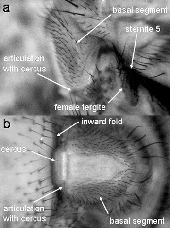
Several behaviour patterns were characterized on the basis of recordings from pairs in which this behaviour particularly was clear due to the angle of view. Some details were only visible when the viewing and lighting angles were favourable, and it was not possible to determine whether they occurred at other times; these are indicated by the phrase ‘in some cases’. We used the articulation between the male's basal segment and his cerci as a reference point to determine the strength of genitalic squeezes (fig. 1).
(a) Lateral and (b) ventral views of male genitalic grasp on the female as seen in video recordings. Most of the length of the cercus is out of sight in the inward fold of the female's abdominal wall (female below in (a), to left in (b)).

Internal events were determined by flash-freezing copulating pairs in liquid N2 at times ranging from 15 to 120 min after copulation began. Details of the copulation behaviour of these pairs were not observed. The container of liquid N2 was placed in a freezer at −20°C and, after the N2 boiled off, the frozen flies were flooded (while still in the freezer) with −20°C absolute ethanol. By leaving the container in the freezer for another week, the flies were fixed by the alcohol without their having thawed, thus guaranteeing that their positions did not change following freezing (it is possible that positions may have changed during the fraction of a second when the pair was being frozen). After being fixed, pairs were held at room temperature until they were dissected. A total of 22 copulating pairs were dissected to determine the positions of male and female genitalia. Some male genitalia from copulating pairs were isolated and examined in the SEM. Designations of the positions of male genitalia during copulation (anterior, ventral, etc.) refer to the axes of the female, unless stated otherwise.
The names used for the male genitalic structures follow recommendations by D.M. Wood (personal communication). Unfortunately, Glossina workers have utilized several ‘private’ terms. Because of the highly derived morphology in this genus, we were able to homologize only some of these with terms used for other Diptera (the terms we use are italicized); the ‘superior claspers’ of previous authors (e.g. Buxton, Reference Buxton1955; Potts, Reference Potts and Mulligan1970; Pollock, Reference Pollock1973) are his highly modified cerci; the ‘eighth dorsal sclerotization’ (Pollock, Reference Pollock1973) is the basal segment; the ‘editum’ (Pollock, Reference Pollock1973) is his surstylus; the ‘penis and harpes’ are his hypandrium and phallus complex; the ‘paramere’ (Patton, Reference Patton1936), the ‘basal plate’ (Jackson, Reference Jackson1952), the ‘ninth tergo-sternum’ (Patton, Reference Patton1934), the ‘inferior clasper’ (Potts, Reference Potts and Mulligan1970), ‘anterior paramere’ (Pollock, Reference Pollock1973) and the ‘ventral bar of sternite nine’ (Pollock, Reference Pollock1973) is either his pregonite or his gonocoxite (in view of this uncertainty, we use the traditional and functionally descriptive term inferior clasper); and the ‘vesicae’ (Patton, Reference Patton1934) are inflatible sacs of the phallus complex. The names we use for other male and female structures are either purely descriptive, with no attempt to indicate homologies, or are as in Buxton (Reference Buxton1955).
Results
Behaviour of male genitalic structures
Morphology
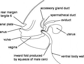
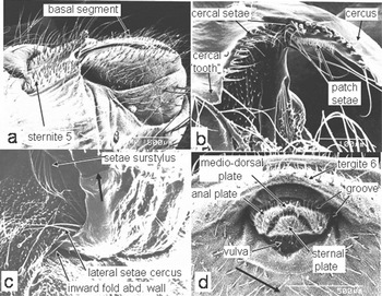
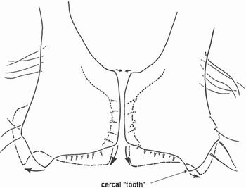
A brief overview of female and male genitalic morphology emphasizing the portions directly involved in copulation is needed to understand the behavioural descriptions below. The female vulva opens ventrally near the tip of her abdomen (fig. 2). The ventral surface of abdominal segments 2–6, anterior to the vulva, is membranous; the last major tergite is that of segment 6 (fig. 2). The anus is bounded by three setose cuticular plates, laterally and dorsally by a pair of anal plates that are connected medially by a medio-dorsal plate and ventrally by a sternal plate (fig. 3d) that is thought to function to keep the vulva (which is just anterior and ventral to the sternal plate) clear of feces (Potts, Reference Potts and Mulligan1970). The anterior margins of the anal and medio-dorsal plates are in a deep, approximately circular, inward fold or groove of the external body wall (fig. 3d).
Schematic layout of the female body wall and reproductive tract (positions approximately as during copulation; details of the folds in the wall of the oviduct near the openings of the uterine gland and spermathecal ducts and the uterus varied in different pairs).

Male and female genitalia and associated structures. (a) tip of male abdomen in resting position in latero-ventral view, showing the array of strong spines on the heavily sclerotized sternite 5 and the basal segment; the cerci are out of sight, folded beneath the basal segment. (b) Distal edges of cerci seen end-on, showing the lateral tooth, their curved shape and their array of strong, pointed setae. (c) Male cerci (largely out of sight) clasp female abdomen during copulation, causing a deep inward fold in the female abdominal wall; the long setae on the surstylus and the lateral edge of the cercus contact female membranes (heavy black arrow indicates articulation of cercus with basal segment). (d) Tip of the female abdomen, showing the groove distal and lateral to her anal plates (‘groove’) into which the setae on the inferior claspers were inserted during copulation. The heavy black arrow indicates approximate site where the male cerci fold the female's body wall inward during copulation.

The male's genitalic and perigenitalic structures can be conveniently divided into those that enter the female's reproductive tract and those that remain outside during copulation. Details of the structures that are introduced into the female (mainly the aedeagus) will be described later in association with the internal events that occur during copulation, while the non-intromittent male structures are reviewed here.
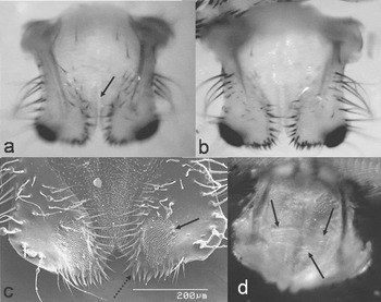
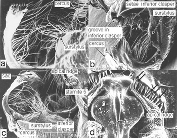
The two cerci curve nearly 90° along their length. There is a large, flattened, dark ‘tooth’ near the lateral-distal corner of each cercus (fig. 4a,b). The two cerci are not ‘fused medially at the distal extremity’ as maintained by Potts (Reference Potts and Mulligan1970). Instead, their medial margins are clearly separated ventrally (fig. 4a,b); their nearly linear (slightly convex) margins are more closely apposed dorsally (arrow in fig. 4a), but are nevertheless separate and capable of independent movement (below). Both the concave curved distal margins of the cerci and the distal portions of their ventral medial margins have multiple robust sharp, dark spines (figs 3–5). There is also a row of especially long, thinner setae along the lateral margin of each cercus (figs 3 and 5a–c), and a patch of very small setae on the dorsal surface (solid arrow in fig. 4c). Bands of muscle fibers originate near the midline, and are inserted along the length of each cercus (arrows in fig. 4d). Otherwise, the cerci have no obvious muscles.
The distal portions of the two isolated male cerci (distal edge downward) in (a) dorsal and (b) ventral view, showing the large, dark setae on their distal tips, the dark setae near their medial margins and the large dark, lateral ‘tooth’ on each. The slightly convex, closely apposed distal dorsal medial margins are visible (arrow) in (a), while the more widely separated ventral medial margins are visible in (b). (c) Ventral view of tips of setae (in SEM) showing areas of very small setae (solid arrow) and large distal setae (dotted arrow). (d) Ventral view of basal portions of the two cerci, showing the multiple bands of muscles (arrows) whose contractions bring the basal portions of the cerci together (see fig. 8), thus causing the tips of the cerci to swing apart (fig. 9).

Male genitalia in positions they assumed during copulation (female removed). (a) Lateral view of the cerci folded away from the basal segment; the intromittent genitalia (phallic complex: large arrow) are not extended. (b) Posterior-lateral view of clamp formed by the distal portions of the cerci and the inferior claspers; the long setae of the surstylus, of the lateral margins of the cercus, and of the inferior clasper are visible, as is the groove at the tip of the inferior clasper into which the posterior edge of the female tergite 6 fits during copulation, while the intromittent genitalia are not extended. (c) Lateral view of cercus folded away from the basal segment, with the intromittent genitalia extended; the tip of the phallus complex (apical ridge) is just beyond the cerci. (d) End-on view of the same genitalia as in (c); the force with which the distal edges of cerci press on the phallus complex is indicated by the deflected tips of several strong setae (arrows).

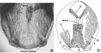
The cerci articulate basally with the ‘basal segment’ (fig. 1), which has a strongly flattened cylindrical shape. The basal segment houses several groups of muscle fibers whose contractions move the cerci (fig. 6) and are, thus, of interest for the behavioural descriptions below. Contraction of muscle A probably causes the cerci to move dorsally (to increase the angle between the ventral surface of the cerci and the basal segment), as pressure on the sides of the basal segment with a forceps caused the cerci to move dorsally (below). Inward (rather than outward) buckling of the cuticle in the central area was visible in some video sequences (below) at moments when the male squeezed with maximum force. Two other muscles (B and C) originate on the walls of the basal segment, and are apparently attached to the cerci themselves, and thus move them directly. We were not able to determine, however, their points of attachment on the cerci. Judging by its more dorsal position, C may cause the cerci to flex ventrally (toward the basal segment), thus implying that the larger muscle band B causes the cerci to extend (these conclusions are only tentative). The function of muscle D may be to close the anus.
Muscles in dorsal views of the basal segment (all but the bases of the cerci are removed; distal end at the top, distal end down). (a) Microphotograph with cuticle partially cleared. (b) Diagrammatic drawing showing different muscle groups mentioned in the text. The approximate sites at which the basal segment was squeezed to mimic the effects of contraction of basal segment muscles A (thus producing extension of the cerci) are indicated with an ‘x’.

The surstyli are lateral and basal to the cerci, and also bear a tuft of long setae (fig. 5a–c). Another, more linear tuft of long setae occurs near the inferior clasper (figs 5b and 7). The paired, flattened ventral processes of the hypandrium (the inferior clasper) are opposite the cerci when they are opened to grasp the female; the processes are smooth and lack setae near their notched distal margins (fig. 5b). Also in this general region and opposing the cerci when they grasp the female is the highly modified male sternite V, which is heavily sclerotized and covered with stout, pointed bristles (figs 1a, 3a and 5c).
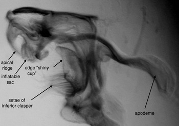
Glycerine preparation of isolated distal portion of male phallic complex and hypandrium dissected from the resting position (the cerci are removed) showing the collapsed inflatable sacs of the phallic complex (preparation courtesy of D.M. Wood).

Genitalic behaviour – units of behaviour
Mean copulation duration in large chambers was 22.2±6.4 min (N=15), and 54.0±17.9 min (N=8) in Petri dishes under a dissecting microscope (Z=3.33, P=0.00086 with Mann-Whitney U Test). The male clamped the tip of the female's abdomen between his cerci and his inferior claspers. The cerci pressed dorsally and posteriorly against the membranous ventral surface of the female's abdomen, and their distal margins bent the female external body wall sharply inward and posteriorly (fig. 2), so deeply that the distal half or more of each cercus was engulfed in this fold (figs 1–3c). The male cerci were, thus, largely out of sight during copulation, but they were not inside the female. Rather, they were pressed on the outer surface of her abdomen and folded it inward. In all 22 pairs in which viewing angles allowed us to see possible male genitalic behaviour, the male's clasp with his cerci was not sustained and immobile. Instead his cerci performed a variety of complex, rhythmic, relatively stereotyped behaviour patterns. The following patterns occurred repeatedly (all data are from the eight pairs studied under the microscope, unless otherwise specified).
(i) Tighten basal segment
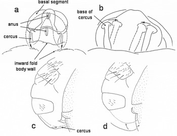
During rhythmic movements, the lateral sclerites of the basal segment came together briefly (presumably due to contractions of muscle A, then separated again. In some cases, the central dorsal cuticle near the tip of the basal segment buckled inward as the sclerites came together, presumably as a result of this contraction. The mean duration (length of the time the sclerites were held together) was 0.64±0.56 s. The mean number of tighten basal segment movements was 12.2±10.0 per copulation (range 0–22). A second, associated movement that was observable only in dorsal view of the cerci and basal segment, was an abrupt movement of the bases of the cerci toward each other (‘cercus base’) (fig. 8b). This movement was presumably due to the contraction of the muscles associated with the cerci (fig. 4d).
Squeezing movements of the cerci (traced from video recordings of copulating pairs). (a) Distal view of tip of basal segment and base of cerci showing movement during a strong squeeze (arrows). (b) Distal view of bases of cerci during cercus base movement (arrows). (c), (d) Lateral view of successive stages of a very strong squeeze movement (arrows) (dotted lines 0.17 s after solid lines in (c) and 0.47 s in (d)).

The probable consequences of tightening and cercus base movements were determined by softening the tip of the abdomen of a male that had been in alcohol by soaking it in water for two days, and then gently pinching the distal sides of the basal segment and the basal portions of the cerci with forceps. With each squeeze on the basal segment, the cerci moved dorsally. Such a movement would be hidden inside the fold of the female's body wall and would move the cerci and the folded female body wall antero-dorsally with respect to the female. Often the completion of a tighten basal segment movement was followed, about 0.07 s later, by the male's cerci emerging partially from the fold in the female's abdomen; this movement may have resulted from cercal extension. However, in some other cases, the cerci clearly did not emerge from the fold immediately after tighten basal segment movements in the same pairs.
The result of pinching the bases of the cerci, thus mimicking the cercus base movement (fig. 8b) that presumably resulted from the contraction of muscle fibers there (fig. 4d), was to cause their tips to tilt and swing laterally away from each other (fig. 9). This movement resulted from their nearly linear dorsal median margins coming together. They folded ventrally, which caused the strong spines on their ventral medial margins to project more ventrally (e.g. against the wall of the female's abdomen). At the same time, the area of contact between the cerci moved basally along their slightly rounded medial margins, thus causing the distal ends of the cerci to swing apart (fig. 9).
Schematic dorsal view of spreading movement of distal portions of the cerci (large arrows) when the basal ends of convex medial margins were brought together (small arrows) when contraction of the muscles attached to the basal portions of the cerci (fig. 4d) was simulated (see also fig. 8b). Dashed lines represent the probable positions of the distal portions when the muscles are contracted; dotted lines show positions of the ventral margins and their setae (traced from a video recording of a pinch at the base).

Although we could directly observe the tighten basal segment and the cercus base movements (often only one or the other at a given time) but could not see the possible lateral swinging movements of the distal portions of the cerci, we suspect that cercus extension and lateral swinging movements occurred at the same time. If so, the resulting movements of the cerci were compound, including an anterior-dorsal movement in the fold of the female's body wall, and a lateral swing of their setose tips that caused their ventral setae to press or scrape laterally against her body wall.
(ii) Small squeeze
The cerci flexed rapidly toward the basal segment (reducing the angle between the basal segment and the cerci) and then back. As far as we could determine, both the cerci and the membranes between them moved as a unit. Small squeezes were short (mean duration was 0.10±0.03 s, N=150 in five mating pairs) and relatively rapid (mean frequency in 161 squeezes in 20 pairs was 2.26±2.69 s−1). The mean number of bursts of small squeeze movements per copulation was 18.9±10.9 (range 7–31), and the mean percentage of copulation spent in small squeezes was 9.2±5.6%. In some cases we registered small squeezing on the basis of vibrations within the basal segment which were of a similar frequency, rather than by direct observation of either the movement of the cerci themselves or movement of the female's abdominal wall.
(iii) Medium squeeze or push
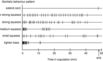
The force exerted during a medium squeeze was not strong enough to cause the line of articulation at the bases of the cerci to disappear in the fold of the female's abdomen. The observed movement appeared to result from both dorso-anterior pushing movements of the basal segment and flexing of the cerci at the articulation with the basal segment, as in small squeezes. Medium squeeze movements were longer (mean was 22.0±9.0 s), and were often repeated in rhythmic bursts (fig. 10) but with a lower frequency (mean in 59 bursts in 20 pairs was 1.53±0.60 s−1). The mean number of medium squeeze movements pre copulation was 7.2±3.7 (range 3–10), and the mean percent of the copulation spent in medium squeezes was 2.6±2.3%.
Graphical representation of different genitalic movements during the course of a 48 min copulation.

(iv) Strong squeeze
The force of a strong squeeze caused the line of articulation at the base of the cerci to nearly disappear in the fold of the female abdomen. The mean number of strong squeeze movements per copulation was 19.6±2.7 (range 17–24), and the mean percent of the copulation spent in strong squeezes was 10.3±6.9%. A strong squeezing movement was complex (fig. 8c,d). It involved both a ventrally directed thrust of the tip of the male's abdomen that pushed the female's body wall dorsally and a ventral flexing at the articulation between the basal segment and the cercus, pushing the fold in the female's body wall posteriorly as the basal segment of the male moved anteriorly (fig. 8d). The two types of movement were combined in different sequences and strengths; typically a strong squeeze began with a quick thrust followed by a slower ventral flex. These details could only be reliably distinguished when viewing angles were favourable and are not included in the behavioural analyses below.
Some strong squeezes were rhythmic, with one often following directly after another (fig. 10). In other cases, strong squeezes occurred individually with other behaviour patterns preceding and following. The mean duration for 119 individual strong squeezes was 50.3±46.3 s.
(v) Very strong squeeze
These squeezes were similar to strong squeezes, except that the line of articulation at the bases of the cerci disappeared from sight in the fold of the female's body wall. The basal segment moved slightly anteriorly and was inclined more perpendicularly to the female's ventral surface (fig. 8c), and it appeared that the angle between the basal segment and the cerci was less than 90°. Very strong squeezes were somewhat longer than strong squeezes (the mean duration of 256 very strong squeezes was 62.6±44.1 s; P<0.01 with Mann-Whitney U Test). They seldom followed directly one after another without other behaviour patterns intervening (fig. 10). The mean number of very strong squeeze movements per copulation was 24.8±9.2 (range 9–32), and the mean percent of the copulation time that was spent in very strong squeezes was 30.0±13.0%.
(vi) Extend cerci
The cerci were extended anteriorly (with respect to the female) so that their angle with the basal segment increased. The mean duration of ten extend cerci movements was 4.64+2.87 s. The angle between the long axis of the basal segment and the base of the cerci increased from about 90° during most of the copulation to approximately 135°. Extend cerci movements occurred just before the end of copulation, usually only once (6 of 8 copulations).
Genitalic behaviour – patterns
One pattern in the durations of different types of squeezing movements was that more intense squeezes lasted longer, both in terms of mean durations (means for small, medium, strong and very strong were 0.10, 22.0, 50.3 and 62.6 s), respectively, and in terms of the fraction of the copulation spent in each type of squeeze (9.2, 2.6, 10.3 and 30.0%, respectively).
There were also patterns with respect to the order in which different genitalic movements occurred (fig. 10). Genitalic movements (including all types) were more common early in copulation (during the first 10 min) than late (last 10 min); the mean total number of movements early was 35.0±17.6 and 10.8±7.1 late (P=0.012 with Mann-Whitney U Test). Small squeeze and (especially) tighten basal segment tended to occur more often early in copulation. They occurred one or more times during the first 10 min in 7 and 8 of the 8 copulations, respectively, and both were entirely absent during the last 10 min of all but one of these same copulations (P=0.012 for both with Fisher Exact Tests). On average, the last tighten basal segment movement occurred very early (on average, 4.6±1.6 min after copulation began) (these copulations lasted on average 54 min). Small squeezes continued until later (the last movement was on average 40.6±17.5 min after copulation began). The other squeezing movements were more evenly distributed. Although all tended to be more common early on, none of these trends was statistically significant. But when strong and very strong squeezes were combined (our criterion for differentiating them was in any case arbitrary), the difference was significant (P=0.0032 with Mann-Whitney U Test).
Positions relative to the female's outer surface
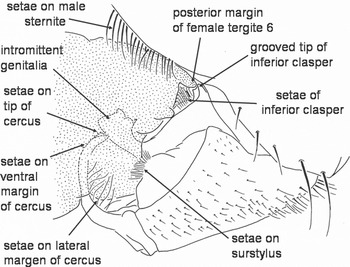
One major morphological pattern was that the six different brushes and arrays of long setae and stout-pointed setae on and near the male's genitalia were all brought into direct contact with the female during copulation; in addition, the male's genitalic squeezing movements caused these setae to move rhythmically against her (fig. 11). As already seen in other species (Vanderplank, Reference Vanderplank1947), the robust bristles covering the male sternite 5 rubbed against the dorsal surface of the female's tergite 6 (figs 1a and 11). The strong bristles on the distal margins of his cerci pressed against the membranous ventral surface of her abdomen (fig. 11). Their ‘stabbing’ action against the female's abdomen must have increased each time the male squeezed, and each cercal ‘tooth’ must have scratched or stretched her body wall laterally (figs 9 and 11) with each cercus base movement. The strong setae on the medial ventral margins of the cerci were directed toward, and probably rubbed or at least pressed laterally against, the female's abdominal wall with each cercus base movement.
Diagrammatic representation of how six groups of modified setae on and near the male genitalia contact the female (and probably rub against them) during copulation (female stippled).

In addition, the long setae on the lateral margins of the male's cerci (fig. 5a) rubbed on the membranous area at the edge of the indentation produced by the cerci (fig. 3c); the long setae of the surstyli contacted the membranous area at the lateral edge of the deep indentation around the female's anal plates or sometimes on the lateral wall of this indentation (fig. 3c); and the curved row of setae on his inferior claspers (figs 5b–c, 7 and 11) probably rubbed against the similarly curved deep membranous groove where the anterior margins of the anal and medio-dorsal plates of the female were embedded (fig. 3d), and possibly also across the setae on these plates (fig. 11). It was not possible to observe any of these setae directly during copulation, but manipulation of the bodies of flash-frozen mating pairs confirmed that the setae on the surstyli and the lateral margins of the cerci rubbed on the female when the cerci squeezed her. The setae on the inferior claspers were more hidden, but it seems highly likely that they too moved against the female with the movements of the male's genitalia, as the tips of these setae were bent in some pairs, indicating that they pressed forcefully against the female.
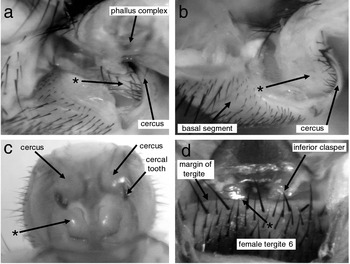
The groove in the flattened inferior claspers (fig. 5b) pressed against the posterior margin of the female's tergite 6 (fig. 12d). The pressure exerted when the male's cerci squeezed the female would press the female tergite against these immobile processes, so they thus formed the dorsal part of the male's dorso-ventral clamp on the tip of the female's abdomen. In addition, a pair of smooth, shiny cupped male sclerites (* in fig. 12c; also fig. 7) received the more posterior portions of the female's anal plates.
Copulating pairs with the female dissected partly (a), (b) or completely (c) away, and with the male dissected partly away (d); (a) and (b) are extremes of the positions of the distal tip of the phallic complex with respect to the cerci after 15 min of copulation. (a) The tip of the phallic complex extends past the cerci, and there is a relatively small amount of female tissue squeezed (*) by the cerci; the tips of the cerci press directly against the surface of the phallic complex. (b) The phallic complex is substantially short of the cerci, and is hidden in the thick mass of female tissue (*) that is grasped by the cerci. (c) Male genitalia in position to clamp the female (female removed), showing the smooth, deep concave surfaces that receive the female's anal plates (*). (d) Dorsal view of the posterior margin of the female tergite pressed into (left) and near (right) the groove in the tip of the inferior clasper (* marks where the margin of the tergite is inside the groove in the inferior clasper).

Positions and events within the female
Dissections of 22 pairs in which the male was clasping the female's abdomen with his cerci showed that the deep indentation of the female's ventral body wall produced by the clamping action of the male cerci caused her uterus and a portion of her median oviduct to fold dorsally in the area anterior to the vagina-uterus junction. The exact site of the fold in the oviduct varied somewhat, but was generally proximal to the junctions of the uterine gland duct, the spermatheca duct and the uterus with the oviduct (fig. 2).
The position of the male aedeagus varied. Of six coupled pairs frozen after 15 min of copulation, the intromittent genitalia of one male were not everted from their resting position (fig. 5b) and were not inside the female. Another had only partially everted his hypandrium and phallus complex, and its distal end reached only the female's vulva. In a total of five other males in the other 16 pairs, the male cerci clamped the female's abdomen (above), but his aedeagus did not penetrate the female. Lack of intromission was seen as late as after 120 min of copulation.
In four pairs lacking intromission, the male's phallus complex was not everted, and the toothed membranous sacs of his phallus complex were not inflated. In the other three, the aedeagus was partially everted into the vulva and vagina, and the sacs of the phallus complex were inflated. In one of these latter pairs, the tip of the aedeagus pressed so forcefully against the wall of the female's vulva that the inflatable sacs and the median apical ridge of the aedeagus indented the dorsal wall of the vulva (or of the distal end of the vagina – there was no clear line separating them). In one other pair lacking intromission, in contrast, the vulva of the female was open, with a diameter approximately that of the male's phallic complex, so the male may have achieved intromission previously and then subsequently withdrawn his intromittent genitalia while retaining his external grasp on the tip of the female's abdomen.
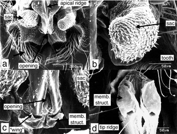
In the other 15 pairs, the hypandrium and phallus complex were more completely extended (fig. 5c), and penetrated deep into the female, with the distal end of the phallus complex positioned at or just anterior to (beyond) the junction of the vagina with the uterus. The toothed membranous sacs of the phallus complex (figs 7 and 13a,b) were inflated to variable degrees, sometimes asymmetrically. Some sacs were directed laterally, and others distally, and they rested near or against a variety of sites in the vicinity of the junction of the vagina with the uterus. In three cases, the sacs ‘embraced’ the ‘oviductal shelf’ or ‘milk gland papilla’ (Roberts, Reference Roberts1973) at the mouth of the oviduct. In others, they were associated with wrinkles in the wall of the vagina and of the uterus. In all pairs with deep penetration, the posteriorly-curving tip of the anterior ridge of the male's phallus complex (figs 5c and 7) bore an everted membranous structure at the opening on its tip (fig. 13c,d) that was snagged in a wrinkle of the dorsal lining of the female's reproductive tract. This snag, presumably partly due to the ‘wings’ on the everted membranous structure (fig. 13c), held the tip of the male's intromittent genitalia firmly attached to the female lining during dissections until it was forcibly pulled free. We were not able to ascertain whether the ‘wrinkle’ where this membranous structure was inserted was the mouth of the spermatheca duct.
Male genitalia in positions assumed during copulation (female removed). (a) Apical portion of the phallic complex seen end-on; the thick arrow indicates the long setae of the inferior claspers, the thinner arrow the opening near the tip of the apical ridge. (b) Inflated form of the toothed inflatable sac of phallic complex. (c) Close-up of the opening at the tip of apical ridge of phallic complex, from which a membranous structure with lateral ‘wings’ has emerged. (d) Close-up of membranous structure everted from the opening at the tip of the apical ridge.

The position of the distal tip of the phallic complex relative to the tips of the male cerci varied substantially. In 5 of 15 pairs with deep intromission, the tip of the phallic complex was short of (posterior to) the tips of the cerci (fig. 12a); in two it was about even with the tips (fig. 5c,d); and in six it was substantially beyond the cerci (fig. 12b) (in two others their relative postions were not determined). There was no correlation between the depth of penetration and the elapsed time in copulation; both very shallow and very deep penetrations were seen after 15 min, and both also after 105 min.
One mechanical consequence of insertion of the male's intromittent genitalia may have been to strengthen his clamp on the tip of the female's abdomen. The substantial pressure the cerci exerted with their periodic squeezing must have severely pinched some female tissues (the wall of her oviduct, her body wall and any tissues between them), especially when the phallic complex had penetrated more deeply. The small separation between the distal margin of the cerci and the surface of the phallus complex (fig. 5c,d) and the bent tips of some of the strong cercal setae (fig. 5d) offer mute testimony to the strong pressures on the female tissues that had been pinched there.
Non-genitalic behaviour
We recognized the following male and female behaviour patterns:
Male behaviour units
(i) ‘Peep’
Vibrate wings rapidly while they were folded over the male's abdomen, producing a high-pitched whine or ‘peep’ (the image of the wings became blurred in video images during each peep). The average duration of 307 peeps in 20 pairs was 1.32±1.92 s.
(ii) Wings forward and buzz
The wings were brought forward to extend laterally and were buzzed rapidly (blurred images of wings were displaced about 20–30° dorsally and ventrally in video images). In 115 bursts of wing buzzing in 20 pairs, the average duration was 0.68±0.77 s.
(iii) Raised legs II
Both middle legs were raised and briefly held extended dorsally and anteriorly, just as the male began a burst of wing forward. The legs were held raised for only a fraction of a second (mean 0.046±0.035 s) and then lowered while the wings buzzed briefly (mean 0.045±0.053 s). Most often, the male moved both his legs and his wings (69% of 59 occasions in 20 pairs); less often he moved his wings but not his legs (24%), and only infrequently did he move his legs but not his wings (7%). Usually the male made two wing-forward movements in each burst of buzzing (mean 1.80±0.82, N=59 in 20 pairs).
(iv) Rub with legs I
The front legs rubbed or tapped repeatedly on the pronotum and/or dorsal surface of the head and eyes of the female. Bursts of rubbing lasted a mean of 0.61±0.37 s (N=20).
(v) Rub with legs II
The middle legs rubbed or tapped repeatedly on the sides of the female's thorax, her head or her abdomen.
(vi) Rub with legs III
The hind legs rubbed or tapped repeatedly on the ventral surface of the female's abdomen. Initiation of leg III rubbing varied; in some cases the male's legs III began rubbing the female's eyes and gradually worked posteriorly until they ended up rubbing her abdomen.
Female units of behaviour
(i) Vibrate wings
The female's wings were vibrated rapidly, causing their images in video recordings to become blurred, while they were held folded over her abdomen. The mean duration of 323 bursts of wing vibration in 20 pairs was 5.2±6.4 s. As we will describe elsewhere, wing vibrations were apparently female signals to induce the male to loosen squeezes with his cerci (Briceño et al., unpublished results).
(ii) Dorso-ventral oscillation
The abdomens of both the male and the female oscillated abruptly dorso-ventrally. This occurred near the end of copulation, and corresponded to the ‘male jerking’ behaviour of previous authors (Jaenson, Reference Jaenson1979a,Reference Jaensonb; Leegwater-van der Linden & Tiggelman, Reference Leegwater-van der Linden and Tiggelman1984). The dorsal movements were not due to the male's legs II or III pushing against the substrate, however; as in some video sequences, they were in the air as the dorsal displacement began.
(iii) Lateral shake
The female wagged her abdomen rapidly from side to side. She did not lean from side to side as she shook (as occurs in some flies; see Baena & Eberhard, in press), keeping her body more or less parallel to the substrate as she shook. The duration of shaking (N=180 in 20 pairs) was 6.90±7.16 s. This is probably also a female signal to induce the male to relax squeezes (Briceño et al., unpublished results).
(iv) Push with legs III
The hind legs were repeatedly extended posteriorly and dorsally, often pushing against the male's abdomen.
Discussion
Genitalic morphology and behaviour
Different stages of copulation – seizing, intromitting and sperm transfer
Genitalic coupling, in the sense that the male cerci grasp the female and fold her ventral body wall deeply inward, is not always accompanied by eversion of the male's hypandrium and phallic complex and intromission in G. pallidipes; in one extreme case, the male's intromittent genitalia were only slightly everted and were pressing forcefully on the female's vulva but had apparently not entered 120 min after copulation began. Vanderplank (Reference Vanderplank1947) also mentions a pair of G. fuscipes that copulated for 24 hrs without accomplishing sperm transfer. This is in accordance with Jaenson (Reference Jaenson1979a), who found substantial rates of copulation that failed to result in insemination in G. pallidipes. Similarly, short and apparently failed ‘copulations’ occur in other Glossina species, including palpalis, morsitans and swynnertoni (Vanderplank, Reference Vanderplank1948), tachinoides and morsitans (Buxton & Lewis, 1934 cited in Buxton, Reference Buxton1955), palpalis fuscipes (Mellanby, Reference Mellanby1936), morsitans (Saunders & Dodd, Reference Saunders and Dodd1972; Chaudhury & Dhadialla, Reference Chaudhury and Dhadialla1976), austeni (Pinhâo & Grácio, Reference Pinhâo and Grácio1973) and morsitans and austeni (Foster, Reference Foster1976). Clasping without intromission could also explain why some females of G. palpalis collected in the field bore mating scars from being clasped by a male, but nevertheless lacked sperm in their spermathecae (Squire, Reference Squire1951). Our finding differs, however, in that it suggests that failure resulted from possible female resistance rather than male inadequacy (Jaenson, Reference Jaenson1979b).
The depth of penetration did not correlate with the time in copulation. Deep intromission, reaching the vicinity of the mouth of the spermathecal duct, was also not tightly linked in G. pallidipes with sperm transfer; such deep intromission frequently occurred minutes earlier in copulation than sperm transfer, which occurs during the last 30 s of copulation.
Genitalic design – restrain the female
Some of the male genitalic structures and behaviour of G. pallidipes are appropriately designed to clamp and physically restrain the female. The powerful ventral clamping action of the cerci, which folds the female's abdominal membrane deeply inward, has long been known in Glossina (Vanderplank, Reference Vanderplank1948; Squire, Reference Squire1951). The strong inferior claspers of the male and their distal groove are well designed to mesh with and hold the posterior edge of the female tergite 6, and were consistently positioned near or against the edge of this tergite in copulating pairs. They, thus, function as the dorsal side of the clamp formed by the male's cerci. The powerful nature of this genitalic clamp has been documented in other species (Vanderplank, Reference Vanderplank1948; Squire, Reference Squire1951). A strong clamp with the cerci is probably advantageous in holding the female securely to prevent premature (from the male's point of view) separation and also perhaps to position the male's intromittent genitalia to reach appropriate sites within the female. Vanderplank (Reference Vanderplank1948) mentioned that the male cerci of G. fuscipes ‘pull the uterus in line and … open the second valve fold’ of the female, but gave no supporting evidence; we saw no sign of these functions in G. pallidipes (in which the cerci are of quite different form).
The male inferior claspers also press the dorsal tip of the female's tergite 6 in G. austeni (Pollock, Reference Pollock1973). They have different forms in different species of the G. morsitans species group (Jackson, Reference Jackson1952). There are apparently no corresponding differences, however, in the morphology of the female tergite 6. This tergite is not mentioned in discussions of characters that are useful in distinguishing species, despite careful attention to the tip of the female abdomen (Potts, Reference Potts and Mulligan1970). We inspected this tergite in female G. pallidipes and G. morsitans but failed to find differences. Such a lack of corresponding evidence coevolution of female body parts contacted by species-specific male structures is common in insects and spiders (Shapiro & Porter, Reference Shapiro and Porter1989; Eberhard, Reference Eberhard2004b). It constitutes evidence against both the lock and key and the sexually antagonistic coevolution hypotheses to explain the rapid evolutionary divergence of these male structures. The probable lack of mobility of the female tergite in Glossina, and thus the low probability that the differences in the male structures (shape of the prominence, shape of gap between them) could have resulted from selection due to the behaviour rather than the morphology of the tergite seems highly unlikely. This constitutes further evidence against the sexually antagonistic coevolution hypothesis. The male inferior claspers apparently have a different function in G. tachinoides (a member of a different subgenus, palpalis), in which they apparently hook tissue just ventral to the female's anus (Vanderplank, Reference Vanderplank1948). The inferior clasper is also apparently softer in this species, as it becomes distorted in dried specimens (Vanderplank, Reference Vanderplank1949a).
Genitalic design – stimulate the female
Some male genitalic traits have no obvious mechanical function, and several lines of evidence imply that they have a stimulatory function instead. Most convincingly, the arrays of abundant setae and bristles on different portions of the male's genitalic and perigenitalic structures are all positioned so that they rub or push against the female during the male's copulatory genitalic movements (fig. 11). These setae include the stout bristles on his sternite 5, the sharp bristles on the distal and medial margins of his cerci, the long setae on the lateral margins of his cerci, the long setae on the basal portion of the inferior claspers and the tufts of long setae on his surstyli. A further indication of a stimulatory function for these setae is the correlation between their size and stoutness and the degree of sclerotization of the female surfaces that they contact: the very strong setae on male sternite 5 contact the female's strongly sclerotized abdominal tergite; the pointed, robust setae on the distal and medial margins of the cerci ‘stab’ or scrape forcefully against the female's ventral body wall; and the weaker setae on the lateral margins of the cerci, the surstyli and on the inferior claspers all rub against membranous surfaces of the female. The patch of very small setae on the ventral surface of the cercus might facilitate the sliding movement of the cercus against the surface of the female's abdomen.
Male behaviour also seems designed to stimulate the female. The male's genitalia moved in persistent, complex and stereotyped ways, including squeezes with different rhythms and amplitudes (and our descriptions ignored modulations in the force of male thrusting and ventral flexion of his cerci, and are thus undoubtedly overly simplified). The rhythmic dorsal cercus extension movements (tighten basal segment), which were deduced from both direct observations and examination of the muscles that move the cerci, reduced rather than strengthened the firmness of the male's clamp on the female's abdomen, arguing against the alternative interpretation of male morphology and behaviour as functioning simply to hold on to the female. The rocking movements of the cerci against each other during cercus base movements (fig. 9) would cause each cercus tooth to scrape laterally against the female (or at least stretch her body wall laterally), again a movement that could stimulate the female but which is not appropriate to hold on more firmly. It is also clear that females can sense squeezes, as supposed by the stimulation hypothesis, because they often give immediate behavioural responses to male squeezes (Briceño et al. unpublished results). A major proportion of the time in a copulation is spent squeezing the female (mean was 52±18%).
Still another set of data in agreement with a stimulatory function concerns interspecific differences. Stimulation is expected to diverge rapidly under sexual selection. All of the arrays of setae, we have argued, function to stimulate the female (except, perhaps, those on sternite 5, which are not featured as characters to distinguish species in taxonomic works such as Potts (Reference Potts and Mulligan1970) ) vary substantially among species of Glossina, even among those in the morsitans subgenus to which G. pallidipes belongs. There are differences in the relative lengths, robustness or density of the setae on the distal and lateral margins of the cerci, the surstyli, and the inferior clasper or paramere (Newstead et al., Reference Newstead, Evans and Potts1924; Patton, Reference Patton1936; Vanderplank, Reference Vanderplank1949b,Reference Vanderplankc; Nash & Jordan, Reference Nash and Jordan1959; Potts, Reference Potts and Mulligan1970; Briceño & Eberhard, unpublished results). Three of the four differences between the male genitalia of G. pallidipes and its close relative, G. longipalpis (which has sometimes been mentioned as a conspecific; Vanderplank, Reference Vanderplank1949b), concern structures that we have argued above (fig. 11) are likely to have stimulatory functions: the position of the cercal tooth, the length of the long setae on the lateral margin of the cercus and the length of the long setae on the surstyli (Newstead et al., Reference Newstead, Evans and Potts1924; Vanderplank, Reference Vanderplank1949b) (the fourth difference concerns the inner margins of the cerci, where they rock against each other during cercus base movements, and thus may also be linked to stimulation behaviour). There are also differences in subspecies of G. morsitans in two cercus structures, the size and shape of the median ‘lobe’ or tooth, and a strong ventral spine (Vanderplank, Reference Vanderplank1949c) that probably scrape against the female's body wall during tighten basal segment and strong and very strong squeeze movements (fig. 9).
The sharp interspecific differences in the morphology of male cerci are not accompanied by corresponding female differences; the area of the female abdomen that the male cerci contact and squeeze is quite uniform. This lack of female change is compatible with the female choice by stimulation of the female hypothesis, but it argues against the mechanical lock and key and the sexual antagonistic coevolution hypotheses because the female morphology fails to show the predicted coevolutionary changes. A possible modification of antagonistic coevolution hypothesis is that antagonistic coevolutionary changes in species-specific female behaviour rather than morphology has selected for divergent male morphology (Eberhard, Reference Eberhard2004a,Reference Eberhardb). But changes of this sort do not appear feasible in Glossina, because the female's abdomen was firmly clasped and basically immobile in the male's clasp in pallidipes and other species (Vanderplank, Reference Vanderplank1948), and was unable to move in any way that would reduce the effectiveness of his clasp.
There may also be differences in the patterns of squeezing behaviour in different species of Glossina, as would be expected if the stimuli produced by squeezing behaviour are under sexual selection (West-Eberhard, Reference West-Eberhard and Lewis1984). Males of G. palpalis apparently squeeze with a rhythmic movement different to those of G. pallidipes, opening and closing about twice per second, and these squeezes may be elicited by female movements (Squire, Reference Squire1951). Huyton & Langley (Reference Huyton and Langley1982) noted that the male genitalia of G. morsitans and G. austeni are moved during copulation, but gave no details. We have observed additional differences in G. brevipalpis, G. fuscipes and G. morsitans (Briceño & Eberhard, unpublished results).
One possible function of male stimulation of the female could be to induce the female not to fly. Squire (Reference Squire1951) suggested this function in G. palpalis, noting that more squeezing occurred immediately after the female flew (he gave no quantitative data, however). Experiments with dead females (Jaenson, Reference Jaenson1979b) also suggested the possibility that genitalic as well as non-genitalic stimulation during copulation could serve to prolong copulation, as females influence the length of copulation (‘presumably, the copulating male regulates the copulatory time in response to stimuli emitted from live females’; p. 5). The length of copulation may be especially important in Glossina, because of the additive effects of copulatory stimulation on triggering ovulation (Saunders & Dodd, Reference Saunders and Dodd1972). It appears that such triggering is especially important in a female's first ovulatory cycle, which is sometimes skipped (Jaenson, Reference Jaenson1979b).
An alternative possibility is that genitalic squeezing serves to mechanically facilitate penetration by the male's intromittent genitalia to the site where he will deposit his spermatophore. Presumably spermatophore deposition is associated with the membranous structure that is everted from the opening at the tip of the apical ridge of the phallic complex (fig. 13c) and is inserted near the mouth of the spermathecal duct. Our failure to resolve the position of the mouth of the spermathecal duct in the dissections of copulating pairs leaves this possibility unresolved. It was clear, however, that copulation does not represent a gradual process of deeper and deeper penetration. As early as 15 min into copulation many males had apparently ‘arrived’, with the opening at the tip of the apical ridge at least very close to the mouth of the spermathecal duct, yet genitalic squeezing movements continued throughout copulation. It is also possible that the squeezing movements of the cerci somehow helped force the membranous structure into the mouth of the spermathecal duct after they had become aligned. Deep penetration also occurs long before the end of copulation in G. austeni (Pollock, Reference Pollock1974). One type of genitalic movement that has a more obvious possible mechanical function was the strong cercus extension near the end of copulation, which might serve to free the wall of the vagina from the tip of the male's phallic complex.
While these possible mechanical functions cannot be excluded, they are not sufficient, in and of themselves, to explain some of our observations. In particular, they leave unexplained why the male movements were so rhythmic and stereotyped, and why male genitalia have so many design features (the groups of setae discussed above) that are appropriate for stimulating the female and that lack any other obvious function.
All the copulations we observed were in captivity rather than in nature, and those in which we made close-up recordings of genitalic movements were in very confined quarters and unusually long (over twice as long as others in a less confined situation). Care must, therefore, be exercised in interpreting our observations. The close-up recordings can be taken to show that males are capable of several different types of genitalic movements, and that these movements show clear patterns, but not that these patterned movements occur during more ‘normal’ copulations. Nevertheless, it is likely that rhythmic genitalic squeezing is typical of this species. Genitalic squeezing movements (probably equivalent to ‘strong’ and ‘very strong’ squeezes) occurred in all 14 pairs that we observed copulating in less constrained circumstances in which viewing angles made it possible to check for this behaviour. The one copulation under the microscope whose duration was more typical of unrestrained flies (29 min) was otherwise typical with respect to male squeezing movements (61 squeezes that occupied 45% of the duration of the copulation). In addition, it seems improbable that males would have evolved the morphological capability (e.g. appropriate muscles, articulations) for executing these movements and the neural ability to pattern them if such capabilities were never used and of no selective advantage.
Genitalic design – inside the female
The functions of the intromittent portions of the male genitalia of G. pallidipes are less obvious than those contacting her external surface. The mobile, inflatable membranous sacs of the phallus complex are covered with sharp, curved spines (fig. 13b). Spines of this sort on male genitalia are sometimes thought to be penetrating or holdfast devices (e.g. Squire, Reference Squire1951; Eberhard, Reference Eberhard1993; Flowers & Eberhard, Reference Flowers and Eberhard2006). Another, non-exclusive possibility is that they function to stimulate the female. An overall holdfast seems superfluous in G. pallidipes, given the male's extremely firm grip with his cerci and inferior claspers. It is possible, however, that the sacs might serve to push through the vulva (as may occur in the ‘foot-in-the-door’ sacs of the beetle, Macrodactylus; Eberhard, Reference Eberhard1993) or to hold the male's phallus complex in the uterus near the mouth of the female's spermathecal ducts. The often asymmetrical positions of the sacs, and their different degrees of inflation in different pairs and even in the same pair, suggest that they move actively and independently during copulation.
The sperm and the spermatophore presumably emerge from or near the membranous structure that was everted from the aperture at the tip of the apical ridge (fig. 13c,d). By analogy with other flies that produce spermatophores (Eberhard & Huber, Reference Eberhard and Huber1998; see also Eberhard, Reference Eberhard1993; Eberhard & Kariko, Reference Eberhard and Kariko1996, Förster et al., Reference Förster, Gack and Peschke1998 on beetles), deposition of the spematophore so that its sperm exit duct is positioned in or near the mouth of the spermathecal duct probably depends on the male first locating the duct mouth with his genitalia.
We saw no indications that the male facilitated intromission by drawing apart the female's anal plates, elevating her sternal plate or drawing taut a membranous fold to expose her vulva, as reported in G. palpalis (Squire, Reference Squire1951). But this is perhaps not surprising, as male G. pallidipes apparently lack the ‘scrolls’ on the inferior claspers that are thought to produce these effects in G. palpalis (Squire, Reference Squire1951).
Non-genitalic behaviour
G. pallidipes
The mating duration we observed in Petri dishes (mean 54.0 min) was similar to the 61 min observed in similarly aged females by Jaenson (Reference Jaenson1979b), while the mean duration we observed in open cages (22.2 min) was similar to durations reported in several other studies. The copulations that Jaenson (Reference Jaenson1979b) observed also occurred in cramped quarters (the male was placed in a vial with a female). Perhaps something about cramped quarters causes longer copulations.
We confirmed many aspects of Jaenson's (Reference Jaenson1979a,Reference Jaensonb) excellent descriptions, including the general division of copulation into three phases, and also several of the behaviour patterns he described, including male wing buzzing, the high-pitched male whine during the first and third phase, male tapping with his front legs during the first phase, female wing vibration and pushing against the male's abdomen with her legs III during the second phase, the gradual change in the male's angle with the female and intermittent male wing buzzing during the final ‘jerking’ stage of copulation.
Nevertheless, other details of our observations differ from Jaenson's descriptions. Most of these differences are probably due to the greater resolution of details that was facilitated by our close focus of the camera on the flies, by filming their behaviour through a dissecting microscope and by our ability to simultaneously record sound and detailed digital visual images. In the first stage, Jaenson failed to realize that the male's high-pitched ‘peep’ sounds are not produced while his wings are directed laterally and are visibly buzzing, but instead while they are held folded over his abdomen (as also occurs in G. morsitans; Huyton & Langley, Reference Huyton and Langley1982). He, thus, did not appreciate that buzzing with laterally directed wings and ‘peeping’ are separate behaviour patterns.
In the third stage, Jaenson's idea that the rapid, rhythmic dorso-ventral movements of the male and female were produced by movements of the male rather than by the female (thus his ‘male jerking’ behaviour) is also open to doubt. This is a difficult distinction, and indeed we only became convinced that the female was responsible for the movement after repeated frame by frame analyses of close-up lateral views showed that sometimes all of the male's legs were off the substrate when the pair began to move dorsally. Nevertheless, Jaenson observed jerking behaviour in copulations with dead females (Jaenson, Reference Jaenson1979a), supporting his idea that jerks must be due to male behaviour. We can only speculate that male rubbing with his legs II and III on the ventral surface of the female's abdomen in copulations with dead females caused his tibiae to push against the substrate and lift both the male and female jerkily off the substrate. Lifting movements would be especially likely to occur with dead females, whose ventral surfaces would be resting on the substrate. Huyton & Langley (Reference Huyton and Langley1982), who analyzed movies of copulation, thought that jerking is produced in G. morsitans and G. austeni when male legs III ‘hit the substrate with sufficient force to cause the pair to rock backwards and forwards’ (p. 170); perhaps forward rocking is produced in this way in these species, but the backward rocks they observed cannot have been produced in this way, and may depend on the female. ‘Jerking’ behaviour is of particular interest because insemination only occurs if jerking occurs, and jerking was shorter when insemination did not occur (Jaenson, Reference Jaenson1979a).
Jaenson was correct that the male's hind legs quiver at the beginning of the last phase, but he failed to note that after these preliminary quivers both the hind and the middle legs of the male rub actively and extensively on the female. He missed the brief raising of the male's second legs at the instant the male began a burst of wing buzzing during the third phase, presumably because the movement is so quick (about 0.1 s). Jaenson was silent regarding the complex behaviour of the male cerci that we have documented, presumably because his focus was on larger structures or because he followed the tradition of ignoring genitalic behaviour (Eberhard, Reference Eberhard1991, Reference Eberhard1994). One difference that is not as easily attributed to differences in the resolution is that Jaenson (Reference Jaenson1979a) did not describe the frequent and obvious lateral shaking by the female. Perhaps he followed the prevailing tradition of focusing on male rather than female behaviour.
Other species of Glossina
Copulation behaviour has been described carefully in three other species of Glossina, two of which are in the same subgenus as pallidipes. Huyton & Langley (Reference Huyton and Langley1982) observed several male behaviour patterns in G. morsitans and G. austeni similar to those of G. pallidipes: ‘wing vibration’ (‘peep’ of this study); stroking the female thorax with legs II and III; ‘male jerking’; stroking or drumming on the female's head and pronotum with legs I; and wing forward and vibration (though this was combined with movements of legs II and III not seen in G. pallidipes).
Functions of behaviour
Several aspects of male behaviour during copulation, including peeping, wings forward and buzz, raised legs II, and rubbing with legs I, II, and III fit the criteria for ‘copulatory courtship’ behaviour (Eberhard, Reference Eberhard1994). Copulatory courtship is thought to function to trigger female behavioural or physiological responses (such as sperm transport, ovulation, inhibition of remating, and others) that are in the male's reproductive interests (Eberhard, Reference Eberhard1994, Reference Eberhard1996). The apparent use of male rubbing in G. morsitans and G. austeni to inhibit female movements (Huyton & Langley, Reference Huyton and Langley1982; they gave no quantitative data, however) is in accord with this idea. In addition, the ‘wing open’ copulatory display of G. morsitans ‘seems to be a response to female rejection’ (Huyton & Langley, Reference Huyton and Langley1982, p. 171). Jaenson (Reference Jaenson1979a) suggested that male wing buzzing at the end of copulation induces the female to ‘facilitate’ insemination.
Wall & Langley (Reference Wall and Langley1993) proposed an alternate hypothesis for the male behaviour during copulation, ‘to reinforce the copulatory union’. But neither their observations nor ours offer any indication that the male's behaviour affects the physical coupling of their genitalia. The only possible exception we can see is rubbing the female's abdomen near her genitalia in G. morsitans and G. austeni that might somehow aid penetration by the male's intromittent genitalia.
General considerations regarding copulatory courtship and genitalia
The hypothesis that genitalic and non-genitalic stimulation in G. pallidipes function as copulatory courtship supposes that these stimuli trigger one or more female processes or types of behaviour that could improve the male's chances of paternity. Previous studies of the closely related species, G. morsitans, have documented just such effects on females. Mechanical stimuli from copulation promoted ovulation (Saunders & Dodd, Reference Saunders and Dodd1972; Chaudhury & Dhadialla, Reference Chaudhury and Dhadialla1976). Female resistance to further mating is also induced by mechanical stimulation from copulation (Gillott & Langley, Reference Gillot and Langley1981). Female resistance to remating in Glossina is apparently under hormonal control (Tobe & Langley, Reference Tobe and Langley1978), so the mechanism by which male stimuli during copulation trigger female responses may involve release of hormonal factors in the female.
The hypothesis that both genitalic and non-genitalic behaviour of male Glossina during copulation have evolved under sexual selection is only tenable if females, at least sometimes, mate with more than a single male in nature. If females are strictly monandrous, there can be no selection on males to stimulate them more effectively during copulation (Eberhard, Reference Eberhard1985; Arnqvist, Reference Arnqvist1998). A single mating in Glossina provides the female with enough sperm for perhaps her entire reproductive life (Jaenson, Reference Jaenson1979b; Wall & Langley, Reference Wall and Langley1993), but there are suggestions (though inconclusive) that female are nevertheless polyandrous in nature. Older field-captured nulliparous females of G. pallidipes had more sperm in their spermathecae than younger nulliparous females that had mated, suggesting multiple copulations (Jaenson, Reference Jaenson1980; Rogers, Reference Rogers1973, cited in Jaenson, Reference Jaenson1980). Female Glossina must feed about three times to mature each larva (Buxton, Reference Buxton1955; Tobe & Langley, Reference Tobe and Langley1978), and males often gather at feeding sites (‘following swarms’ near large mammals). Females are, thus, exposed repeatedly to males in the field, although it seems to be the female which controls whether or not copulation occurs in nature (Wall & Langley, Reference Wall and Langley1993). In captivity, females of G. pallidipes sometimes remate (as do those of G. palpalis, G. austeni and G. morsitans) (Jordan, Reference Jordan1958; Curtis, Reference Curtis1968; Dame & Ford, Reference Dame and Ford1968; Pinhão & Grácio, Reference Pinhâo and Grácio1973; Jaenson, Reference Jaenson1980; Gillott & Longley, Reference Pinhâo and Grácio1981; Leegwater-van der Linden & Tiggelman, Reference Leegwater-van der Linden and Tiggelman1984). Remating behaviour in captivity is generally a poor indicator, however, of female remating frequencies in nature (Eberhard, Reference Eberhard1996).
Male genitalic squeezing occurs in other Diptera, utilizing a variety of different structures (e.g. Wood, Reference Wood, Weismann, Orszagh and Pont1991; Sinclair et al., Reference Sinclair, Cumming and Wood1994; Cumming et al., Reference Cumming, Sinclair and Wood1995). Rhythmic genitalic squeezing behaviour is also known in sepsid flies (Eberhard, Reference Eberhard2001a, Reference Eberhard2005) and sciarid flies (Eberhard, Reference Eberhard2001b). Further studies will be needed to determine whether apparent copulatory courtship behaviour involving squeezing, such as that described here, is widespread in Diptera.
Acknowledgements
We thank D.M. Wood for crucial help regarding homologies of male genitalic structures, Angel Aguirre of the Smithsonian Tropical Research Insitute Library for help obtaining references and Maribelle Vargas for help producing SEM images. We thank the International Atomic Energy Agency, the Smithsonian Tropical Research Institute and the Universidad de Costa Rica for financial support.