INTRODUCTION
Amphibian populations have seen a dramatic global decline (Blaustein and Wake, Reference Blaustein and Wake1990, Reference Blaustein and Wake1995; Houlahan et al. Reference Houlahan, Findlay, Schmidt, Meyer and Kuzmin2000; Roelants et al. Reference Roelants, Gower, Wilkinson, Loader, Biju, Guilaume, Moriau and Bossuyt2007; Blaustein et al. Reference Blaustein, Gervasi, Johnson, Hoverman, Belden, Bradley and Xie2012), and amphibian infectious diseases are key factors implicated in amphibian population declines (Berger et al. Reference Berger, Speare, Daszak, Green, Cunningham, Goggin, Slocombe, Ragan, Hyatt, McDonald, Hines, Lips, Marantelli and Parkes1998, Reference Berger, Speare, Hyatt and Campbell1999, Lips, Reference Lips1999; Daszak et al. Reference Daszak, Cunningham and Hyatt2000). Most studies of amphibian infections focus on two pathogen groups – chytridiomycete fungi (Lips et al. Reference Lips, Brem, Brenes, Reeve, Alford, Voyles, Carey, Livo, Pessier and Collins2006; Densmore and Green, Reference Densmore and Green2007; Smith et al. Reference Smith, Lips and Chase2009) and ranaviruses (Daszak et al. Reference Daszak, Cunningham and Hyatt2000; Duffus and Cunningham, Reference Duffus and Cunningham2010). However, a rising number of reports of amphibian infectious skin diseases have been attributed to a relatively poorly studied group of organisms belonging to the order Dermocystidia (Class Mesomycetozoea: Pascolini, et al. Reference Pascolini, Daszak, Cunningham, Tei, Vagnetti, Bucci, Fagotti and Di Rosa2003; Raffel et al. Reference Raffel, Bommarito, Barry, Witiak and Shackelton2008; González-Hernández et al. Reference González-Hernández, Denoël, Duffus, Garner, Cunningham and Acevedo-Whitehouse2010; Rowley et al. Reference Rowley, Gleason, Andreou, Marshall, Lilje and Goslan2013). The order Dermocystidia consists of pathogens known to infect mammals and birds (Rhinosporidum sp.) (Herr et al. Reference Herr, Ajello, Taylor, Arseculeratne and Mendoza1999; Vilela and Mendoza Reference Vilela and Mendoza2012), fish (Dermocystidium spp. and Rosette agent) (Ragan et al. Reference Ragan, Goggin, Cawthorn, Cerenius, Jamieson, Plourde, Rand, Soderhall and Gutell1996; Mendoza et al. Reference Mendoza, Taylor and Ajello2002) and amphibians (Amphibiocystidium and Amphibiothecum spp.) (Pascolini et al. Reference Pascolini, Daszak, Cunningham, Tei, Vagnetti, Bucci, Fagotti and Di Rosa2003; Feldman et al. Reference Feldman, Wimsatt and Green2005). Due to their similar morphology and explicit affinity to amphibian hosts, the amphibian-infecting Dermocystids were considered a single genus (Pascolini et al. Reference Pascolini, Daszak, Cunningham, Tei, Vagnetti, Bucci, Fagotti and Di Rosa2003). However, the use of more advanced molecular phylogenetics recently saw the reclassification of these pathogens into two distinct genera, Amphibiocystidium and Amphibiothecum (Feldman et al. Reference Feldman, Wimsatt and Green2005), which include some of the first pathogens described in amphibians (Perez, Reference Perez1907, Reference Perez1913), and are now known to be associated with both caudate and anuran species (Pascolini et al. Reference Pascolini, Daszak, Cunningham, Tei, Vagnetti, Bucci, Fagotti and Di Rosa2003; Feldman et al. Reference Feldman, Wimsatt and Green2005; Densmore and Green, Reference Densmore and Green2007; Raffel et al. Reference Raffel, Bommarito, Barry, Witiak and Shackelton2008).
In amphibians, Dermocystid infections manifest as spore-filled cysts (Pascolini et al. Reference Pascolini, Daszak, Cunningham, Tei, Vagnetti, Bucci, Fagotti and Di Rosa2003; Pereira et al. Reference Pereira, Di Rosa, Fagotti, Simoncelli, Pascolini and Mendoza2005; Raffel et al. Reference Raffel, Bommarito, Barry, Witiak and Shackelton2008; González-Hernández et al. Reference González-Hernández, Denoël, Duffus, Garner, Cunningham and Acevedo-Whitehouse2010) that macroscopically present as small discrete cysts (~1 mm) to larger multi-focal nodules. The limited data available suggest that Dermocystid infection rarely causes mortality in amphibian hosts or population-level responses (Densmore and Green, Reference Densmore and Green2007; González-Hernández et al. Reference González-Hernández, Denoël, Duffus, Garner, Cunningham and Acevedo-Whitehouse2010). However, an association between the presence of Amphibiocystidium spp. and declines in populations of Notophthalmus viridescens (Raffel et al. Reference Raffel, Bommarito, Barry, Witiak and Shackelton2008) and Pelophylax esculentus (Rana esculenta; Pascolini et al. Reference Pascolini, Daszak, Cunningham, Tei, Vagnetti, Bucci, Fagotti and Di Rosa2003) has been suggested.
In 2006, an unusual skin disease was reported in an isolated population of palmate newts (Lissotriton helveticus) on the Isle of Rum, Scotland (Gray et al. Reference Gray2008), although anecdotal reports precede this (Isle of Rum Rangers, personal communication, 2006). There are two described Amphibiocystidium sp. in Europe; A. ranae known to infect green frogs (Pelophylax lessonae and P. esculentus) and a Dermocystid infection affecting L. helveticus in France (González-Hernández et al. Reference González-Hernández, Denoël, Duffus, Garner, Cunningham and Acevedo-Whitehouse2010). Skin lesions affecting newts on Rum bear strong resemblance to the latter. In both cases, newts of the same species exhibited raised cystic to nodular cutaneous lesions. However, on Rum gross manifestations of disease can appear more severe than those reported in France, and therefore, despite the similarity, the pathogenesis and impact of this disease on the palmate newt population of Rum remains poorly understood. Anecdotal reports from Rum suggest that, whilst some newts succumb to severe infection, some populations are consistently free from infection (Anderson Reference Anderson2010). Anti-microbial peptides (AMPs) produced by granular glands present in the skin of amphibians (Rollins-Smith et al. Reference Rollins-Smith, Doersam, Longcore, Taylor, Shamblin, Carey and Zasloff2002; Zasloff, Reference Zasloff2002) – part of the innate immune response – play an important role in the first defence against microorganisms, and are increasingly being linked to the resistance of some species to skin infecting diseases, such as Chytridiomycosis (Woodhams et al. Reference Woodhams, Rollins-Smith, Alford, Simon and Harris2007; Rollins-Smith, Reference Rollins-Smith2009). It is conceivable that gland function could extend to other fungal-like organisms and gland structure should reflect any such interaction (Zasloff, Reference Zasloff2002).
The classification of amphibian Dermocystids has largely been based on pathogen morphology (Perez, Reference Perez1907; Granata, 1919; Poisson, 1937; Jay and Pohley, 1981, Pascolini et al. Reference Pascolini, Daszak, Cunningham, Tei, Vagnetti, Bucci, Fagotti and Di Rosa2003). The unusual fungal-like nature of these organisms, and differences in the terminology used in earlier pathology descriptions, has led to uncertainty in their taxonomic placement. For example, pathogens now considered to be from the same genus have previously been classified as both protozoans (Dermosporidium spp.) and fungi (Dermocycoides spp.) (Perez, Reference Perez1907; Granata, 1919; Poisson, 1937; Jay and Pohley, 1981). DNA sequencing and molecular phylogenetics have been important in resolving the taxonomic relationships of these similar, Dermocystid-like pathogens described from amphibian hosts (Feldman et al. Reference Feldman, Wimsatt and Green2005; Pereira et al. Reference Pereira, Di Rosa, Fagotti, Simoncelli, Pascolini and Mendoza2005). Pereira et al. (Reference Pereira, Di Rosa, Fagotti, Simoncelli, Pascolini and Mendoza2005) first used these techniques to analyse a small region of 18SrRNA from infected P. esculenta and P. lessonae. Recovered topologies confirmed the pathogen to be a member of the Mesomycetozoeans, and further supported the creation of a single genus, Amphibiocystidium (Pascolini et al. Reference Pascolini, Daszak, Cunningham, Tei, Vagnetti, Bucci, Fagotti and Di Rosa2003), to incorporate amphibian-infecting pathogens previously classified as Dermocystidium, Dermocoides and Dermosporidium (Pereira et al. Reference Pereira, Di Rosa, Fagotti, Simoncelli, Pascolini and Mendoza2005). Feldman et al. (Reference Feldman, Wimsatt and Green2005) later reclassified this group into two genera, Amphibiothecum and Amphibiocystidium, based on the placement of A. penneri as a member of the Mesomycetozoeans but distinct from other Amphibiocystidium spp. However, he emphasized the limitations of the small gene region used for analysis and the small number of known and sequenced Mesomycetozoeans, stating that increased sequence data for the Dermocystids might improve the resolution and low bootstrap support (Feldman et al. Reference Feldman, Wimsatt and Green2005).
Here the combined results of gross, histological and molecular investigations are presented, characterizing a novel dermocystid-like infection causing cutaneous disease in palmate newts on the Isle of Rum, and providing phylogenetic support for the consideration of a new species within the genus Amphibiothecum; Amphibiothecum meredithae. We conclude that disease on Rum represents a severe case of Dermocystid-infection, and we examine the preliminary investigation into the presence or absence of Amphibiothecum sp. and possible morphological differences in granular glands (linked to AMP production and host innate immunity) that may play a role in the susceptibility of skin infection.
MATERIALS AND METHODS
This study was carried out in May and June 2014 on the Isle of Rum (N57°00′55·1″, W006°16′53·3″) Scotland, the largest of the Small Isles in the Inner Hebrides. The island is of volcanic origin and contains numerous natural, dystrophic lakes and ponds that are the natural habitat of the palmate newt (L. helveticus), the sole amphibian species present on the island.
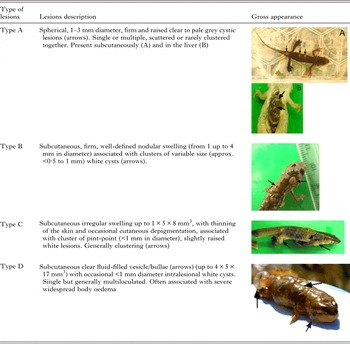
One hundred and sixteen live adult newts were dip netted from three static water bodies. The selection of sample sites was based on a previous study (Anderson Reference Anderson2010) and included one pond (control site) where infection had not previously been recorded and no macroscopic cutaneous lesions were observed during our sampling (n = 12 newts). The remaining 104 newts were captured from the other two water bodies (n = 51 and n = 53). In addition, 23 dead newts were observed around the banks of one infected site. Forty newts (28 from infected ponds and 12 from a control site) were retained and transported live to the field station where they were euthanized. To do this, newts were fully immersed in an aqueous solution of tricaine methane sulphonate (MS-222, solution of 0·2% buffered with sodium bicarbonate) in accordance with the schedule 1 method under the Animals (Scientific Procedures) Act 1986 and the American Veterinary Medical Association guidelines and recommendations for ethical euthanasia of amphibians (AVMA, 2007). Dermocystid lesions have previously been described on the skin of amphibian hosts, and on rare occasions on the liver (Pascolini et al. Reference Pascolini, Daszak, Cunningham, Tei, Vagnetti, Bucci, Fagotti and Di Rosa2003; Feldman et al. Reference Feldman, Wimsatt and Green2005; Raffel et al. Reference Raffel, Bommarito, Barry, Witiak and Shackelton2008; González-Hernández et al. Reference González-Hernández, Denoël, Duffus, Garner, Cunningham and Acevedo-Whitehouse2010). A detailed external examination was performed where macroscopic dermal lesions were recorded, detailing the size, colour and texture (Table 1), as well as noting their abundance and distribution across the newt body. In order to retain the anatomical location of visceral organs, a partial necropsy examination was performed and the coelomic cavity was accessed via a ventral midline incision. The liver was fully exposed and, if present, cystic lesions were recorded. Newts were then individually fixed by full body immersion in neutral buffered 10% formalin. A further six newts with suspected Dermocystid-like lesions were humanely euthanized by anaesthetic overdose as previously described and were immediately stored in 100% ethanol for molecular analysis.
Description and gross lesions of Amphibiothecum sp. infection in L. helveticus on Rum.

A subsample of 30 formalin-fixed newts, were processed for histological examination: 20 from infected ponds (18 with lesions and two without) and ten from the control site. Six axial sections of the whole body were taken at predefined intervals; carcasses were sliced across the head (rostral to the eyes and at the base of the skull), and across the trunk (at the level of the pectoral and pelvic girdles and with two extra sections in between). The proximal third section of the tail, including the cloaca, and fore- and hind-limbs were longitudinally oriented. Tissues were sectioned at 5 µm thick and stained with haematoxylin and eosin (H&E) and examined by a light microscope for the presence of parasitic cysts.
For transmission electron microscopy (TEM) analysis, one formalin-fixed skin tissue sample containing multiple subcutaneous cystic lesions was deparaffinized, post-fixed in 1% osmium tetroxide and processed routinely by dehydration through graded acetones prior to embedding in araldite resin. Ultrathin sections (60 nm thick) were counterstained with uranyl acetate and lead citrate and viewed with a Philips CM120 TEM. Images were taken on a GatanOrius CCD camera.
To investigate the distribution and condition of cutaneous granular glands, across diseased (n = 16) and control newts (n = 8), a standardized position was located in the tail dorsum (a segment just caudal to the cloaca), that provided clear visibility of epidermal and dermal tissue in all newts and adequate coverage of granular glands. These histological sections were photographed with a digital camera (Olympus DP72) at 4 × magnification (approximately 2 mm long segment). The number and diameter of all cutaneous granular glands in this section were assessed using Cell^D software (Olympus Soft Imaging Solutions). Granular glands were considered mature when more than ¾ of the gland alveolus was filled with bright eosinophilic granular material.
Data analysis
A logistic regression analysis was performed in R version 3.2.1 (R Core Team, 2015) to test the hypothesis that cutaneous granular glands of the dorsal tail were different in those animals with disease. The analysis was based on the infection status of the ponds (control, n = 8; infected, n = 16) using a forward stepwise approach where the independent variables were: the number of cutaneous granular glands, their diameters and the relative percentages of full glands, were entered into the model with P ⩽ 0·1 and excluded with P⩾0·2 (Hosmer and Lemeshow, Reference Hosmer and Lemeshow2000).
Molecular phylogenetics
A sample of oedematous tissue (1 mm2), a single dermal cyst or a single liver cyst was excised from each of the six ethanol-preserved newts, respectively. Excised tissue and cysts were washed in deionized water and dried. DNA was extracted using DNeasy® Blood & Tissue Kit (Qiagen, Crawley, UK) according to the manufacturer's instructions.
Primers specific to the mesomycetozoean clade were designed, targeting a 1400 bp region of 18s rRNA, from an alignment of five Dermocystidia and Ichthyophonida 18s rRNA sequences sourced from GenBank (Dermocystidium sp. CM-2002, AF533950; Rhinosporidium seeberi, AF118851; Ichythophonus irregularis, AF232303; Ichthyophonus hoferi, U25637, Pseudoperkinsus tapestis, AF192386). Sequences were aligned using ClustalW (Larkin et al. Reference Larkin, Blackschiled, Brown, Chenna, McGettigan, McWilliam, Valentin, Wallace, Wilm, Lopez, Thompson, Gibson and Higgins2007). Conserved regions were identified across the sequences to act as primers, ensuring enough variability in the target amplicon to provide phylogenetic information. Both primers were compared with the online Basic Local Alignment Search Tool (BLAST) (NCBI, online), confirming 100% homology with the Mesomycetozoan species listed above and weaker homology with L. helveticus (84%, e-value 0·026). Polymerase chain reactions (PCRs) were performed in 25 µL volume containing 1 µg DNA, 0·3 µ m of each forward (5′-GTAGTCATATGCTTGTCTC-3′) and reverse (5′-TATTGCCTCAAACTTCCAT-3′) primer, 200 µ m dNTPs (Bioline, London, UK), and 2·5 units of HotStarTaq Plus (Qiagen, Crawley, UK). Two μL of each PCR product were electrophoresed on 0·6% agarose gel, stained with GelRed™ Nucleic Acid Gel Stain (Biotium) alongside 2·5 µL of a 1 kb DNA Hyperladder™ (BioLine) to assess amplicon size. Amplification products of the correct size were cleaned using polyethylene glycol precipitation, and commercially sequenced by GATC Biotech. Sequences were manually edited in BioEdit (Hall, Reference Hall1999) by trimming the outermost 5′ and 3′ ends near the sequencing primer site, where read quality was poor or ambiguous.
Edited sequences were aligned with all 26 18S rRNA mesomycetozoean sequences submitted in GenBank. Non-Mesomycetozoean outgroup sequences were chosen based on high query cover (98% ident.) but weaker similarity to the Rum isolate than the mesomycetozoan sequences (<93%); JN054684·1, DQ9958071·1, KC488361·1. Sequences were aligned using multiple sequence alignment in ClustalX2·1 (Thompson, Reference Thompson1997).
Maximum-likelihood (ML) analysis and Bayesian analysis have different strengths and may return different topologies due to random errors in tree reconstruction (Svennbland et al. Reference Svennbland, Erixon, Oxelman and Britton2006; Yang and Rannala, Reference Yang and Rannala2012). For that reason, phylogenetic relationships were generated using both ML and Bayesian analysis to compare the topologies recovered and therefore offer more support to the relationships and clades recovered by both analyses. MrModelTest (Posada and Crandall, Reference Posada and Crandall1998) was performed in PAUP* 4·0 (Swofford, Reference Swofford2002), testing 56 different models of DNA evolution against a starting Neighbour-joining tree. The best-fit nucleotide model as determined by the Bayesian information criterion (Schwarz, 1978; Ripplinger and Sullivan, Reference Ripplinger and Sullivan2010) was Hasegawa–Kishino–Yano (Hasegawa et al. 1985) with gamma distributed rate variation and invariable sites (HKY + G + I). ML analysis was performed in Paup*4·0 (Swofford, Reference Swofford2002) setting parameter values as detailed above, and specifying the outgroup. Analysis was run with 1000 bootstrap iterations of a heuristic search and tree-bisection–reconnection branch swapping (Posada and Crandall, Reference Posada and Crandall1998). So as not to restrict model selection to named (i.e. Jukes and Cantor) or pre-specified models, model averaging was implemented for Bayesian analysis, sampling among the 203 general time reversible (GTR) models (Huelsenbeck et al. 2004). This was achieved by running analysis using the reversible-jump Markov chain Monte Carlo (rj-mcmc) in MrBayes 3·1·2 (Ronquist and Huelsenbeck, Reference Ronquist and Huelsenbeck2003), specifying gamma distributed rate variation, running two independent metropolis-coupled MCMC with four chains each, terminating after 10 000 replications when the standard deviation between the split frequencies reached <0·01 (indicating that the trees being sampled have converged), sampling every 100. Final consensus trees were edited in FigTree v1.4.2 (Rambaut, Reference Rambaut2014) adding bootstrap values or bipartition posterior probabilities.
RESULTS
Gross pathology
A total of 66 newts with macroscopic cutaneous lesions, suggestive of Dermocystid-like infection as described by González-Hernández et al. (Reference González-Hernández, Denoël, Duffus, Garner, Cunningham and Acevedo-Whitehouse2010), were recovered from two ponds; prevalence of 47% [95%CI (34, 60)] and 76% [95%CI (64, 87)], respectively. Of the 23 dead newts found, six were sufficiently well preserved to see that they had extensive dermal lesions, similar to those seen in live newts described hereafter.
Ninety-three per cent (26/28) of newts (15 males and 13 females) subject to detailed external examination had macroscopic lesions of all types (e.g. A, B, C and D; Table 1). The shape of skin lesions varied depending on the number and size of parasitic cysts and the associated subcutaneous oedema, whereas the shape of parasitic cysts was consistently spherical and pale grey/white. Additionally, subcutaneous haemorrhage and circular skin ulcers (up to 6 mm in diameter) were observed affecting 14 (54%) newts; these were clustered over the tail, limb insertions and subgular regions. Parasite cysts and skin lesions were most frequently observed on the dorsal surface of the body (n = 23). More specifically, cysts were located on the heads (n = 16), tails (n = 20), limbs (n = 20) and subgular regions (n = 18) along with solitary or multiple type A lesions on the liver (n = 11). Four newts (two females and two males) (15%) had additional, severe and diffuse subcutaneous oedema associated with cutaneous depigmentation and presence of numerous type D lesions. These animals were moribund or showed limited body movements prior to euthanasia.
Histology and TEM
Histopathological examination confirmed the presence of parasite cysts, or related pathological changes in newts (n = 20) from infected ponds including the two individuals that had no macroscopic skin lesions.
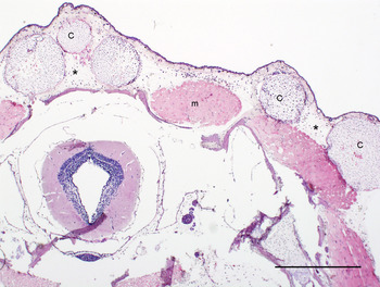
Single or multiple, intact or ruptured spheroid parasite cysts were consistently present expanding the strata spongiosum and compactum of the dermis (Fig. 1). Cysts were also present in the subjacent skeletal muscular layers (epi-and perimysium; n = 10 newts), oral mucosa (n = 5 newts), gastrointestinal lumen (n = 1 newt), liver (n = 7 newts) and cloaca stroma (n = 3 newts). See Supplementary Fig. 1A–D.
Intradermal Amphibiothecum sp. cysts in palmate newt (L. helveticus). (A) Cross-section of the trunk. Microscopic appearance of multiple spheroid cysts expanding and distorting the strata spongiosum and compactum of the dermis. Cysts are surrounded by diffuse and moderate subcutaneous oedema (*) associated with mild mixed inflammatory infiltrates. Scale bar = 500 µm. Specific features: (C) cyst; (m) muscular fibres.

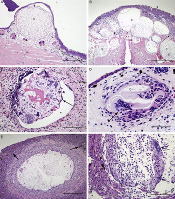
Based on cyst morphology and the associated host's inflammatory response (Fig. 2A–D), we could identify three different developmental stages: (1) developing (intact), (2) mature (intact and ruptured) and (3) degenerating and degenerated parasite cysts. All stages were observed concurrently affecting the same individual. Developing cysts were associated with no, or a mild, host cellular response (Fig. 2A), whereas ruptured and mature cysts were associated with tissue oedema and moderate to severe focal chronic-active inflammatory cell infiltrate. In addition, multinucleated giant cells (foreign body-type cells) and focal tissue necrosis (Fig. 2B) were occasionally present. In cases of severe subcutaneous swelling, numerous and generally ruptured parasitic cysts were present (up to 55), surrounded by a moderate chronic-active inflammatory cell infiltrate and tissue oedema. Degenerating cysts (Fig. 2C) were comparatively smaller and were characterized by a corrugated and convoluted cyst wall, partially or fully detached from the host connective tissue. A focal chronic-active inflammatory host response surrounded degenerating cysts and their lumen contained an amphophilic to brightly eosinophilic granular matrix admixed with irregular islands of pale basophilic material reminiscent of endospore formations. The advanced stage of cyst degeneration (degenerated cyst) (Fig. 2D) was characterized by a focal granulomatous lesion formed by packed mononuclear cells admixed with multinucleated giant cells, centred on a remnant of collapsed cyst wall. The majority of the parasitic cysts observed in the liver (84%) were either ruptured or degenerated, and were surrounded by a dense chronic inflammatory cell infiltrate admixed with several multinucleated giant cells (foreign body-type cells) (Fig. 2E, F).
(A) Longitudinal section of the tail. A large single developing Amphibiothecum sp. cyst expands the stratum spongiosum of the dermis. The cyst lumen is filled with a myriad of basophilic IE. Note the absence of inflammatory cell infiltrate. (B) Cross-section of the trunk. The dermis and axial musculature are markedly expanded by the presence of multiple coalescing cysts. Focal necrosis and moderate chronic inflammatory cell infiltrate accumulates around (long arrow) or within (short arrow) the cysts. There is focal epidermal hyperplasia (arrowhead). (C) Degenerating intradermal cyst surrounded by chronic active inflammation (arrows). The cyst wall is distorted and partially detached from the host tissue by the presence of a clear space. Within the cyst lumen, embedded in amorphous eosinophilic matrix, there are isolated islands of polymorphic basophilic endospores and scattered endospores (arrowhead) which still retain the morphological features observed in developing and mature cysts. Scale bar = 200 µm. (D) Advanced stage of an intradermal degenerated cyst characterized by a granulomatous lesion. Multinucleated giant cells (arrow), plasma cells and reactive fibroblasts surround a collapsed, empty ellipsoid cyst. (E) Liver. Large hepatic granuloma consisting of a central empty cyst surrounded by concentric layers of proliferating fibroblasts forming a fibrous wall. Admixed there are macrophages, scattered lymphocytes and occasional multinucleated giant cells. There are subjectively increased numbers of melanomacrophages (arrows) within the surrounding hepatic parenchyma. (F) Liver. Hepatic granulomatous lesion. The cyst lumen is partially replaced by moderate number of foamy macrophages along with numerous multinucleated giant cells (arrows) and occasional granulocytes. Few granular mature spores are observed within the cyst lumen or within local macrophages. Inflammatory mixed cell infiltrate expands the surrounding oedematous hepatic parenchyma. Specific features: (C) Cyst; (ep) Epidermis; (g) Cutaneous granular gland; (ss) Stratum spongiosum of the dermis; (w) Cyst wall; (m) Skeletal muscular fibres. Scale bar A, B, E = 500 µm; C, D, F = 200 µm.

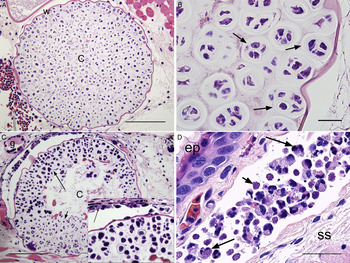
Intradermal developing cysts appeared as spherical sporangia ranging in size from 250 µm to 1·7 mm in diameter separated from the host connective tissue by 2–6 µm thick eosinophilic cyst walls (Fig. 3A). These contained a myriad of basophilic developing immature endospores (IE). IE were polygonal to crescent shaped, measured approx. 6 µm in diameter and had amphophilic to basophilic, occasional vacuolated, protoplasm depending on the stage of development. IE were packed within round to oval and refractile (‘mucoid-like’) chambers ranging in size from 10 to 40 µm in diameter (average approx. 24 µm), formed by clusters of dividing and budding elements further separated by internal faintly distinguishable septa (Fig. 3B). Depending on the stage of cyst maturation, IE were present along with a variable number of mature endospores (ME); clusters of IE or ME were observed either at the centre or at the inner periphery of the cyst wall within the same cyst (Fig. 3C). ME were located within less well-demarcated and comparatively smaller internal chambers, were round to ovoid measuring on average approx. 15 µm in diameter and were stained deeply basophilic. In the later stage of cyst maturation, ME (hereafter granular mature spore) were characterized by eosinophilic protoplasm swamped by numerous sub-spherical (approx. 1 µm in diameter) basophilic granular bodies. Granular mature spores were found within the lumen of mature cysts or were noted as free forms or engulfed by macrophages in the contiguous host tissues (Fig. 3C and D). When observed at higher magnification, granular mature spore resembled clusters of merozoite elements budding from the surface of mature shizonts (Fig. 3D). No flagellae or other motility organelles were observed.
(A) Cross-section of a developing intradermal Amphibiothecum sp. cyst containing myriad IE (IE). (B) High-power magnification of the inner lumen of a mature cyst. IE contained in septate chambers (arrows) clustering at the inner periphery of the cyst wall. (C) Cross-section of intradermal Amphibiothecum sp. cyst containing both IE and ME. Clusters of ME (long arrow) are opposite to IE (short arrow). Insert: ME and granular mature spores (circled). Note few macrophages containing intracytoplasmic granular mature spores surrounding the outer cyst wall (arrow). (D) Subcutaneous dilated lymphatic vessel adjacent to a ruptured cyst. Granular mature spores are free within the lumen (short arrows) or within macrophages (long arrows). Specific features are indicated with lower case letters: (c) Cyst; (ep) Epidermis; (g) Cutaneous granular gland; (ss) Stratum spongiosum of the dermis; (w) Cyst wall. Scale bar A, C = 200 µm; B = 20 µm; D = 50 µm.

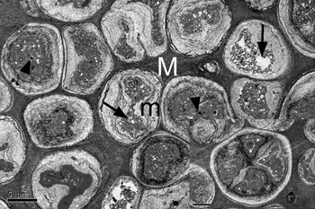
Ultrastructural (TEM) examination of an intradermal cyst revealed endospores as an individual unit enclosed in a thick electron dense fibrous and granular matrix delimiting outer endospore capsule (Fig. 4). The endospore protoplasm was further enclosed by an additional, non-uniformly distributed granular coat formed by convoluted membranes. The protoplasm of larger endospores was further subdivided through a process of invagination of these internal delimiting membranes, where the outer matrix eventually enclosed them into distinct subunits. Dividing endospores contained between two to four daughter cells (Fig. 4A, insert).
Transmission electron microscope microphotograph of Amphibiothecum sp. intradermal cyst from an infected palmate newt. Multiple endospores embedded in a thick electron dense fibrous and granular matrix (endospore capsule) (M). Variable sized food vesicles (long arrows) and multiple dense round coarsely osmiophilic granular inclusion bodies (short arrows) are occasionally present in cells protoplasm. Membranous granular–fibrillar membranes (endospore membranes) forming concentric rings around the protoplasm of each individual endospore (m). The right lower insert shows one endospore dividing into four ‘daughter’ cells. Encircled one visible nucleus with prominent nucleolus.

Tail granular glands
The number of cutaneous granular glands of the dorsal tail skin in the control group (n = 8) ranged from 7 to 14 (mean 10·4 ± 2·5). On average, 79% of tail glands were mature. Gland diameters ranged from 53·3 to 401·0 µm (average 204 ± 80 µm). The number of glands in infected newts (n = 16) ranged from 8 to 28 (mean 13·4 ± 4·3) with their diameter ranging from 27·1 to 489·8 µm (mean 147 ± 90). On average 50% of glands in the tail skin of infected animals were classified as mature. Stepwise regression analysis indicated that infected newts had marginally smaller glands than non-infected newts [(β = −0·025, 95% CI (−0·047, −0·023), P < 0·031].
Molecular analysis
Successful amplification of a 1400 bp DNA fragment was achieved from all DNA extractions. Retrieved sequences from samples representative of liver and dermal cysts and subcutaneous oedema were identical, confirming that each of these distinct pathologies is associated with the presence of the same pathogen. These sequences shared high nucleotide similarity (>94% similarity, >81% query cover) to members of the Mesomycteozoeans. Upon alignment with all Mesomycetozoean 18srRNA sequences archived in GenBank, nearly complete consensus was observed (1 bp difference) between Rum and two Dermocystid sequences isolated from infected L. helveticus in France (Dermocystid-Larzac; accession numbers GU232542·1 and GU232543·1).
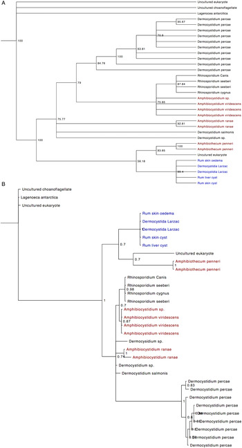
Overall the topologies obtained by Bayesian and ML techniques were congruent, although some differences in the internal relationships between Dermocystidium sp. and Amphibiocystidium sp. were recovered. Both Bayesian and ML analysis confirmed the Rum parasite to be a member of the Dermocystids, forming a well-supported clade with sequences from infected L. helveticus sampled in Larzac (Fig. 5: BS = 99·4%; PP = 0·7.) Whilst the Rhinosporidium sp. formed a monophyletic clade, the amphibian-infecting Mesomycetozoeans were polyphyletic. Under both analyses a clade was recovered suggesting a sister relationship between A. penneri and sequences from Rum and Larzac. Despite relatively weak confidence in this relationship (PP = 0·7; BS = 58·2%), support for a split between this clade and the rest of the Mesomycetozoeans was extremely high (BS = 100%; PP = 1·0.).
Consensus trees representing phylogenetic relationships of Mesomycetozoeans based on 18srRNA sequences. (A) Maximum likelihood analysis implementing HKY + G + I with node support shown using bootstrap support values; (B) Bayesian inference run using reversible-jump MCMC to average over the GTR models, with node support displayed as posterior probabilities; Amphibian-infecting species are highlighted in red, whilst sequences from Rum and Larzac are coloured blue.

DISCUSSION
This study reports combined gross, histopathological, ultrastructural (TEM) and molecular findings that characterize Amphibiothecum sp. infection in a geographically isolated population of palmate newts on the Isle of Rum (Scotland). Whilst we can say little about the prevalence of infection across the island from our sample of three water bodies, 63·5% of sampled live newts from infected ponds showed signs of disease, indicating that infection is likely to be common in palmate newts on the island.
As observed microscopically and ultrastructurally, intradermal parasitic cysts shared many common features with other Dermocystic organisms (Broz and Privora, Reference Broz and Privora1952; Jay and Pohley, 1981; González-Hernández et al. Reference González-Hernández, Denoël, Duffus, Garner, Cunningham and Acevedo-Whitehouse2010), in particular with that described in palmate newts in France (Gonzalez-Hernández et al. 2010). Similar to their observation, we also noted the presence of numerous endospores organized in internal, septated chambers enclosed by a cyst wall. Within the same cyst, clusters of endospores were observed at different developmental stages where the smallest compartmented chambers enclosed 2–4 single endospore elements. However, the arrangement of IE within mature cysts differed from that described by González-Hernández et al. (Reference González-Hernández, Denoël, Duffus, Garner, Cunningham and Acevedo-Whitehouse2010) where the authors propose a centrifugal fashion of endospores maturation. Instead, in this study, IE were present in clusters closer to the inner cyst wall of developed cysts and not exclusively at the centre of the cyst (Fig. 2C). This finding therefore suggests a different pattern of endospore maturation and a potential mechanism of endospores release. One possibility is that fully matured endospores escape in a ‘programmed’ pattern as described for R. seeberi (Mendoza et al. Reference Mendoza, Herr, Arseculerante and Ajello1999), where endospores might develop toward the cyst's pore from where they are discharged. However, we were unable to identify a cyst pore in any of the histological sections.
In agreement with other amphibian-specific Dermocystid infections (Pascolini et al. Reference Pascolini, Daszak, Cunningham, Tei, Vagnetti, Bucci, Fagotti and Di Rosa2003; Pereira et al. Reference Pereira, Di Rosa, Fagotti, Simoncelli, Pascolini and Mendoza2005; Raffel et al. Reference Raffel, Bommarito, Barry, Witiak and Shackelton2008; González-Hernández et al. Reference González-Hernández, Denoël, Duffus, Garner, Cunningham and Acevedo-Whitehouse2010 and Courtois et al. Reference Courtois, Cornuau, Loyau and Schmeller2013), diseased newts on Rum had macroscopic distinctive, raised, pale-white cystic skin lesions. In contrast to what was observed from Dermocystidim spp. infection of Rana temporaria and P. esculenta (Guyénot and Naville, 1922; Pascolini et al. Reference Pascolini, Daszak, Cunningham, Tei, Vagnetti, Bucci, Fagotti and Di Rosa2003) and Amphibiocystidium sp. infection in Eastern red-spotted newt (Raffel et al. Reference Raffel, Bommarito, Barry, Witiak and Shackelton2008), here no ‘U’ or bent ‘C’ shaped cysts were seen. In addition, we frequently observed unusually large subcutaneous fluid filled vesicles/bulla (e.g. type D lesion) accompanied with full body oedema as a result of Amphibiothecum sp. infection.
Similarly to Amphybiocystidium sp. infected newts in France (González-Hernández et al. Reference González-Hernández, Denoël, Duffus, Garner, Cunningham and Acevedo-Whitehouse2010), we observed skin lesions primarily distributed over the newt dorsum, limbs and tail, and only occasionally over the ventral trunk. This is in contrast to other Amphibiocystidium spp. infections where cyst distribution was often concentrated on the ventral surface (Broz and Privora, Reference Broz and Privora1952; Pascolini et al. Reference Pascolini, Daszak, Cunningham, Tei, Vagnetti, Bucci, Fagotti and Di Rosa2003; Pereira et al. Reference Pereira, Di Rosa, Fagotti, Simoncelli, Pascolini and Mendoza2005; Densmore and Green, Reference Densmore and Green2007; Raffel et al. Reference Raffel, Bommarito, Barry, Witiak and Shackelton2008).
The presence of parasitic cysts at different developmental stages and the related pathological changes (as observed grossly and histologically) are together suggestive of infection progression. While intact developing cysts were characterized by absent or a mild host inflammatory response, fully mature and ruptured cysts were always associated with a discrete inflammatory response. In the latter stages of cyst degeneration, granuloma formation resulted in reduced tissue inflammation and the restoration of surrounding tissues. Similarly, following the hepatic dissemination, granulomatous lesions commonly occurred. Although not the primary focus of this study, we suggest that the cyst wall plays a crucial role in protecting the parasite from the host immune system during its development. In fact, the virtual absence of an inflammatory response surrounding intact developing cysts is noteworthy in comparison with the inflammation surrounding ruptured and degenerating cysts. Clinical manifestation of Amphibiothecum spp. infection in palmate newts on Rum varied from subclinical (e.g. apparently healthy individuals with only few microscopic subcutaneous parasitic cysts), up to severe generalized infection. While the processes causing different clinical outcomes remain unclear, these observations suggest that whilst some newts may recover from Dermocystid infection (e.g. presence of microscopic dermal resolving lesions), other individuals develop a generalized and potentially fatal disease.
Here we described animals with microscopic parasite cysts, without the presence of gross lesions. González-Hernández et al. (Reference González-Hernández, Denoël, Duffus, Garner, Cunningham and Acevedo-Whitehouse2010) found no evidence of asymptomatic or carrier-state individuals from infected palmate newts in France; however, the presence of A. viridescens cysts was observed on the livers of apparently uninfected Eastern red-spotted newts (Raffel et al. Reference Raffel, Bommarito, Barry, Witiak and Shackelton2008). This suggests that subclinical infection might be more common than expected and the observation of macroscopic skin lesions alone may not be a good proxy to determine infection prevalence. The detection of pathogen genomic DNA from toe or tail clippings, and skin, oral or cloacal swabs, offer alternative detection methods extensively employed for Bd and Ranavirus surveillance (Annis et al. Reference Annis, Dastoor, Ziel, Daszak and Longcore2004; Hyatt et al. Reference Hyatt, Boyle, Olsen, Boyle, Berger, Obendorf, Dalton, Kriger, Hero, Hines, Phillott, Campbell, Marantelli, Gleason and Colling2007; Skerratt et al. Reference Skerratt, Berger, Speare, Cashins, Mcdonald, Phillott, Hines and Kenyon2007; Goodman et al. Reference Goodman, Miller and Ararso2013). However, the validity and accuracy of these methods are being questioned. Whilst swabbing techniques have been found to underestimate both infection prevalence and parasite burden in Chytridiomycosis (Shin et al. Reference Shin, Bataille, Kosch and Waldman2014; Clare et al. Reference Clare, Daniel, Garner and Fisher2016), they often miss subclinical Ranvirus infections (Greer and Collins, Reference Greer and Collins2007; Gray et al. Reference Gray, Miller and Hoverman2012). Detailed histopathological examination therefore represents an important diagnostic tool, particularly in cases of seemingly healthy individuals with only subclinical disease.
Severe full body oedema was microscopically associated with a high parasite burden along with a generalized form of infection evidenced by concurrent presence of cysts in the liver. To the authors’ knowledge, this is the first published report of a generalized Dermocystid spp. infection, which results in severe subcutaneous oedema as confirmed by both histopathological and molecular analysis. Subcutaneous oedema would likely result from osmotic and electrolytic imbalances caused by extensive breaches in skin integrity due to numerous presence of parasite cysts. Parasite cysts in the liver might also result in hepatic insufficiency and systemic disease.
In addition to skin (mainly present within the dermis) and hepatic lesions, parasite cysts were also found in previously unreported body sites such as oral mucosa, intestinal lumen, cloaca and infiltrating within the skeletal muscle bundles. Secondary skin lesions were also occasionally observed from infected newts and consisted of skin ulcers and haemorrhages as previously reported (González-Hernández et al. Reference González-Hernández, Denoël, Duffus, Garner, Cunningham and Acevedo-Whitehouse2010). We consider that secondary lesions resulted from self-induced trauma, and from weakening or breaches of the epidermis associated with parasite cysts.
Altogether these findings suggest that the intensity of infection may be critical in determining the outcome of disease. Severe infection could increase mortality either directly, or indirectly by compromising newt fitness (e.g. reducing foraging and motility capabilities) or by rendering diseased newts more vulnerable to predation by compromising locomotion (Lindstrüm et al. Reference Lindström, van der Veen, Legault and Lundstroöm.2003). The presence of lesions in the oral mucosa could impact food intake, whereas cysts and oedema on the cloaca could have an impact on breeding and courtship. If cysts and oedema on the cloaca are a common finding the ability of male to produce spermatophores may be compromised. Similarly breeding success may also be reduced in other ways; male newts rely on tail fanning to entice females and lead them over deposited spermatophors (Halliday, Reference Halliday, Peter, Slater and Colin1990; Griffiths, Reference Griffiths1996), behaviours that may be hindered considerably by the presence of tail oedema or significant lesions.
Since the majority of Amphibiothecum spp. cysts were found within the dermis or adjacent tissues (e.g. skeletal muscles bundles), parasite transmission is likely to occur by direct skin exposure to contaminated sediment/water or infected newts. Infectious elements are likely to be released onto the skin surface and/or surrounding environment after mature cysts rupture as suggested by others (Perez, Reference Perez1913; Broz and Privora, Reference Broz and Privora1952; Jay and Pohley, 1981). The transmission mechanisms of Dermocystidium spp. between their fish hosts are well documented, where all species produce zoospores to facilitate waterborne transmission (Perkins, Reference Perkins1976; Olson et al. Reference Olson, Dungan and Holt1991). However, in this study, both histological and TEM examinations consistently showed no sign of parasite spores developing flagellae. In addition to direct skin exposure, the possibility of oral transmission cannot be ruled out due to the microscopic observation of parasite cysts within the digestive system. This might also explain the presence of liver cysts, which could pass through the bile duct following ingestion as hypothesized previously (Raffel et al. Reference Raffel, Bommarito, Barry, Witiak and Shackelton2008). However, due to the anatomical location of the liver, it is possible that parasite spores migrate through the sub-adjacent dermal-muscle layers and into the liver.
Further studies are necessary to confirm the mode of parasite transmission and also to investigate whether multiple infections, characterized by different developmental cyst stages within the same individual, resulted from intra-tissue spread of mature infectious elements (i.e. merozoite-type elements released from mature cysts), or repeated external exposure where each cyst represents a ‘discrete infection event’ as proposed by Raffel et al. (Reference Raffel, Bommarito, Barry, Witiak and Shackelton2008).
To explore the variation in disease susceptibility several studies have considered the role played by the innate immunity provided by AMPs (Zasloff, Reference Zasloff2002). Amphibian AMPs, released from granular cutaneous glands are increasingly recognized as a first-line of defence against pathogens that use the skin as their route of infection (Rollins-Smith et al. Reference Rollins-Smith, Doersam, Longcore, Taylor, Shamblin, Carey and Zasloff2002; Woodhams et al. Reference Woodhams, Rollins-Smith, Carey, Reinert, Tyler and Alford2006, Reference Woodhams, Rollins-Smith, Alford, Simon and Harris2007; Rollins-Smith, Reference Rollins-Smith2009). We speculate that the reduced diameter of granular glands in infected newts, along with a partial depletion of these glands, could have a negative impact on the production of AMPs, resulting in a partially compromised innate immune response and increased susceptibility/severity to infection. Whilst we cannot allude to the mechanisms leading to this difference, or the order of cause and effect, it is an interesting observation that may deserve more investigation.
Based on the high-sequence identity and phenotypic similarities, the pathogen observed here is the same pathogen described from L. helveticus in Southern France (González-Hernández et al. Reference González-Hernández, Denoël, Duffus, Garner, Cunningham and Acevedo-Whitehouse2010). Phylogenetic analysis not only emphasizes their affiliation, but also highlights their distinctiveness from other species of Dermocystid. The well-supported and distinct clade containing the Rum and France sequences, and the short branch lengths recovered under Bayesian analysis, are indicative of a single species. Whilst the internal relationships between Amphibiocystidium, Dermocystidium and Rhinosporidium species were not consistent across ML and Bayesian analysis, the amphibian and fish infecting pathogens are not monophyletic. Rhinosporidium sp. formed a discrete clade suggesting one evolutionary host-shift to mammalian hosts. However, the nested arrangement of Dermocystidium sp. and Amphibiocystidium spp. suggests that pathogens can undergo host-shifts, resulting in several, independent amphibian-specific lineages. Whilst host shifts between the lower invertebrates are not uncommon (Densmore and Green, Reference Densmore and Green2007; Bandín and Dopazo, Reference Bandín and Dopazo2011, Price et al. Reference Price, Garner, Nichols, Balloux, Ayres, Mora-Cabello de Alba and Bosch2014), the presence of multiple host-shifts from fish to amphibians, but not the converse, is atypical and appears to contradict previous theories on host-shifts (Jancovich et al. Reference Jancovich, Bremont, Touchman and Jacobs2010). In agreement with Feldman et al. (Reference Feldman, Wimsatt and Green2005) our analysis distinguished A. penneri from other Mesomycetozoeans with high confidence, supporting its consideration as a separate genus. Sequences from Rum and Larzac were also distinct from Amphibiocystidium sp., instead forming a clade with A. penneri, a pathogen of B. americanus in Northern America. The L. helveticus infecting pathogens are therefore not members of Amphibiocystidium, but instead should be consider a novel species within the genus Amphibiothecum; Amphibiothecum meredithae.
SUPPLEMENTARY MATERIAL
The supplementary material for this article can be found at https://doi.org/10.1017/S0031182016001943
ACKNOWLEDGEMENTS
We would like to thank SNH for their support and guidance during field research. We gratefully acknowledge all the staff of the Easter Bush Pathology unit at he Royal (Dick) School of Veterinary Studies for their technical assistance; in particular Ms Jennifer Harris for performing microbiology tests and Dr. Sionagh Smith. We are also grateful to Stephen Mitchell (Kings Building) for assistance in TEM sample preparation and for support with TEM.
FINANCIAL SUPPORT
Work was supported by The Natural Environment Research Council (Grant number S055) and funding from the Morris Animal Foundation. Funding for microbiology analysis came from The Royal (Dick) School of Veterinary Studies.