Syncope refers to a sudden-onset and temporary loss of consciousness along with the loss of body muscle tone in an individual secondary to insufficient cerebral perfusion.Reference Myung 1 The most common type of syncope is vasovagal syncope.Reference Mcload 2 , Reference Driscoll, Jacobsen and Porter 3
The pathophysiology of vasovagal syncope has not yet been elucidated. One of most controversial issues is the rule of cerebral autoregulation. Cerebral autoregulation refers to the intrinsic mechanisms that stabilise blood flow despite changes in cerebral perfusion pressure. When an individual goes from a supine position to an upright position, cerebral blood flow is regulated by autonomic, neurocardiogenic, vascular, and humoral mechanisms.Reference Schondorf, Benoit and Wein 4 , Reference Albina, Cisneros and Laino 5 For healthy subjects, cerebral blood supply is maintained against changes in the systemic arterial pressure up to 50 mmHg. When the blood pressure of cerebral vessels decreases, arteriolar vasodilatation occurs; conversely, vasoconstriction occurs when blood pressure increases.Reference Strandgaard, Olesen, Skinhoj and Lasson 6 – Reference Schondorf, Stein, Roberts, Benoit and Cupples 7
The head-up tilt test induces a strong orthostatic effect by using a mildly tilted upright position, which ensures maximal venous pooling. Syncope is induced with Bezold Jarisch reflex in patients with vasovagal syncope. The head-up tilt test is used to evaluate a patient’s symptoms including heart rate, blood pressure, and cardiac rhythm response during orthostatic stress. During the test, symptomatic patients may be identified even though there may be no hypotension and/or bradycardia.Reference Sung, Du, Yu, Yam and Fok 8 , Reference Grubb, Samoil and Kosinski 9 Isoproterenol, adenosine, nitroglycerine, and edrophonium can be used to increase the sensitivity of the test.
Transcranial Doppler is a non-invasive and reliable test that enables the evaluation of both cerebral blood supply and cerebral vessel response by monitoring cerebral blood flow rate.
In the current study, orthostatic stress is induced by head-up tilt test, and blood flow rate is simultaneously measured with transcranial Doppler to better understand the mechanism of syncope in children with a history of recurrent vasovagal syncope, and the results were compared with a control group. Our study aimed to evaluate changes in the cerebral blood supply in children during vasovagal syncope and to clarify the diagnostic value of transcranial Doppler for vasovagal syncope.
Materials and methods
Our study enrolled 52 children and adolescents, who were in the age group of 7–17 years of age and had recurrent vasovagal syncope characterised by loss of consciousness and muscle tone with spontaneous recovery within a short time. The study was conducted at Pediatric Cardiology Department, School of Medicine, Osmangazi University, Eskişehir, Turkey. Written approval was obtained on October 27, 2010 from the Ethics Committee, School of Medicine, Osmangazi University, Eskişehir.
Patients were divided into three groups. Group 1 consisted of 31 patients who were symptomatic and whose head-up tilt test was positive. In this group, 19 patients indicated that they had one or more syncopal episodes, whereas 12 patients had a minimum of three presyncopal episodes associated with symptoms, excluding loss of consciousness, such as dizziness, excess sweating, blurred vision, and fatigue. Group 2 comprised 21 patients who were symptomatic but whose head-up tilt test was negative. Of those patients, 11 had one or more than one syncope episode, whereas 10 patients had a minimum of three presyncopal episodes. Group 3 included 22 healthy children, who were asymptomatic and had normal physical examinations, were of similar age and gender distribution with that of patient groups, had no presyncopal or syncopal episodes, and had normal neurological and cardiovascular evaluations.
All patients had screening cardiac and neurological physical examinations, electrocardiogram, EEG, and cardiac ECHOs. A detailed history was obtained regarding past medications, which may lead to cardiologic and neurologic clinical findings. Patients with congenital or acquired heart disease, arrhythmia, history of seizure with abnormal EEG findings, systemic diseases that may affect the autonomic nervous system, and having a history of taking medications that were potentially confounding were excluded.
For the diagnosis of vasovagal syncope, head-up tilt test was applied for 20 minutes using a hydraulic tilt table that may be inclined up to 80° from the supine position. The test was performed ∼2 hours after breakfast – no tea or coffee – or lunch and the patients were allowed to rest for 20 minutes at room temperature. Pulse and arterial blood pressure were monitored throughout the test period. Haemodynamic response was identified as follows: cardio-inhibition includes patients who had at least a 40 beats/minute decrease in their heart rate unassociated with bradycardia; vasodepression indicates patients with a decrease of 25 mmHg in mean blood pressure; and mixed type refers to patients who had decrease in both heart rate and arterial blood pressure. Those patients were rapidly placed supine. Pre-syncope symptoms involved dizziness leading to anxiety, pale skin, blackout, nausea, anxiety, myalgia, and respiratory distress. Electrocardiogram traces were recorded before and after the test.
For the subjects of the patient and control groups, head-up tilt test was repeated on the hydraulic test table, which may be inclined up to 60°, at room temperature. It was established that the patients were not hungry. 2-Mhz transcranial Doppler (TCD) probe was fixed on each patient’s bilateral temporal bones using a bandage. Flow rates of bilateral middle cerebral arteries were continuously and simultaneously recorded with temporal window transcranial Doppler. Pulse and arterial blood pressure were monitored throughout the test period. Measurements were made and recorded at 2-minute intervals. In all, three measurements, at 2-minute intervals, were made at the supine position, and thus baseline values were obtained. Later, the bed was inclined upwards to 60°. A recording was taken at the upright position for 20 minutes, and later patients were placed in a supine position. The test was stopped after measurements were made at 2-minute intervals. The symptomatic patients were rapidly placed supine, and the test was discontinued.
SPSS for Windows 15.0 was used for statistical analyses; Shapiro–Wilk’s test was used for evaluating normal distribution of data. Among parametric tests, t-test and variance analysis were used for analysing the data with normal distribution for the inter-group comparisons, whereas Tukey test was used, among multiple-comparison tests (post hoc tests), for identifying the mean of different groups in the variance analysis. For comparison of the values obtained at different measurement time points, repeated measures analysis of variance was used, whereas Pearson’s χ2 analysis was used for analysing comparison tables. For the head-up tilt test-positive group, receiver operating characteristic analysis method was used to determine the cut-off values of the cerebral blood flow variables. The data were expressed as the mean±standard deviation, and p-values <0.05 were considered significant.
Results
Of the patients, 27 were girls (51%) and 25 were boys (49%), with a female/male ratio of 1.08/1. The age range of the female subjects was 7–17 years (mean: 13.7±2.5 years), whereas the age range of male subjects was 8–17 years (mean: 13.8±2.5 years). There were no statistically significant differences between the three groups with respect to age and gender distribution (p>0.05; Table 1).
Age and gender distribution of the study groups.

Of the symptomatic patients who had head-up tilt test at 80°, 31 were considered head-up tilt test positive (Group 1) and 21 were considered head-up tilt test negative (Group 2). In Group 1, 19 out of 31 patients presented with a history of syncope, whereas the remaining 12 patients presented with a history of presyncope. In Group 2, 11 out of 21 patients presented with a history of syncope, whereas the remaining 10 patients presented with a history of presyncope. There were no differences between Groups 1 and 2 with respect to history of presyncope and syncope and application rates (p>0.05).
Symptoms developed at 8 minutes (mean) during the test in Group 1. Of all cases, three had bradycardia (10%), 10 had hypotension (32%), and 13 developed hypotension and bradycardia (42%). There were five patients (16%) who had intact vital findings; however, the test was discontinued because they developed presyncope findings such as pale skin, dizziness, and nausea. The test results of those patients were deemed positive. The symptoms developed at a mean of 8 minutes.
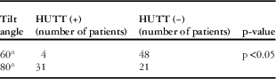
Head-up tilt test was reapplied on the tilt table for Group 1, Group 2, and the control group to evaluate cerebral blood flow rate with transcranial Doppler. Of 52 symptomatic patients (head-up tilt test at 80° inclination), 31 patients had a positive response, whereas only four patients had a positive response when the tilt test was repeated at 60° (Table 2).
Comparison of HUTT angles and resultant responses.

HUTT=head-up tilt test
For head-up tilt test applied at 60°, pulse, systolic blood pressure, and diastolic blood pressure values of the three groups were considered as baseline data. There were no differences between the three groups with respect to the systolic and diastolic blood pressures, as well as pulse values (p>0.05).
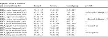
For measurements obtained on right middle cerebral artery with transcranial Doppler, no differences were found between blood flow rates of the three groups, which were measured at the supine position (p>0.05). When the bed was at an upright position, the maximum blood flow rate of the right middle cerebral artery was lower in Group 1 than in Group 2, although the decrease was more significant in comparison to the healthy control group (p<0.05). The maximum blood flow rate of Group 2 was numerically, although not significantly (p>0.05), lower than that of the healthy control group at the upright position. For measurements obtained during the resting period, when the tilt table was at the supine position, no differences were found between the three groups with respect to maximum blood flow rates of the right middle cerebral artery (p>0.05; Table 3).
Maximum and minimum blood flow rate values of right and left MCA.

MCA=middle cerebral artery; LMCA=left middle cerebral arteries; RMCA=right middle cerebral arteries
At the supine position, there were no differences between the three groups with respect to minimum flow rates (p>0.05). When the bed was at an upright position, the minimum blood flow rate of the right middle cerebral artery was lower in Group 1 than in Group 2, although the decrease was more significant in comparison with the healthy control group (p<0.05). A minimum blood flow rate in Group 2 was numerically, although not significantly (p>0.05), lower than that of the control group. For measurements obtained during the resting period, when the tilt table was at the supine position, no differences were found between the three groups with respect to minimum blood flow rates of right middle cerebral artery (p>0.05; Table 3).
For measurements obtained on the left middle cerebral artery with transcranial Doppler at the supine position, no statistically significant differences were found between the maximum blood flow rates of the three groups (p>0.05). For measurements obtained at the upright position, the maximum blood flow rate of the left middle cerebral artery was numerically, although not significantly (p>0.05), lower in Group 1 than in Group 2. However, maximum blood flow rate of the left middle cerebral artery was significantly lower in Group 1 than in the control group (p<0.05). There were no differences between Group 2 and the control group with respect to the maximum flow rates (p>0.05). For measurements obtained during the resting period, when the tilt table was at the supine position, no differences were found between the three groups with respect to maximum blood flow rates of the left middle cerebral artery (p>0.05; Table 3).
For measurements obtained on the left middle cerebral artery with the transcranial Doppler at the supine position, no statistically significant differences were found between three groups with respect to minimum blood flow rates (p>0.05). For measurements obtained at the upright position, the minimum blood flow rate of the left middle cerebral artery was numerically, although not significantly (p>0.05), lower in Group 1 than that of the head-up tilt test symptom (+) in Group 2. However, minimum blood flow rate of the left middle cerebral artery was significantly lower in Group 1 than that of the control group (p<0.05). There were no statistically significant differences between Group 2 and the control group with respect to the minimum flow rates of the left middle cerebral artery (p>0.05). For measurements obtained during the resting period, when the tilt table was at the supine position, no differences were found between the three groups with respect to the minimum blood flow rates of left middle cerebral artery (p>0.05; Table 3).
Discussion
Syncope is a common disease, which is associated with sudden loss of consciousness and posture.Reference Hugh 10 However, presyncope is defined as the presence of warning symptoms such as a blackout or excessive sweating and dizziness that are not associated with loss of consciousness. The most common type of syncope is vasovagal or neural syncope in both the paediatric and the adult population.Reference Mcload 2 , Reference Driscoll, Jacobsen and Porter 3 The age range of patients in our study was 7–17 years, although 36.6% of the patients were in the age range of 15–17 years. As reported in the literature, we found that vasovagal syncope is more common in the adolescent population.Reference Sheldon and Killam 11
In the current study, positive head-up tilt test response was obtained in 31 of 52 patients (60%) in which vasovagal syncope was considered. Symptoms developed at 8 minutes (mean). This finding is consistent with the onset time of the findings, which were identified by another study conducted with head-up tilt test.Reference Schondorf, Benoit and Wein 4
In the previous studies,Reference Zhang, Du and Li 12 – Reference McGavigan and Hood 15 it was seen that rate of vasovagal syncope types may vary according to the haemodynamic response. In the present study, we had obtained the response of cardioinhibition in 3 out of 31 (10%) patients, whereas vasodepression was identified in 10 out of 31 (32%) patients and mixed type in 13 out of 31 (42%) patients.
There many factors influencing positive test results. Test duration is the most remarkable factor, which influences the positivity. Many articles advocate that the longer the test duration is (mostly 45 minutes), higher the sensitivity and specificity are.Reference Raviele, Menozzi, Brignole, Gasparini, Alboni and Musso 16 However, there are also views supporting the hypothesis that false positivity increases as the tests last longer than 20 minutes.Reference Benditt, Lurie, Adler and Sakaguchi 17 Shortening the time can be shorter in order to increase cooperation of children. Considering the head-up tilt test protocol of our paediatric cardiology department, patients are placed in the upright position for 20 minutes. The tilt angle should be a minimum of 60° in order to evaluate the physiological effects of placing the patient in the passive upright posture in the head-up tilt test. Zaidi et alReference Zaidi, Benitez, Gaydecki, Vohra and Fitzpatrick 18 and Matalon et alReference Matalon and Fahri 19 reported that cardiac output, diastolic blood pressure, and heart rate linearly increase when the angle is increased in the range of 30° to 60°, although there is a small angle change between 60° and 80°, resulting in no significant haemodynamic change. It was reported that there was a minor difference between the two angles, which did not lead to significance.Reference Voice, Lurie and Sakaguchi 20 It was demonstrated that for lower angles (30°–45°) orthostatic stress is not effectively induced.Reference Abi-Samra, Maloney, Fouad-Tarazi and Castle 21 – Reference Aydın, Salukhe, Wilke and Willems 22
In the current study, 52 patients had head-up tilt test at 80°. Positive response was obtained in 31 patients. For the patient group who had the test at 60° in the upright position in order to make evaluations with transcranial Doppler, positive result could be obtained only in four patients. The positivity rate of head-up till test was significantly lower relative to head-up tilt test at 80°. In the current study, lower values obtained at angle of 60° are consistent with the finding that sensitivity of the test increased at 80°.
Studies were conducted with healthy control groups, who do not have a history of syncope, in order to reveal out the sensitivity of the head-up tilt test. There are variations between studies investigating positivity rate of head-up tilt test in healthy subjects.Reference Raviele, Menozzi, Brignole, Gasparini, Alboni and Musso 16 , Reference DeMey and Enterling 23 , Reference Fitzpatrick, Theodorakis and Varda 24 In the current study, head-up tilt test (80°) was positive in two of 22 healthy subjects (9%; the control group) who had no history of syncope or presyncope. The variation may result from the difference between protocols, test durations, and test angles.
Cerebral autoregulation refers to all intrinsic mechanisms that stabilise blood flow despite changes in cerebral perfusion pressure, and it is involved in the pathophysiology of the vasovagal syncope. In 1982, Aaslid et alReference Aaslid, Markwalder and Nomes 25 developed the transcranial Doppler method, which allows the measurement of blood flow rate for cerebral arteries. Using this method, it was ensured that the response of patient against acute changes of blood flow rate could be monitored in a non-invasive manner.Reference Tiecks, Lam, Aaslid and Newell 26 In the current study, the patients with vasovagal syncope were divided into head-up tilt test (+), head-up tilt test (−), and a control group, and transcranial Doppler was performed to evaluate cerebral blood circulation. Our aim was to understand the pathophysiology of vasovagal syncope and to clarify the diagnostic value of transcranial Doppler for vasovagal syncope. In the supine position, there were no differences between head-up tilt test (+) and head-up tilt test (−) and the control group with respect to minimum and maximum flow rates of the right and left middle cerebral artery (p>0.05). This result was consistent with the results obtained by Sung et al,Reference Sung, Du, Yu, Yam and Fok 8 who evaluated cerebral blood flow rate of 32 children with vasovagal syncope with transcranial Doppler. Minimum and maximum blood flow rates of the middle cerebral arteries were compared with the cerebral blood flow rates of 23 healthy children, with similar age and gender distribution. Under these conditions, no differences were found between groups with respect to the basal data obtained at the supine position. The cerebral blood flow rate was evaluated in an adult population with transcranial Doppler in association with head-up tilt test, and it was found that the cerebral blood flow rates measured in the supine position were similar to those of the control group. It was concluded that cerebral autoregulation is preserved if there is no orthostatic stress.Reference Carey, Manktelow, Panerai and Potter 27 In the current study, no differences were found between groups with respect to the cerebral blood flow rates measured when orthostatic stress was not present, and this finding further supports the view that autoregulation is maintained until orthostatic stress develops.
The maximum flow rate of the right middle cerebral artery was lower in the head-up tilt test (+) than the head-up tilt test (−) group when this parameter was measured at the upright position (p<0.05). It was observed that this decrease was more significant in comparison with the control group (p<0.05). On the contrary, maximum blood flow rate of the right middle cerebral artery was lower in the head-up tilt test (−) group when compared with the control group, although the difference was not statistically significant (p>0.05). The maximum blood flow rate of the left middle cerebral artery (in the upright position) was lower in the head-up tilt test (+) group relative to the head-up tilt test (−) group; however, the difference was not statistically significant (p>0.05). The maximum blood flow rate of the left middle cerebral artery was lower in head-up tilt test (+) in comparison with that of the control group and the difference was statistically significant (p<0.05). The maximum blood flow rate of the left middle cerebral artery was numerically, although not significantly (p>0.05), lower in the head-up tilt test (−) group compared with the control group. In the study conducted by Sung et al,Reference Sung, Du, Yu, Yam and Fok 8 the maximum flow rate of middle cerebral artery did not differ between the head-up tilt test (+) and the control groups when the parameter was measured in an upright position. The study concluded that maximum blood flow rate does not play a role in the pathophysiology of syncope. However, paediatric studies are scarce. Schondorf et alReference Schondorf, Benoit and Wein 4 conducted a study where the cerebral blood flow rate of 12 adult patients with vasovagal syncope was simultaneously evaluated with head-up tilt test (80°) and transcranial Doppler, and the minimum flow rate of the bilateral middle cerebral arteries were lower in an upright position relative to the control group, although there were no differences in the maximum blood flow rate. In different studies examining adult patients with vasovagal syncope, no similar change was found in the maximum blood flow rate.Reference Hugh 10 , Reference Fitzpatrick, Theodorakis and Varda 24 In the current study, we identified a reduction in the maximum blood flow rate in the head-up tilt test (+) group in an upright position; however, this finding contradicts the data reported in adult studies and in the paediatric study conducted by Sung et al.Reference Sung, Du, Yu, Yam and Fok 8
The maximum flow rate of right middle cerebral artery was lower in the head-up tilt test (+) than the head-up tilt test (−) group when the parameter was measured in the upright position (p<0.05), and it was observed that the decrease was more significant in comparison with the control group. No statistically significant differences were found between the head-up tilt test (−) group and the control group in the upright position (p>0.05). There were no differences between head-up tilt test (+) group and the head-up tilt test (−) group with respect to the minimum flow rate of the left middle cerebral artery (p>0.05). However, the minimum blood flow rate of the left middle cerebral artery was lower in Group 1 than the control group, and the difference was statistically significant (p<0.05). For minimum blood flow rate of the left middle cerebral artery in the upright position, no statistically significant or numerical difference was found between the head-up tilt test (−) group and the control group (p>0.05). In the current study, the minimum blood flow rate of the right middle cerebral artery was lower in the symptom (+) and head-up tilt test (+) group than that of the symptom (+) and head-up tilt test (−) group and the control group. However, the minimum blood flow rate of the left middle cerebral artery was lower in the symptom (+) and head-up tilt test (+) group only when compared with the control group (p<0.05). The findings of the study are consistent with the data on minimum blood flow rate in studies where cerebral blood flow is monitored with transcranial Doppler. The reduction of cerebral blood flow rate in the head-up tilt test (+) group during orthostatic stress supports the view that a loss of consciousness is secondary to the cerebral hypoperfusion.Reference Stephenson 28
In the current study, patients were placed in a supine position during the third stage of the head-up tilt test, and measurements were taken at 2-minute intervals before the test was completed. When the patients were once again placed in the supine position during the rest period at the end of the study, there were no differences between the three groups with respect to arterial blood pressure, pulse, and maximum and minimum blood flow rates (p>0.05). These data support the view that cerebral auto-regulation is impaired during orthostatic stress, and cerebral perfusion rapidly recovers when stress is eliminated. Symptoms are due to acute changes that occur during orthostatic stress.Reference Sung, Du, Yu, Yam and Fok 8 , Reference Carey, Manktelow, Panerai and Potter 27
This study aims pathologyphysiology of vasovagal syncope. However, transcranial Doppler can be clinically used in the differential diagnosis of vasovaganl syncope as cranial blood flow decreased in head-up tilt test-positive patients with psychogenic syncope during head-up tilt test. In addition, for patients who have suspected findings, such as dizziness and nausea, although there is no cardioinhibitory or vasodepressor response in head-up tilt test, demonstration of decreased cranial blood flow enables final diagnosis of vasovagal syncope.
In conclusion, we believe that the positivity rate increases when the head-up tilt test is applied at 80° rather than 60°. In addition, vasovagal syncope may develop without systemic hypotension and bradycardia, and minimum and maximum blood flow rates are significantly decreased in patients head-up tilt test (+) patients with vasovagal syncope during orthostatic stress. Finally, in addition to minimum blood flow rate, maximum blood flow rate plays a role in the cerebral autoregulation.
Limitations of the study
This study examined the pathophysiology of vasovagal syncope and it enrolled 52 patients. Large-scale studies are surely required. Transcranial Doppler was studied on the head-up tilt table, which can be adjusted to 60° on vertical axis. Transcranial Doppler test can be repeated at different inclinations (45° and 80°) to evaluate differences between cranial blood flows. Cranial blood flow can be evaluated in other types of syncope.
Acknowledgement
The authors would like to thank Cengiz Bal who made statistics this prospective study possible.
Financial Support
This research received no specific grant from any funding agency, commercial, or not-for-profit sectors.
Conflicts of Interest
None.