I. INTRODUCTION
Dimethylarsinic acid (DMAV), also known by the trivial name cacodylic acid (CAS number 75-60-5), is a common form of organic arsenic with two known crystalline polymorphs having triclinic (Trotter and Zobel, Reference Trotter and Zobel1965) and monoclinic (Betz et al., Reference Betz, McCleland and Marchand2011) symmetry. Until fairly recently, DMAV and other organoarsenicals were used as pesticides for crops (Quazi et al., Reference Quazi, Sarkar and Datta2013; Nagar et al., Reference Nagar, Sarkar, Makris and Datta2014), despite abundant evidence of the toxicity of arsenic (Tchounwou et al., Reference Tchounwou, Patlolla and Centeno2003; Capitani, Reference Capitani, Deschamps and Matschullat2011). Arsenic (As) is generally considered to be the most impactful environmental toxin (ATSDR, 2020), based on the frequency of occurrence (at American National Priority List sites), toxicity, and potential for human exposure. In addition to the widespread distribution of DMAV in pesticide polluted soils, arsenic content is a common concern with rice, and DMAV had been demonstrated to cause straighthead disorder in rice (Limmer et al., Reference Limmer, Wise, Dykes and Seyfferth2018). Methods of mitigating DMAV contamination in the environment are an active area of research (Adamescu et al., Reference Adamescu, Hamilton and Al-Abadleh2011; Tofan-Lazar and Al-Abadleh, Reference Tofan-Lazar and Al-Abadleh2012). DMAV has been observed in some seafood and fish species (Borak and Hosgood, Reference Borak and Hosgood2007) and is one of the primary metabolites produced in the body from the ingestion and inhalation of inorganic arsenic compounds (Hughes and Kenyon, Reference Hughes and Kenyon1998).
The monoclinic polymorph of DMAV was only recently characterized by single-crystal X-ray diffraction (SC-XRD) at 200 K (Betz et al., Reference Betz, McCleland and Marchand2011), and there is no Powder Diffraction File (Gates-Rector and Blanton, Reference Gates-Rector and Blanton2019) entry for the compound in the standard PDF-4+ database. The structure has been refined in this work with room temperature powder X-ray diffraction (PXRD) data and compared with a density functional theory (DFT)-optimized structure (plus the original single-crystal structure). The original triclinic polymorph structure of DMAV was also determined by single-crystal X-ray diffraction (Trotter and Zobel, Reference Trotter and Zobel1965), but without solution of the hydrogen atom positions. An experimental entry for triclinic DMAV with no structure is present in the database, PDF 00-029-1588 (Gates-Rector and Blanton, Reference Gates-Rector and Blanton2019). DFT geometry optimization has been used to add hydrogen positions to triclinic DMAV in this work. The experimental PXRD data for monoclinic DMAV and the DFT-optimized structure for triclinic DMAV have been used to generate Bragg reflection lists for inclusion in the PDF.
II. EXPERIMENTAL
A specimen of DMAV obtained from Sigma-Aldrich was lightly ground with a mortar and pestle. The specimen was mounted in a 0.3 mm ID polyimide capillary, sealed at both ends with Loctite adhesive.
PXRD patterns were collected using a Canadian Macromolecular Crystallography Facility beamline (08B1-1, Fodje et al., Reference Fodje, Grochulski, Janzen, Labiuk, Gorin and Berg2014) at the Canadian Light Source (CLS). 08B1-1 is a bending magnet beamline with a Si (111) double-crystal monochromator. Two-dimensional (2D) data were obtained using a Rayonix MX300HE detector with an active area of 300 mm × 300 mm. The patterns were collected at an energy of 18 keV (λ = 0.68919 Å) and a sample-detector distance of 250 mm.
The 2D PXRD patterns were calibrated and integrated using the GSASII software package (Toby and Von Dreele, Reference Toby and Von Dreele2013). The sample-detector distance, detector centering, tilt, and wavelength were calibrated using a series of patterns obtained from a lanthanum hexaboride (LaB6) standard reference material (NIST SRM 660a LaB6), and the calibration parameters were applied to all patterns. The 2D patterns for DMAV exhibited minor graininess, bordering on an ideal powder, and based on the minimal grinding, it is believed that the monoclinic polymorph of DMAV was present in the as-received material. After calibration, the 2D patterns were integrated to obtain standard point detector powder diffraction patterns.
Rietveld refinement was performed starting with the single-crystal structure of Betz et al. (Reference Betz, McCleland and Marchand2011). Rietveld refinements of the crystal structure were performed with the GSASII program (Toby and Von Dreele, Reference Toby and Von Dreele2013) using a Thompson-Cox-Hastings modified pseudo-Voigt peak shape function (Thompson et al., Reference Thompson, Cox and Hastings1987). The background was modeled using a 12-term log interpolation function. Positional parameters were refined with soft restraints on bond distances and angles for the As, C, and O atoms in the DMAV molecule obtained using Mogul 1.8.3 (Bruno et al., Reference Bruno, Cole, Kessler, Luo, Motherwell, Purkis, Smith, Taylor, Cooper, Harris and Orpen2004). Hydrogen positions were left fixed during refinement but were periodically optimized using Avogadro (Hanwell et al., Reference Hanwell, Curtis, Lonie, Vandermeersch, Zurek and Hutchison2012). The isotropic displacement parameters of the hydrogen atoms were constrained to be 1.3 times the value of the chemically bonded non-hydrogen atom.
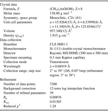
The crystal data, data collection, and refinement details are summarized in Table I.
The crystal data, data collection, and refinement parameters obtained for monoclinic DMAV.

DFT geometry optimizations were performed on both the monoclinic and triclinic polymorphs of DMAV using CRYSTAL17 (Dovesi et al., Reference Dovesi, Erba, Orlando, Zicovich-Wilson, Civalleri, Maschio, Rerat, Casassa, Baima, Salustro and Kirtman2018). Basis sets were obtained from the literature for the C, H, O (Gatti et al., Reference Gatti, Saunders and Roetti1994), and As atoms (Peintinger et al., Reference Peintinger, Vilela Oliveira and Bredow2013). The calculations used the B3LYP functional (Becke, Reference Becke1993) and D3 dispersion correction (Grimme et al., Reference Grimme, Antony, Ehrlich and Krieg2010) with eight k-points.
For the DFT-optimized structure of triclinic DMAV, the PXRD pattern was calculated for Bragg–Brentano geometry with CuKα 1 (λ = 1.54059 Å) radiation using the PowderCell program (Kraus and Nolze, Reference Kraus and Nolze1996).
III. RESULTS AND DISCUSSION
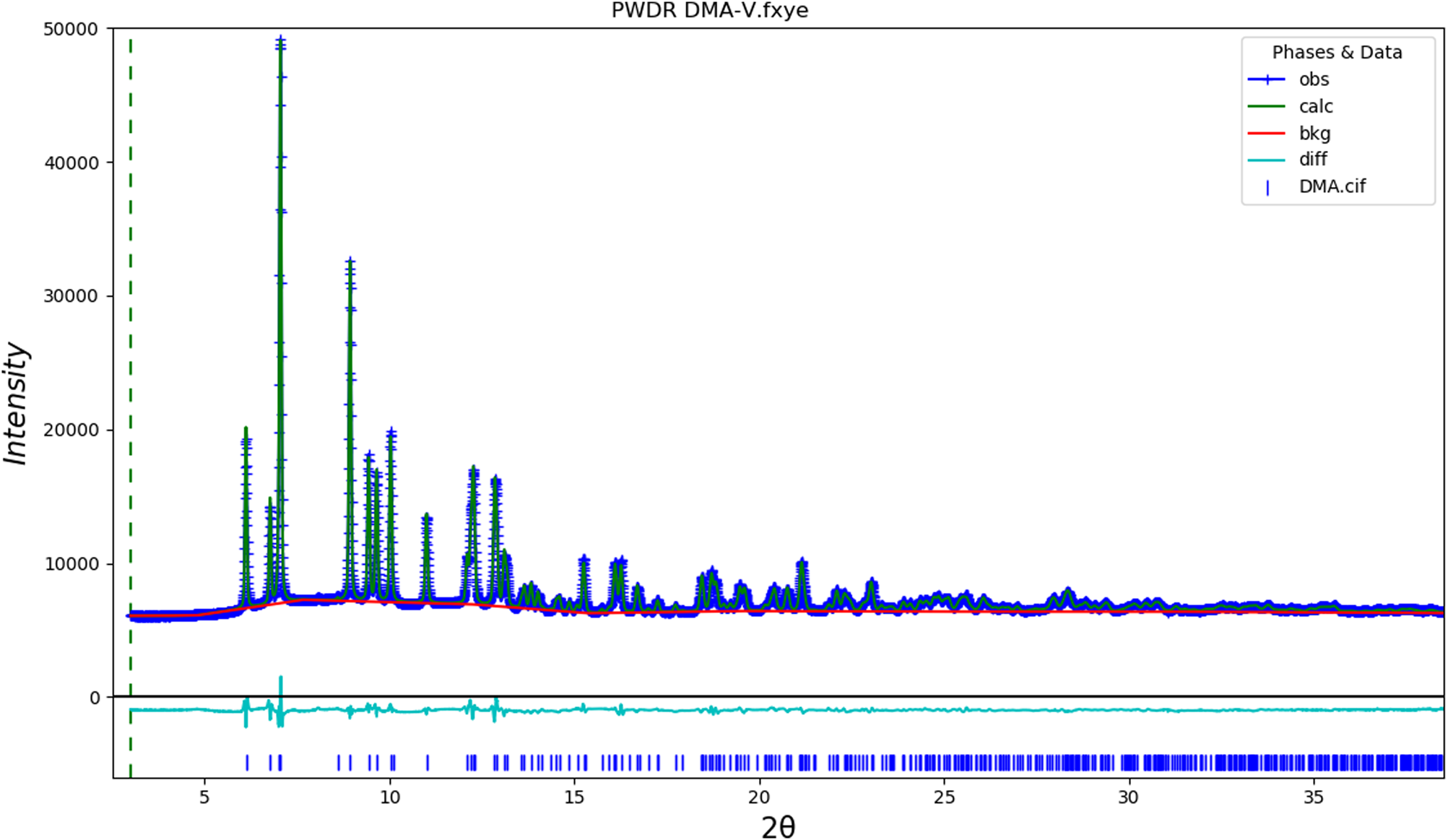
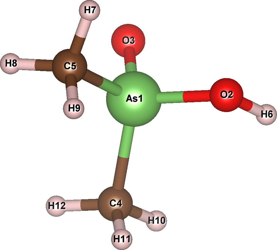
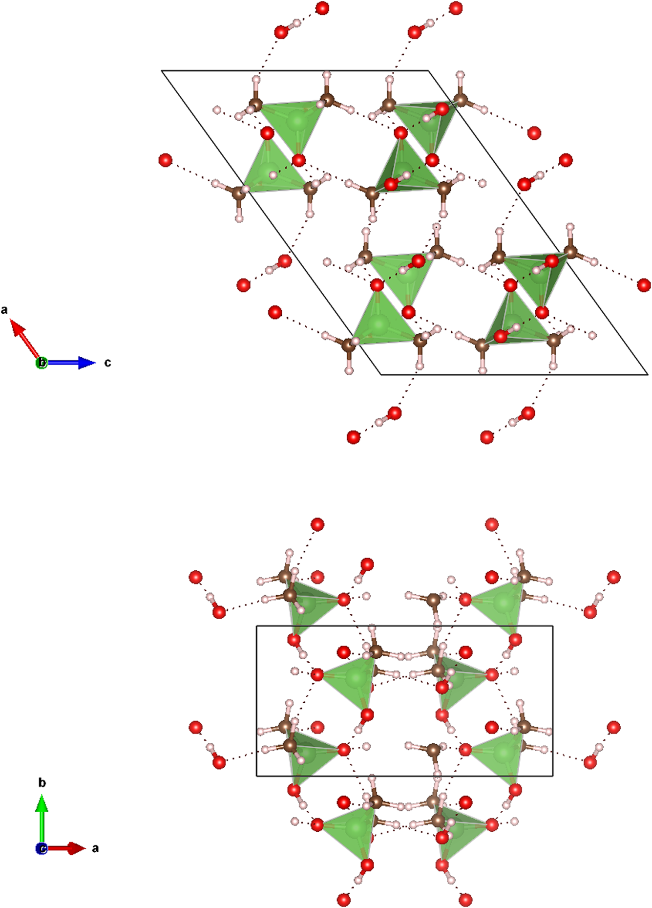
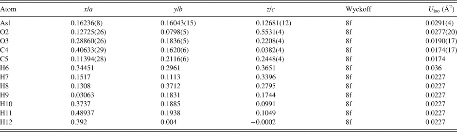
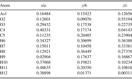
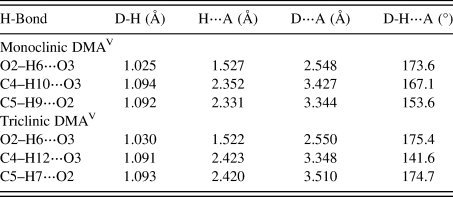
The final Rietveld refinement obtained for the synchrotron PXRD data from the monoclinic DMAV sample is illustrated in Figure 1, whereas the refined atomic coordinates and DFT-optimized atomic coordinates are shown in Tables II and III, respectively. Visually, the refinement looks excellent, and the root-mean-square (RMS) Cartesian displacements between the PXRD, DFT-optimized, and SC-XRD structures are all below 0.1 Å, suggesting excellent agreement between the structures (Van de Streek and Neumann, Reference Van de Streek and Neumann2014). The atom labels used for the structural models are illustrated in Figure 2, and a view of the DFT-optimized structure along the b- and c-axes are illustrated in Figure 3. The structure is well described by Betz et al. (Reference Betz, McCleland and Marchand2011) as infinite zigzag chains parallel to the b-axis defined by the strong O–H⋯O hydrogen bonds between the H6 atom of the hydroxyl group (donor) and the double-bonded O3 (acceptor) in the neighboring molecule along the chain. The hydrogen bonds obtained from the DFT optimization have comparable values to the SC-XRD refinement (Betz et al., Reference Betz, McCleland and Marchand2011) and are shown in Table IV.
A plot illustrating the final Rietveld refinement for monoclinic DMAV obtained with GSASII.

The atom labels used for structure description of DMAV. The figure was prepared with VESTA (Momma and Izumi, Reference Momma and Izumi2011).

The DFT-optimized crystal structure of monoclinic DMAV, viewed along the b-axis (top) and c-axis (bottom). The atom types can be identified by color including arsenic (green tetrahedra), carbon (brown), oxygen (red), and hydrogen (pink). Hydrogen bonds are represented by the dotted lines. The unit cell is outlined in black. The figure was prepared with VESTA (Momma and Izumi, Reference Momma and Izumi2011).

The Rietveld-refined crystal structure of monoclinic DMAV with lattice parameters: a = 15.9264(15) Å, b = 6.53999(8) Å, c = 11.3401(9) Å, and β = 125.8546(17)°.

All atoms were refined with fixed site occupancies of 1.
The DFT-optimized crystal structure of monoclinic DMAV with fixed lattice parameters: a = 15.9263 Å, b = 6.5402 Å, c = 11.3402 Å, and β = 125.856°.

The most prominent hydrogen bonds observed in the two DMAV structures and their parameters obtained from the DFT modeling.

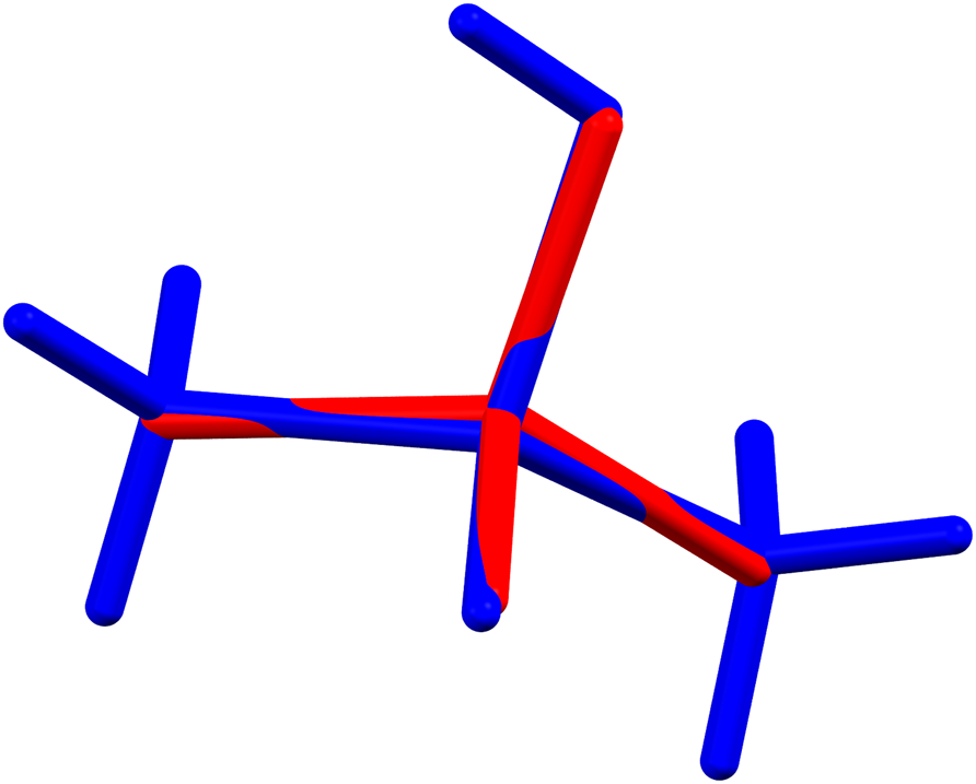
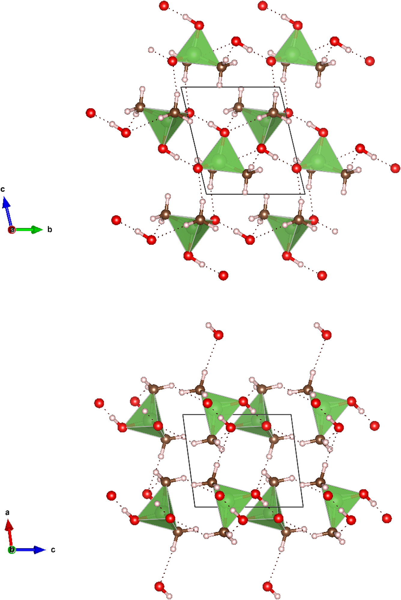
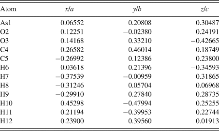
The DFT-optimized atomic coordinates of triclinic DMAV are shown in Table V. A molecular overlay of the DFT-optimized structure and the original SC-XRD structure (Trotter and Zobel, Reference Trotter and Zobel1965) is illustrated in Figure 4. The RMS Cartesian displacement of the non-hydrogen atoms is 0.13 Å, suggesting excellent agreement between the DFT-optimized and original SC-XRD structures (Van de Streek and Neumann, Reference Van de Streek and Neumann2014). The DFT-optimized triclinic DMAV structure is illustrated in Figure 5 along both the a- and b-axes. Similar to the monoclinic structure, the strongest hydrogen bond is the intermolecular O–H⋯O bond, but in contrast to the monoclinic DMAV structure, molecules in triclinic DMAV form dimers with pairs of O–H⋯O bonds between them. Weaker intermolecular C–H⋯O hydrogen bonds provide connections between the dimers. The hydrogen bonds are tabulated in Table IV.
A molecular overlay of the original SC-XRD structure (Trotter and Zobel, Reference Trotter and Zobel1965) of triclinic DMAV, with no hydrogen atoms, in red, and the DFT-optimized structure, with hydrogen atoms, in blue. The RMS Cartesian displacement of the non-hydrogen atoms is 0.13 Å. The figure was prepared with Mercury (Macrae et al., Reference Macrae, Bruno, Chisholm, Edgington, McCabe, Pidcock, Rodriquez-Monge, Taylor, van de Streek and Wood2008).

The DFT crystal structure of triclinic DMAV, viewed along the a-axis (top) and b-axis (bottom). The atom types can be identified by color including arsenic (green tetrahedra), carbon (brown), oxygen (red), and hydrogen (pink). Hydrogen bonds are represented by the dotted lines. The unit cell is outlined in black. The figure was prepared with VESTA (Momma and Izumi, Reference Momma and Izumi2011).

The DFT-optimized crystal structure of triclinic DMAV with fixed lattice parameters: a = 6.1852Å, b = 6.5300 Å, c = 6.6100 Å, α = 101.240°, β = 91.884°, and γ = 115.193°.

For the experimental PXRD data for monoclinic DMAV, a Bragg reflection list based on the GSASII refinement was prepared by summing reflections closer than 0.02°, 2θ as multiple reflections and assigning a weighted average reflection position, then including all reflections with relative integrated intensities of 0.2% or greater, up to 30° in 2θ. For the DFT-optimized structure of triclinic DMAV, the Bragg reflection list calculated using PowderCell (CuKα 1, λ = 1.54059 Å) was edited by summing reflections closer than 0.03° in 2θ as multiple reflections and assigning them a weighted average reflection position, then including all reflections with relative integrated intensities of 0.2% or greater up to 75° in 2θ. The Bragg reflection lists and raw data are contained in a crystallographic information file (CIF), which have been deposited with ICDD, along with individual CIF files for the Rietveld-refined and DFT-optimized structures.
IV. DEPOSITED DATA
The Crystallographic Information Framework (CIF) files containing the results of the Rietveld refinement (including the raw data) and the DFT geometry optimizations were deposited with ICDD. The data can be requested at info@icdd.com.
ACKNOWLEDGEMENTS
Many thanks are due to Jim Kaduk for helpful discussions regarding density functional theory and the CRYSTAL software package, and for providing a constant source of inspiration.
Research described in this paper was performed using beamline 08B1-1 at the Canadian Light Source, which is supported by the Canadian Foundation for Innovation, the Natural Sciences and Engineering Research Council of Canada, the National Research Council Canada, the Canadian Institutes of Health Research, the Government of Saskatchewan, Western Economic Diversification Canada, and the University of Saskatchewan.