Introduction
Head and neck sarcomas represent a relatively rare entity, accounting for 4–10 per cent of all soft tissue sarcomas and less than 1 per cent of head and neck neoplasms.Reference Yamaguchi, Nagasawa, Suzuki, Fujii, Iwaki and Takagi 1 , Reference Chang, Rosenberg, Glastein, Antman, DeVita, Rosenberg and Hellman 2 Sarcomas are of mesenchymal origin, and more than 50 pathological subtypes have been described. The histological type, such as chondrosarcoma, osteosarcoma and angiosarcoma, depends on the cell type of origin. The majority of head and neck sarcomas arise from soft tissues (80 per cent).Reference Greene, Page, Fleming, Fritz, Balch and Haller 3 Bone and cartilage sarcomas represent around 20 per cent of head and neck sarcomas and less than 0.2 per cent of malignant head and neck tumours.Reference Jemal, Murray, Ward, Samuels, Tiwari and Ghafoor 4 Head and neck sarcomas occur more frequently in men (male-to-female ratio of 2:1) and the mean age of presentation is around 50–55 years. In most cases, the patient is asymptomaticReference Colville, Charlton, Kelly, Nicoll and McLean 5 or presents with a painless neck mass (80 per cent).Reference Wanebo, Koness, Macfarlane, Eilber, Byers and Elias 6
Although head and neck sarcomas are not common, they are associated with high rates of recurrence and mortality. Surgical resection with wide margins may be associated with significant morbidity. There is a paucity of evidence in the head and neck surgical literature concerning the treatment options, and no clear guidelines exist to guide management. This relates not only to the current controversy about classification and grading, but also to difficulties confirming histological diagnosis, as some lesions may be confused with benign head and neck lesions.Reference Patel, Shaha and Shah 7
This paper aimed to report the experience of a UK tertiary referral head and neck oncology department in terms of head and neck sarcoma management. In addition, we report the oncological outcomes of head and neck sarcomas, and highlight the management dilemmas that arise during diagnosis, treatment and follow up.
Materials and methods
All patients who underwent treatment for head and neck sarcomas at the Head and Neck Unit at Guy's and St Thomas’ NHS Foundation Trust, London, from 1996 to 2012, were identified. A retrospective review of patients’ data, obtained from electronic computer records and case notes, was conducted. Data on the patients’ age, gender, histological type, disease site, treatment, adjuvant therapy, overall survival, disease-specific survival and recurrence (local, regional and distal) were collected.
This study did not require ethical approval according to current research and development hospital guidelines, but was registered and approved as an audit of service improvement.
Patients who were diagnosed but not completely treated under the care of our teams because of early referral to the regional sarcoma centre were excluded. All patients with human immunodeficiency virus (HIV) or acquired immunodeficiency syndrome (AIDS) in whom Kaposi sarcoma was a further feature of their underlying disease were also excluded.
All patients were evaluated clinically, and staged using either computed tomography (CT) or magnetic resonance imaging (MRI) or both. All histological diagnoses were confirmed by at least two senior dedicated head and neck histopathologists.
All patients were discussed at the multidisciplinary tumour board meeting where the management strategies were agreed. Moreover, all sarcomas were jointly reviewed at the regional sarcoma multidisciplinary meeting at University College London as per regional guidance.
The data collected were analysed using SPSS® statistical software. Variables were compared using Pearson's chi-square test, and survival outcomes were calculated using the Kaplan–Meier method. A p-value of less than 0.05 was considered significant.
Results
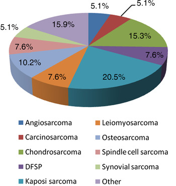
Twenty-eight males and 11 females were identified. Patients’ median age was 45.6 years (range, 13–88 years). The histopathological diagnosis was Kaposi sarcoma in eight patients (20.5 per cent), chondrosarcoma in six (15.3 per cent), osteosarcoma in four (10.2 per cent), leiomyosarcoma in three (7.6 per cent), dermatofibrosarcoma in three (7.6 per cent), spindle cell sarcoma in three (7.6 per cent), angiosarcoma in two (5.1 per cent), carcinosarcoma in two (5.1 per cent) and synovial sarcoma in two (5.1 per cent). A variety of other sarcomas were diagnosed in the remaining patients, including pleomorphic rhabdomyosarcoma, malignant fibrous histiocytoma, Ewing sarcoma, myofibroblastic sarcoma, liposarcoma and sarcoma not otherwise specified. Each of those subtypes was found in 2.5 per cent of the study group (Figure 1).
Frequency of different histological subtypes. DFSP = dermatofibrosarcoma protuberans

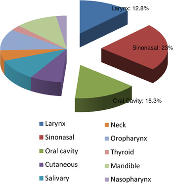
The site of disease was most commonly sinonasal (23.0 per cent), followed by the oral cavity (15.3 per cent). The larynx (12.8 per cent), oropharynx (10.2 per cent), mandible (10.2 per cent) and salivary glands (10.2 per cent) were also frequently involved. The remaining patients had disease involving the skin, cervical lymph nodes, thyroid and nasopharynx. The sites of disease are summarised in Figure 2.
Frequency of various disease sites.

Oncological follow-up details were available for all 39 patients. The median follow-up duration was 31 months (range, 0.10–185.6 months). Nine patients died during follow up.
The five-year overall and disease-specific survival rates were 25.64 per cent and 25.64 per cent, respectively.
Six patients developed local recurrence, three of whom went on to have distant recurrence. Two patients had regional recurrence, both of whom went on to have distant recurrence. Overall, 6 patients had distant metastases during follow up. Five-year local, regional and distant recurrence rates were 15.38 per cent, 5.12 per cent and 15.38 per cent, respectively.
A poor outcome was observed in patients who developed recurrence. Sixty-seven per cent, 100 per cent and 83 per cent of those patients who had local, regional and distant recurrence, respectively, died during follow up.
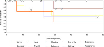
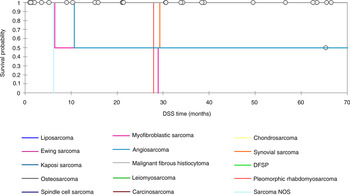
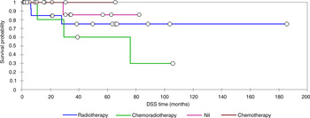
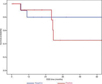
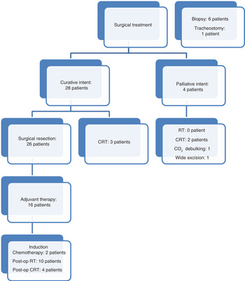
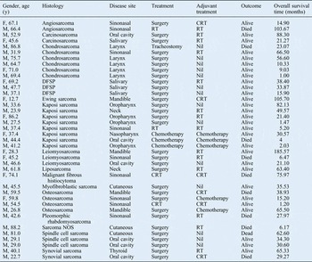
Table I summarises the histology, disease sites, treatments, adjuvant therapies, outcomes and overall survival times of the cohort. Figures 3–6 present the Kaplan–Meier disease-specific curves for disease site, histological subtype, therapy and excision margins, respectively. The different treatment modalities employed for the study patients are illustrated in Figure 7.
Kaplan–Meier curve for disease site and disease-specific survival time. DSS = disease-specific survival

Kaplan–Meier curve for histological subtype and disease-specific survival time. DSS = disease-specific survival; DFSP = dermatofibrosarcoma protuberans; NOS = not otherwise specified

Kaplan–Meier curve for adjuvant therapy and disease-specific survival time. DSS = disease-specific survival

Kaplan–Meier curve for excision margins and disease-specific survival time. DSS = disease-specific survival

Treatment modalities. CRT = chemoradiotherapy; RT = radiotherapy; CO2 = carbon dioxide

Summary of study group findings

Y = years; F = female; CRT = chemoradiotherapy; M = male; RT = radiotherapy; DFSP = dermatofibrosarcoma protuberans; NOS = not otherwise specified
Discussion
Head and neck sarcomas are rare tumours; they account for 1–11 per cent of all head and neck neoplasms.Reference Patel, Shaha and Shah 7 Regarding age of presentation, a trimodal distribution has been described. Peak ages are: less than 10 years, between 11 and 40 years, and over 40 years.Reference Kraus, Dubner, Harrison, Strong, Hajdu and Kher 8 Bentz et al., in 2004, reported a mean age of presentation of 47 years, with a male-to-female ratio of almost 1:1.Reference Bentz, Singh, Woodruff, Brennan, Shah and Kraus 9 In our cohort, the mean age was 62 years, and the male-to-female ratio approached 2:1. A significant characteristic of head and neck sarcomas is their heterogeneity, associated with their diverse origin. As a result, trials with large groups and long-term follow up are rare, and therefore clear management guidelines are not available.Reference Ketabchi, Kalavrezos and Newman 10
Clinical presentation can be relatively obscure and indolent in nature. The majority of patients (up to 80 per cent) present with a painless mass, but are otherwise asymptomatic.Reference Wanebo, Koness, Macfarlane, Eilber, Byers and Elias 6 Less frequent clinical manifestations, as they are described in the literature, include: neurological dysfunction such as sensory and/or motor disturbances, epistaxis, visual impairment, chronic sinusitis, and otalgia. Pain is mainly associated with bone sarcomas.Reference Wanebo, Koness, Macfarlane, Eilber, Byers and Elias 6 , Reference Aljabab, Nason, Kazi and Pathak 11 Stridor and laryngeal obstruction are associated with laryngeal chondrosarcomas.Reference Rinaldo, Howard and Ferlito 12
A crucial step in the diagnostic ladder is imaging of the tumour and the surrounding tissues, which allows accurate staging. Taking into consideration the proximity of vital structures in the head and neck, imaging is essential not only for diagnostic and staging purposes but it also helps the surgeon to plan therapy. Magnetic resonance imaging is generally considered to be superior for soft tissue imaging. On the other hand, CT scanning is of higher value in depicting bony structures. Because of the complex anatomy of the head and neck, a combination of MRI and CT scanning is preferred on many occasions. Chest CT is also recommended for staging purposes.Reference Patel, Shaha and Shah 7 Positron emission tomography is a valuable tool in diagnosis, follow up and diagnosis of recurrence.Reference Nieweg, Hoekstra and Pruim 13
The importance of adequate tissue biopsy and histological examination is paramount. Fine needle aspiration cytology, Tru-cut needle biopsy, and incisional or excisional biopsy may all be considered. They can provide valuable information about histopathology and tumour grade (high or low grade).Reference Patel, Shaha and Shah 7 This is crucial for management planning, not only for the surgery but for the post-operative treatment too. Incisional biopsy is preferred as it allows adequate sampling and optimal intra-operative haemostasis. It can be performed directly or under microlaryngoscopy, depending on tumour accessibility.Reference Patel, Shaha and Shah 7
Confirming a histological diagnosis is frequently problematic. In the literature, there are several reports illustrating cases where benign tumoursReference Allen and Allen 14 or unrelated malignant neoplasms, such as melanoma, lymphoma and carcinoma,Reference Flens, Valk, Tadema, Huysmans, Risse and Van Tol 15 can be misdiagnosed as sarcomas.
Early recognition is of vital importance for a better outcome. This is supported by National Institute for Health and Care Excellence (NICE) guidance, published in 2006, which states that ‘In patients with an unexplained lump in the neck which has recently appeared or a lump which has not been diagnosed before that has changed over a period of 3 to 6 weeks, an urgent referral should be made’. 16
There is controversy in the literature regarding the association between tumour site and survival. Zagars et al., in 2003, reported that head and neck sarcomas have a significantly poorer outcome in terms of both recurrence and survival.Reference Zagars, Ballo, Pisters, Pollock, Patel and Benjamin 17 In contrast, Van Damme et al., in 2010, concluded that anatomical location does not affect prognosis.Reference Van Damme, Schmitz, Machiels, Galant, Grégoire and Lengelé 18 Our study suggests that tumour subsite may affect outcome; however, with a limited cohort size, this effect failed to reach statistical significance. This can be attributed to the fact that such tumours are diagnosed late (as they do not produce early symptoms) and their excision involves radical surgery, which is associated with difficulty in achieving clear surgical margins given the proximity of vital structures.
Thirty-two of our patients (82 per cent) were treated with wide local surgical excision of the tumour, with clear margins whenever it was possible. Only seven patients (17.9 per cent) were treated with chemotherapy, radiotherapy (RT) or combined chemotherapy and RT. Some patients with low-grade sarcomas (e.g. of the larynx) were treated with transoral carbon dioxide laser surgical debulking and follow up. Select patients, particularly those with low-grade histology and a favourable disease site, can have resection with regular monitoring and access for repeat procedures. Such patients can achieve long-term, symptom-free survival, with minimal morbidity. This modality of treatment should always be considered in this patient subgroup.Reference Rinaldo, Howard and Ferlito 12
Patients with higher-grade, operable tumours underwent wide excision with clear margins whenever possible. In those cases where clear margins were not achieved, further surgery was considered on a case-by-case basis in order to complete surgical excision. Such an approach is widely accepted and, according to the literature,Reference Clark, Unni, Dahlin and Devine 19 – Reference Singh, Grimer, Bhujel, Carter, Tillman and Abudu 21 is associated with better outcomes;Reference Granados-Garcia, Luna-Ortiz, Castillo-Oliva, Villavicencio-Valencia, Herrera-Gómez and Mosqueda-Taylor 22 , Reference August, Magennis and Dewitt 23 it may even avoid the need for adjuvant RT.Reference Ketabchi, Kalavrezos and Newman 10 It is accepted that wide excision and clear margins are paramount to achieve sustained disease-free survival. In aggressive histological tumours, even close margins are not acceptable, as they have a similar recurrence rate as in patients with positive margins.Reference Kraus, Dubner, Harrison, Strong, Hajdu and Kher 8 , Reference Bentz, Singh, Woodruff, Brennan, Shah and Kraus 9 , Reference Van Damme, Schmitz, Machiels, Galant, Grégoire and Lengelé 18 , Reference de Bree, van der Valk, Kuik, van Diest, Doornaert and Buter 24 – Reference Kraus 27 Unfortunately, this is not always achievable because of the close proximity of vital anatomical structures and the surgical difficulty. That is why there is often residual disease with a negative impact on prognosis, in contrast to sarcomas of other parts of the body which have the same natural history but generally better outcomes.Reference Barrera, López, Gómez and Meneses 28 Thirteen of our patients were treated only with surgery, with average survival of 2.5 years.
In select head and neck sarcoma cases, post-operative RT has proven benefits in terms of local recurrence and survival. According to Tran et al. (2006), adjuvant RT has significant value for low-grade lesions with close or positive margins, and for all high-grade tumours. If wide excision is carried out in negative, clear margins, adjuvant RT is not necessary.Reference Tran, Mark, Meier, Calcaterra and Parker 29 Surgery combined with RT not only increases the disease-free survival time but also decreases the risk of local recurrence.Reference Tran, Mark, Meier, Calcaterra and Parker 29 In our study, patients who underwent combined therapy appeared to have a lower recurrence rate. Sixteen patients had post-operative RT (four of them had chemoradiotherapy), with average survival of 4.1 years. Of note, six patients survived more than five years, which supports the belief that RT may improve local control of the disease and prolong survival time.Reference Eeles, Fisher, A'Hern, Robinson, Rhys-Evans and Henk 30
Chemotherapy was not used in the majority of cases. According to a meta-analysis published in 1997, chemotherapy does not prolong survival in head and neck sarcoma. 31 It is mainly used as palliative treatment for metastatic disease. Pre-operative chemotherapy was used in three cases in this cohort. Neo-adjuvant chemotherapy may downsize primary tumours, because of the intact blood supply, making clear surgical margins achievable, and it may also address micro-metastatic distant disease.Reference Salvati, Ciappetta and Raco 32 , Reference Delgado, Maafs, Alfeiran, Mohar, Barrera and Zinser 33
The malignant nature of Kaposi sarcoma is under debate. It is defined as an angioproliferative disorder characterised by spindle-shaped cell proliferation, neoangiogenesis and oedema.Reference Ramírez-Amador, Martínez-Mata, González-Ramírez, Anaya-Saavedra and De Almeida 34 , Reference Feller, Wood and Lemmer 35 According to Fletcher (2006), Kaposi sarcoma is an intermediate neoplasm because of the absence of conventional malignancy features.Reference Fletcher 36 Although Kaposi sarcoma is frequently found in HIV-positive patients and more specifically when they have developed AIDS, it can also develop in HIV-negative patients, as demonstrated in our series.
In a review by Patrikidou et al., published in 2009, non-AIDS Kaposi sarcoma was described in 251 patients, most commonly affecting the palate and oropharynx. Two other cases of non-AIDS oral and palate Kaposi sarcoma have been reported.Reference Patrikidou, Vahtsevanos, Charalambidou, Valeri, Xirou and Antoniades 37 The management of these patients varies, and depends on the stage of disease. Stages I and II with slow progress may not need intervention, or they can be treated with surgical excision or RT. Stages II with rapid progress, and stages III and IV, need chemotherapy.Reference Di Lorenzo 38 , Reference Brambilla, Boneschi, Taglioni and Ferrucci 39 Patients with AIDS-related Kaposi sarcoma need to take antiretroviral therapy.Reference Di Lorenzo, Konstantinopoulos, Pantanowitz, Di Trolio, De Placido and Dezube 40 – Reference Krown 43 In the study group, one non-HIV-related Kaposi sarcoma was identified. The patient was an 86-year-old female with repeatedly negative HIV tests. Kaposi sarcoma was located in the oropharynx (tonsil) and she underwent tonsillectomy. The diagnosis was confirmed histologically and the excision margins appeared infiltrated. The patient had post-operative RT and regular follow up, and at the time of writing had been disease-free for 21 months.
The local recurrence rate was 15.3 per cent, and recurrence became apparent 4 to 65 months after treatment. In comparison to the literature, where the local recurrence rate is up to 50 per cent,Reference Mendenhall, Mendenhall, Werning, Riggs and Mendenhall 44 – Reference O'Sullivan, Gullane, Irish, Neligan, Gentili and Mahoney 46 we had a lower rate of recurrence. This could be because some of our patients had only recently been diagnosed and treated. Eighteen of 39 patients had adjuvant therapy (not just surgical excision), and only 3 of them were found to have high-grade soft tissue sarcoma.
We acknowledge that this paper has clear limitations. Head and neck sarcomas are a very heterogeneous group of tumours, with different clinical presentations, natural histories and management strategies, and therefore very different outcomes. Nevertheless, the authors feel that this is an important report of the clinical and histopathological presentations, management strategies, and outcomes, in one of the largest series collected in a UK tertiary head and neck unit.
According to NICE guidelines, it is crucial to refer patients with head and neck sarcomas to sarcoma centres. 16 There is no doubt that regional specialised units are necessary, particularly in order to centralise expertise in histological assessment. However, the vast majority of adult patients with head and neck sarcomas will require surgical resection whenever possible, and post-operative adjuvant RT. We therefore believe that the majority of patients can be treated adequately, with comparable outcomes, in tertiary units with a multidisciplinary expertise in head and neck sarcoma management, with adequate access to and support from specialised sarcoma multidisciplinary units.
-
• Head and neck sarcoma is a rare condition that poses significant challenges in management
-
• The most prevalent diagnoses were Kaposi sarcoma, chondrosarcoma and osteosarcoma; the most common disease sites were sinonasal, oral cavity and larynx
-
• Median follow-up period was 31 months; 9 patients died
-
• Only a few multicentre randomised, controlled trials are available to guide management
-
• Wide local excision with clear resection margins is essential to achieve local control and long-term survival
-
• Cross-specialty collaboration will help accrue evidence necessary to improve long-term outcomes
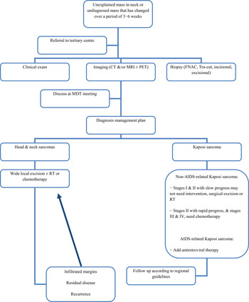
An algorithm of management is presented, which has been included in the unit's Standard Operational Policy, which we think will be helpful to streamline the management of these patients (Figure 8).
Guidelines for head and neck sarcoma management. CT = computed tomography; MRI = magnetic resonance imaging; PET = positron emission tomography; FNAC = fine needle aspiration cytology; MDT = multidisciplinary team; RT = radiotherapy; AIDS = acquired immunodeficiency syndrome

Conclusion
Head and neck sarcomas are rare clinical entities. They are challenging to treat, with few multicentre randomised, control trial data available to guide multidisciplinary teams. Wide local excision with clear resection margins is essential to achieve adequate local control and longer survival in those with aggressive tumours, with a select group of patients benefiting from the lower modality of debulking approaches. There is a need for cross-specialty collaboration in order to accrue the evidence necessary to improve the long-term outcomes for this select group of patients.