In adults with congenitally corrected transposition, the systemic ventricle is morphologically the right ventricle, which is not optimally designed to maintain systemic cardiac output in the long term. In addition, due to malalignment between the atrial and ventricular septums in congenitally corrected transposition, the atrioventricular conduction depends on an anomalous atrioventricular node. Variable degrees of atrioventricular block are encountered in up to half the patients, who frequently present with symptomatic systolic heart failure and complete atrioventricular block, even when not afflicted with other intra-cardiac defects.
As systolic dysfunction is common in adults with congenitally corrected transposition and they frequently require pacemaker implantation, cardiac re-synchronisation therapy has emerged as a potential treatment option in this population.Reference Diller, Okonko, Uebing, Ho and Gatzoulis 1 The anatomy of the coronary sinus and its tributaries, however, is not comparable to that found in the normal heart. Typically, the cardiac veins such as the great cardiac vein do not drain into the coronary sinus, being mirror-imaged in the usual variant of congenitally corrected transposition. This imposes several limitations to cardiac re-synchronisation therapy in these patients. In this study, we sought to re-evaluate the anatomy of the coronary sinus and its tributaries in hearts from patients with congenitally corrected transposition, establishing potential implications for cardiac re-synchronisation therapy, and comparing the features with those found in the normal heart.
Methods
We obtained 10 hearts from patients with congenitally corrected transposition, all held in the anatomic collection of the Heart Institute of São Paulo University Medical School (InCor), comparing the findings with eight matched specimens obtained from patients with no congenital cardiac defects who died from known cardiac causes. All procedures were performed according to the institutional guidelines and the study was approved by the Ethical Committee of the Heart Institute (CAPPesq n.o 1081/08).
All the hearts with congenitally corrected transposition, as expected, had discordant atrioventricular and ventriculo-arterial connections. In two of the specimens, we could not identify the orifice of the coronary sinus. In these patients, all the ventricular veins opened directly to either the right or the left atrium via small Thebesian openings. In the remaining eight specimens, the orifice of the coronary sinus was patent. These hearts were dissected, and the findings were compared with the ones from the control specimens, so as to determine the anatomic course of the epicardial coronary veins, the coronary sinus, and the oblique vein of the left atrium, also called the vein of Marshall.
The origin of the coronary sinus was taken as the point of union between the left atrial oblique vein and the left circumflex venous system. In normal hearts, this circumflex vein is known as the great cardiac vein. The great vein, however, accompanies the anterior interventricular artery, which is right-sided in the setting of the usual variant of congenitally corrected transposition. The vein joining the oblique vein in the setting of congenitally corrected transposition accompanies the morphologically right coronary artery, and drains the parietal wall of the morphologically right ventricle. Because of this difference compared with the normal situation, we have described the vein as a left-sided circumflex venous channel.
We also ascertained the sites of drainage of all the additional veins draining the walls of the morphologically right and left ventricles, using the nomenclature previously suggested by Uemura et al.Reference Uemura, Ho and Anderson 2 In addition to the anatomic course of the veins, we measured the linear distance between the right atrial orifice of the coronary sinus and the site of drainage of the left atrial oblique vein, along with the maximal diameters of the right-sided morphologically mitral and left-sided morphologically tricuspid atrioventricular valves.
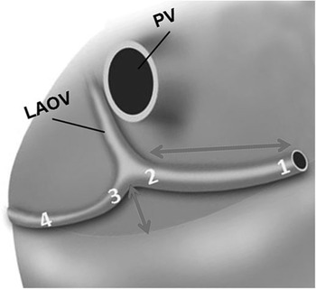
In specimens where the coronary sinus deviated from its expected location within the atrioventricular groove, as will be described, we also measured the linear orthogonal distance between the atrioventricular groove and the maximal point of deviation of the coronary sinus on the postero-inferior left atrial wall, the diameter of the right atrial orifice of the coronary sinus, the diameter at the point of union between the oblique vein and the left circumflex venous system, and the diameter of the left circumflex vein immediately proximal to its union with the oblique vein (Fig 1).
Schematic representation of the deviated course of the coronary sinus in the majority of our patients with CCT in our series. The long double-headed arrow shows the length of the sinus, whereas the small arrow shows the distance from the atrioventricular groove to the maximal deviation of the sinus. The numbers correspond to the sites of measurement of the diameters within the venous channels: 1 representing the right atrial opening, 2 the diameter at its origin formed by the confluence of the left atrial oblique vein and the circumflex vein, 3 the circumflex vein immediately proximal to the drainage of Marshall’s vein, and 4 the circumflex vein within the AV groove. AV=atrioventricular; CCT=cardiovascular computed tomography; LAOV=left atrial oblique vein; PV=pulmonary vein.

In order to allow comparisons between hearts of different sizes, which were obtained from patients of different ages, the linear distances were indexed against the largest diameter of the morphologically mitral valve, this being the right-sided valve in hearts from patients with congenitally corrected transposition and the left-sided valve in the control hearts.
Results
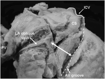
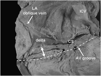
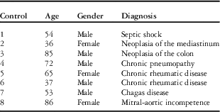
Clinical and morphological data are shown in Tables 1 and 2. Of the eight malformed hearts, five (62.5%) showed deviation of the initial course of the coronary sinus compared with control hearts (Fig 2). In three of these five hearts, the venous channel showed a delta-shaped enlargement of the junction between the oblique vein of the left atrium and the circumflex venous channel (Fig 3). The site of union between the venous channels was deviated away from the atrioventricular junction by distances varying from 0.67 to 2.4 cm in the hearts of different sizes. These distances corresponded to 28–74% of the diameter of the morphologically tricuspid valve (mean=56%), and corresponded to 47–72% of the diameter of the morphologically mitral valve (mean=61%).
This image shows the typical displacement of the coronary sinus (CS) away from the atrioventricular (AV) groove and the course of Marshall’s vein in one of our specimens with CCT. The double arrow shows the length of deviation from the AV groove. CCT=cardiovascular computed tomography; ICV=inferior caval vein.

This specimen from a patient with CCT, viewed from the diaphragmatic wall, shows the coronary sinus (CS) deviated from its normal course, and with a delta-shaped dilation at the site of drainage of Marshall’s vein. The double arrow shows the deviation away from the AV groove. AV=atrioventricular; CCT=cardiovascular computed tomography; ICV=inferior caval vein; LA=left atrium.

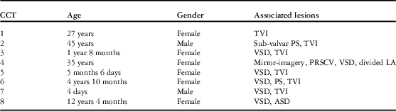
Demographic data and associated lesions of the patients with congenitally corrected transposition (CCT).

ASD=atrial septal defect; LA=left atrium; PS=pulmonary stenosis; PLSCV=persistent right superior caval vein to coronary sinus; TVI=tricuspid valve incompetence; VSD=ventricular septal defect
Demographic data of the control patients.

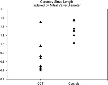
The length of the coronary sinus in the malformed hearts, judged as the distance from its mouth to the point of drainage of the oblique vein, correlated positively with the largest diameter of the morphologically tricuspid valve (R2=0.88, p=0.02). Moreover, when normalised by the largest diameter of the morphologically mitral valve, the sinus was shorter in the malformed hearts when compared with their controls (p<0.001, Fig 4).
Graphical representation of the length of the coronary sinus indexed by the diameter of the morphologically mitral valve, in hearts with CCT and in the controls. CCT=cardiovascular computed tomography.

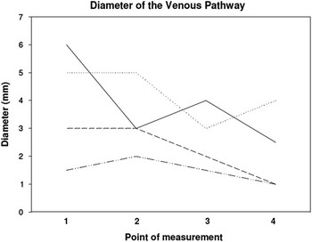
The mean diameter of the mouth of the coronary sinus in the normal hearts was 0.82 cm, whereas in the malformed hearts it varied from 0.15 to 3.0 cm, the smaller dimension being from the youngest patient and the largest corresponding to the adult case with mirror-imagery of the organs (situs inversus). This patient had persistence of a right-sided superior caval vein, which had drained to the coronary sinus. Figure 5 shows the diameters of the venous pathway at the points schematically presented in Figure 1. The valve guarding the orifice of the coronary sinus was absent in four of the malformed hearts, and of semi-lunar shape in the remaining four. None of them was considered to be large, covering no more than one-third of the orifice of the sinus.
Graphical representation of the diameters of the venous channels at points 1 through 4 (1: right atrial orifice; 2: the diameter at the confluence of the left atrial oblique vein and the circumflex vein; 3; the circumflex vein immediately proximal to the drainage of Marshall’s vein; and 4: the circumflex vein within the atrioventricular (AV) groove) and performed in four out of the five specimens with displacement of the coronary sinus. The omitted case is the specimen with the dilated coronary sinus receiving drainage of a superior caval vein (case 4).

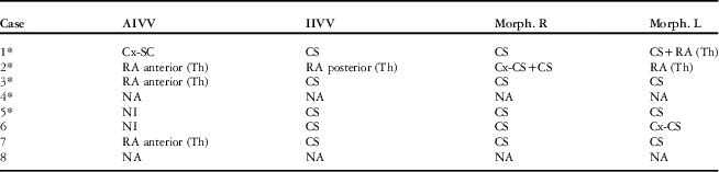
In Table 3, we show the sites of drainage of the major cardiac veins in the eight congenitally corrected transposition hearts. In summary, in three of the four cases in which it could be evaluated, the anterior interventricular vein drained directly to the morphologically right atrium via a Thebesian vein. The inferior interventricular vein, along with the morphologically right ventricular veins, drained predominantly to the coronary sinus. The morphologically left ventricular veins drained to the morphologically right atrium, either through the coronary sinus via a circumflex vein or via Thebesian veins.
Sites of drainage of the main coronary veins in TCGA patients.

AIVV=anterior interventricular vein; Cx-CS=drainage via circumflex vein to coronary sinus; IIVV=inferior interventricular vein; Morph. L=morphologically left ventricular veins; Morph. Th=drainage via Thebesian veins; NA=vein not available for analysis due to previous dissection or slicing; NI=vein not identified despite previously untouched specimen; R=morphologically right ventricular veins; TCGA=The Cancer Genome Atlas
*Cases with deviated course of the coronary sinus (CS)
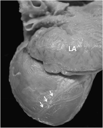
In all but one of the hearts with corrected transposition, a large marginal vein was observed on the epicardial surface of the morphologically right ventricle (Fig 6). In the remaining specimen, a postero-inferior vein was present on the diaphragmatic surface of the morphologically right ventricle, originating at the cardiac apex.
The arrows show the course of the marginal vein on the epicardial surface of the left-sided morphologically right ventricle. The left atrium (LA) is dilated.

Discussion
Re-synchronisation therapy is now recognised as a viable therapeutic option for patients with congenitally corrected transposition who present with systolic dysfunction.Reference Krishnan, Avramovitch, Kim and Trohman 3 , Reference Małecka, Bednarek and Tomkiewicz-Pajak 4 Although, in the absence of associated malformations, the circulation is physiologically corrected by the combination of discordant atrioventricular and ventriculo-arterial connections, the high prevalence of associated lesions, such as “Ebstein-like” malformations of the morphologically tricuspid valve, deficient ventricular septation, and pulmonary stenosis, usually forms the substrate for developing right and left ventricular failure. Moreover, the distorted anatomy of the abnormally located atrioventricular conduction axis is associated with an observed high incidence of conduction disturbances, with up to half manifesting with some degree of atrioventricular block.Reference Rutledge, Nihill, Fraser, Smith, McMahon and Bezold 5 Thus, patients with congenitally corrected transposition are increasingly likely to be referred for re-synchronisation therapy. This mandates a clear understanding of the coronary venous anatomy.Reference Kanter 6
The coronary sinus is the most important component of the coronary venous system. In patients with congenitally corrected transposition, when there is also usual atrial arrangement, the sinus receives the venous return from the left-sided morphologically right ventricle, rather than the morphologically left ventricle as in the normal heart. This means that, unlike the usual situation, it is not the great vein that serves as the major tributary draining to the sinus; this is because it is the morphologically right coronary artery, rather than the circumflex artery, that occupies the left-sided atrioventricular groove. Perhaps, because the anterior interventricular vein is unable to reach the left atrioventricular groove and drain to the coronary sinus, the sinus was deviated from its expected site in five of the eight hearts we examined. The maximal displacement corresponded with the origin of the sinus at the point of union between the left-sided circumflex venous channel and the left oblique vein.
By normalising our measurements relative to the diameter of the morphologically mitral valve, which is not usually distorted or malformed in congenitally corrected transposition, we also showed that the coronary sinus is shorter compared with the normal heart, and was dilated, showing a “delta shape” dilation at its origin in one-quarter of the cases. This enlargement could itself produce technical difficulties in accessing the coronary sinus during electrophysiological procedures.
Our findings concerning the venous drainage to the coronary sinus are in accordance with those reported previously.Reference Uemura, Ho and Anderson 2 Thus, it is the veins from the morphologically right ventricle that drain via the coronary sinus. Those from the morphologically left ventricle reach the heart cavities either via Thebesian veins or via the circumflex venous system, and thence the coronary sinus. It is the veins related to the morphologically right ventricle, therefore, that will be reached directly via the coronary sinus. In three of the four cases suitable for study, the anterior interventricular vein drained directly to the right atrium via Thebesian openings. Moreover, atresia of the right atrial orifice of the coronary sinus was found in one-fifth of our cases.
Atresia of the orifice of the sinus was found in less than one-twentieth of the series studied by Uemura,Reference Uemura, Ho and Anderson 2 with similar proportions noted by Bottega et al.Reference Bottega, Kapa and Edwards 7 Hornung and Calder,Reference Hornung and Calder 8 nonetheless, when reporting their clinical experience, noted a high incidence of problems with catheterising the right atrial orifice of the sinus. We identified the vein of Marshall in all our specimens, as did Uemura et al.Reference Uemura, Ho and Anderson 2 Similar to their findings, we took its union with the circumflex venous channel as the origin of the coronary sinus. Bottega et al,Reference Bottega, Kapa and Edwards 7 however, failed to find this vein in one-quarter of their cases. These investigators, nonetheless, did observe the deviation of the coronary sinus away from the atrioventricular groove, although in only two of their larger series of 54 cases. They did not go on to examine the magnitude of its deviation from the atrioventricular groove, nor did they direct attention to the dilation, or delta shape, of the initial segment. In our hearts, the mean deviation of the venous pathway from the atrioventricular groove corresponded to more than three-fifths of the diameter of the morphologically mitral valve. Thus, it is evident that patients with congenitally corrected transposition show unusual findings with regard to the course, shape, and length of the coronary sinus. These almost certainly reflect the difference in structure of the cardiac venous circulation in congenitally corrected transposition compared with normal hearts.
Clinical implications
Coronary venous anomalies are frequent in adults with congenitally corrected transposition, and may impose important limitations, even preventing electrode implantation. Pre-procedural image screening such as coronary venous tomography may allow for adequate planning and adequate selection of patients with anatomy of the coronary sinus amenable to cardiac re-synchronisation therapy. It appears that the optimal “target vein” for re-synchronisation would be right ventricular marginal vein, which was present in all but one of the specimens studied.
Moreover, the direct drainage of some large morphologically left ventricular veins to the right atrium means that such channels could potentially be used as pathways for ablation procedures on the morphologically left ventricular myocardium.
Conclusion
In adults with congenitally corrected transposition, anomalies in the coronary venous system may impose important limitations to cardiac re-synchronisation therapy. Pre-procedural imaging of the cardiac venous system by techniques such as coronary tomography is, therefore, advisable.
Acknowledgement
Authors’ Contributions: VDA designed the investigation, collected and analysed data, prepared the manuscript; FCNF collected and analysed data; RHA analysed data and edited the manuscript; MIS made a critical review of the manuscript; AD designed the investigation and edited the manuscript.
Financial Support
Flávia C. N. Ferreira received a scholarship from FAPESP during the development of this project (Grant #2008/09748-6).
Conflicts of Interest
None.
Ethical Standards
All procedures were performed according to the institutional guidelines and the study was approved by the Ethical Committee of the Heart Institute (CAPPesq n.o 1081/08).