Introduction
For many years, both mechanical and surgical castration techniques have been utilized in livestock breeding to sterilize animals, enhancing livestock management, increasing business profits, and facilitating the propagation and selection of superior breeds. However, it is crucial to recognize that traditional surgical castration can lead to negative consequences for animals and potentially endanger both animals and humans (Wassie et al., Reference Wassie, Zeng, Jiang, Liu, Kasimu, Ling and Girmay2020). In the late 1960s, a new approach called hormone immunization emerged. This method involves using hormones as antigens to activate the immune system, prompting the production of anti-hormone antibodies. This immunological response aims to decrease hormone levels in the body (Meloen et al., Reference Meloen, Turkstra, Lankhof, Puijk, Schaaper, Dijkstra, Wensing and Oonk1994). Immunological castration is being considered as a potential substitute for traditional surgical castration. Recent studies on immunization vaccines targeting gonadotropin-releasing hormone (GnRH) and kisspeptin have yielded promising results, suggesting that this method could be an effective way to achieve castration (Lents et al., Reference Lents, Barbosa, Santana, Pinheiro, Mugabe, Biscarde, Kiya, Machado and Souza2018; Wassie et al., Reference Wassie, Liu, Jiang, Tesema, Han, Zhao, Girmay and Ahmad2019; Xu et al., Reference Xu, Xu, Liu, Shen, Meng, Chen, Yang, Zhou, Gao and Gan2018).
GnRH is essential for stimulating the production of follicle-stimulating hormone (FSH) and luteinizing hormone (LH) from the pituitary gland, playing a key role in various reproductive processes, such as the initiation of puberty (Han et al., Reference Han, Zhou, Zeng, Sui, Liu, Tan, Cao, Du, Meng and Zeng2017). Positioned as a pivotal hormone in the hypothalamic-pituitary-gonad axis (HPGA), GnRH is a primary target for immune castration. GnRH vaccines are employed in animal immunocontraception to manage reproductive functions. These vaccines stimulate the production of anti-GnRH antibodies, which counteract endogenous GnRH. By suppressing the release of FSH and LH, the immune response effectively regulates the normal operation of the HPGA (Lents et al., Reference Lents, Barbosa, Santana, Pinheiro, Mugabe, Biscarde, Kiya, Machado and Souza2018), aiding in the control of reproductive processes. GnRH is commonly regarded as an incomplete antigen with restricted immunogenicity. Subsequently, Oook et al. (Reference Oonk, Turkstra, Schaaper, Erkens, Weerd, Nes, Verheijden and Meloen1998) administered a vaccine containing GnRH tandem dimer to immunize boars and observed an enhanced castration effect. It is important to note that some vaccines are developed by chemical synthesis, which can lead to higher costs, unstable immunogenicity and significant differences in coupling efficiency between batches. Utilizing a genetic engineering approach, Fang et al. (Reference Fang, Li, Liu, Zhang, Tao, Li, Cao, Wang, Wang and Zhang2010) developed MBP-GnRH-I6 and immunized boars with this construct, eliciting a robust immune response and leading to a significant increase in anti-GnRH-I antibody levels. This immunization resulted in a notable reduction in serum testosterone concentration and androsterone levels. In a separate study, the efficacy of a recombinant GnRH vaccine was demonstrated, expressed using an Escherichia coli expression system. This vaccine, comprising multiple copies of GnRH fused with T helper epitopes, induced testicular shrinkage and impaired sperm quality in mice successfully, without any adverse effects observed. (Chang et al., Reference Chang, Chen, Hou, Ke and Lee2021). The improved immunogenicity of the multi-copy GnRH vaccine as indicated by the findings suggests that the presence of multiple copies enhances the immune response in comparison to the single-copy vaccine. This augmentation in immunogenicity is likely attributable to the perception of the multi-copy GnRH as an exogenous substance with a higher molecular mass by the immune system, leading to a more robust immune reaction. Consequently, the incorporation of multiple copies of GnRH plays a pivotal role in elevating the overall immunogenicity of the vaccine.
Kisspeptin, an upstream protein in the reproductive axis, plays a crucial role in stimulating the release of GnRH from the hypothalamus via its receptor, GPR54 (Oakley et al., Reference Oakley, Clifton and Steiner2009). This action consequently impacts the secretion of downstream reproductive hormones. Research has demonstrated that kisspeptin can elevate levels of LH and FSH in adult mice, cattle, and humans (Ahmed et al., Reference Ahmed, Saito, Sawada, Yaegashi, Yamashita, Hirata, Sawai and Hashizume2009; Gottsch et al., Reference Gottsch, Cunningham, Smith, Popa, Acohido, Crowley, Seminara, Clifton and Steiner2004; Jayasena et al., Reference Jayasena, Nijher, Comninos, Abbara, Januszewki, Vaal, Sriskandarajah, Murphy, Farzad, Ghatei, Bloom and Dhillo2011). Notably, a study conducted in 2015 revealed that the DNA vaccine targeting Kiss1 effectively impeded testicular development in rams, underscoring the potential of kisspeptin as a target for immune castration (Han et al., Reference Han, Liu, Jiang, Ijaz, Tesema and Xie2015). Immunization with kisspeptin-54 DNA vaccine by Teketay Wassi et al. (Reference Wassie, Zeng, Jiang, Liu, Kasimu, Ling and Girmay2020) resulted in the production of antibodies against kisspeptin-54 in goats, leading to the inhibition of fertility and sexual behaviour in the immunization group. Furthermore, studies have shown that immunization of rams with kisspeptin recombinant vaccine resulted in a significant decrease in testosterone levels and damage to testicular tissue (Tesema et al., Reference Tesema, Zhao, Jiang, Liu, Han and Wassie2019). It can be seen that the active immunization of kisspeptin also has castration effect. Therefore, it is hypothesized that combining multiple GnRH and kisspeptin as antigens could potentially enhance the immune response significantly. Recent research findings indicate that the chemical synthesis of GnRH-kisspeptin immunogen has been shown to effectively suppress testicular growth and testosterone production in male rats (Junco et al., Reference Junco, Fuentes and Millar2021). Nevertheless, it is worth noting that the GnRH monomer used in the above study was obtained by chemical synthesis. The chemical synthesis process usually yields by-products and involves multiple purification steps to achieve consistent proteins, which can be expensive (Fang et al., Reference Fang, Yang, Liu, Zhang, Tao, Wang, Pu and Zhang2009; Ulker et al., Reference Ulker, Kanter, Gokdal, Aygun, Karakus, Sakarya, deAvila and Reeves2005). As a result, there is a notable potential for improving the immunogenicity and reducing the production costs associated with chemical synthesis technology.
In conclusion, both GnRH and kisspeptin active immunization have castration effect and the combination of GnRH-kisspeptin also has obvious immunogenicity as an antigen. However, research on fusion proteins containing kisspeptin and multiple GnRH copies remains unexplored. To enhance the immunogenicity of the vaccine, this study will use the genetic engineering method to develop GnRH6-kisspeptin protein, aiming to explore its castrated effect of immunization on rats.
Materials and methods
Vaccine formulation
The synthesized GnRH hexamer cDNA (Fang et al., Reference Fang, Li, Liu, Zhang, Tao, Li, Cao, Wang, Wang and Zhang2010) and the Kiss1 gene sequence obtained from the PubMed library (https://www.ncbi.nlm.nih.gov/) were inserted into pET-21a plasmid using BamHI and XhoI restriction enzymes sites for prokaryotic expression, resulting in the production of the GnRH6-kisspeptin fusion protein (85% purity after purification). The lyophilized GnRH6-kisspeptin was then emulsified in white oil adjuvant.
Purification of recombinant GnRH6-kisspeptin
Recombinant GnRH6-kisspeptin protein was expressed in E. coli BL21. The process began with sonication of the cells using a sonication apparatus at 150 W for 3 min. Subsequently, the sonicated cells were centrifuged at 4°C for 20 min at 14,000 × g. Following centrifugation, the supernatant underwent purification through affinity chromatography using straight chain starch resin. The final step in the process was the freeze-drying of the purified GnRH6-kisspeptin, which was then stored at −80°C until needed.
Reactogenicity of recombinant GnRH6-kisspeptin
The reactogenicity of GnRH-kisspeptin was analysed by western blot analysis (WB). The experimental procedure was as described previously (Wang et al., Reference Wang, Gu, Xiong, Shen, Cao, Li, Li, Wu and Liu2009). Firstly, the recombinant protein GnRH-kisspeptin and pre-stained markers were electrophoresed on a 12.5% SDS-polyacrylamide gel and transferred to nitrocellulose membranes (PVDF) (Pulilai, China). Then the PVDF membrane was blocked with TBST (Pulilai, China) for 2 h at 25°C. Thereafter, the PVDF membrane was rinsed and incubated with 1:2000 diluted standard GnRH antibody for 2 h. Next, the membrane was rinsed again before being incubated with enzyme-labelled goat anti-rabbit IgG (Doctoral, China) diluted 1:2000 for 2 h. After rinsing again, the membrane was developed using a DAB developer.
Animals
In this experiment, twenty healthy male SD rats (20 days of age) were utilized, and they were reared in an environment with controlled temperature (20°C) and light (12:12 h light: dark cycle). The animals had ad libitum access to water and food. Approval for the study was obtained from the Animal Care and Use Committee of Anhui Agricultural University, ensuring that all experimental procedures involving the rats were conducted in compliance with the Regulations for the Administration of Affairs Concerning Experimental Animals (Ministry of Science and Technology, China; revised in June 2004).
Immunization
The rats were randomly allocated into one of the following two groups with 10 rats per group: immunization and control groups. All rats were immunized every two weeks starting from week 0, for a total of three immunizations. In the immunization group, rats received GnRH6-kisspeptin (150 μg) mixed with 250 μl of normal saline and 250 μl of white oil adjuvant via multi-point subcutaneous injections on the back. For the control group, a combination of 250 μl of normal saline and 250 μl of white oil adjuvant was injected. All rats were sacrificed in week 12 by decapitation following intraperitoneal injection of 2% pentobarbital sodium (Sinopharm, China).
Collection and processing of samples
Orbital blood samples were collected at 0, 2, 4, 6, 8, 10, and 12 weeks after the initial immunization. The samples were then left standing at 4°C overnight and subsequently centrifuged at 3000 r/min for 20 min, resulting in serum collection for preservation at -20°C. This serum was later utilized for evaluating serum levels of titre and hormone analysis through enzyme-linked immunosorbent assay (ELISA). Following this procedure, both testes were excised, weighed, and various measurements – including length, width, longitudinal circumference, and transverse circumference – of the left testes were taken using a vernier caliper. Meanwhile, the right testes were promptly submerged in 10% paraformaldehyde for histological analysis.
ELISA assays for GnRH and kisspeptin antisera titre determination
The concentrations of anti-GnRH antibody and anti-kisspeptin antibody in serum were determined by ELISA (Biosharp, China). The samples were tested according to the corresponding ELISA kits instructions. The kits and samples were thawed to room temperature after being removed from the refrigerator. 50 μl of standard material was added to the standard well, and 50 μl of serum (1:5 dilution) was added to each sample well. Subsequently, 100 μl of horseradish peroxidase (HRP)-conjugated antibody (Biosharp, China) was added to both the standard and sample wells, and the plate was then incubated at 37°C for one hour to enable binding. Following incubation, the liquid from the wells was removed by drying. The plates were then washed three times with phosphate buffered saline (PBS). Next, substrates A and B (50 ml each) were added to the wells and incubated for 15 min at 37°C in the dark. The reaction was terminated by adding H2SO4, and the optical density (OD) value of each well at 450 nm was promptly measured using a microplate reader.
Testosterone determination
The serum testosterone levels were measured using chemiluminescence immunoassay (CLIA) (MINDRAY, China). The assay had a minimum detectable dose of 0.1 ng/ml and a detection range spanning from 0.1 to 16.0 ng/ml. Intra-assay and inter-assay coefficient of variation (CV) values were recorded at 5.82% and 4.22%, respectively. To conduct the analysis, 25 μl of serum samples were directly added to the testosterone assay reagent cartridge. This analysis was conducted using the Beckman Kurt Unicel Dxl 800 immunoassay system (Beckman Coulter Inc, USA).
Histological analysis of the testes
The right testis samples were removed from paraformaldehyde. Subsequently, the samples were cut into flakes of approximately 5 mm in thickness and placed in an embedding box with water overnight. Following this, the tissue was dehydrated using a series of graded ethanol solutions, cleaned in xylene, and embedded in paraffin wax before being stored at 4°C. The embedded tissue was then cut into sections of 5 μm thickness and stained using haematoxylin and eosin (H-E) staining technique. Lastly, all sections were examined and documented using a microscope (Motic, Hong Kong, China).
Statistical analysis
All data were analysed using SPSS version 17.0. and expressed as means ± standard error. The data were analysed by t-test. For all statistical analyses, it is considered statistically significant when P < 0.05.
Results
Identification of recombinant plasmids pET-21a-GnRH6-kisspeptin
Fig. 1A shows the structure of recombinant plasmid. Fig. 1B displays the electrophoretic profile of recombinant plasmids pET-21a-GnRH6-kisspeptin post-treatment with BamHI and XhoⅠ. Bands measuring 1600 and 3000 bp correspond to the DNA markers of the target genes and the expression plasmids, respectively.
The structure (A) and identification (B) of recombinant plasmids pET-21a-GnRH6-kisspeptin. Lane 1, plasmid DNA; lane 2, digested with BamHI and XhoI; lanes M, DNA Marker; the reclaim production of plasmid and GnRH6 gene digested by BamHI and XhoI, respectively.

The reactogenicity of recombinant GnRH6-kisspeptin
The reactogenicity of the recombinant fusion protein was assessed using WB (Fig. 2). The result indicated a strong reaction between the fusion protein and the GnRH standard antibody. It shows that the recombinant fusion protein successfully matches the desired target protein.
Western blot analysis of recombinant GnRH6-kisspeptin protein. Lane 1, standard molecular weight markers; lane 2 was reacted with rabbit polyclonal to GnRH (dilution 1:2000).

Individual reaction differences
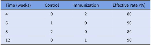
Non-effective individuals appeared at different times in both the immunized and control groups based on serum antibody and testosterone concentrations and histologic analysis of rat testes (Table 1). In the immunized group, 2 rats were ineffective at week 4, with an effective rate of 80%; 1 rat was ineffective at week 12, with an effective rate of 90%. In the control group, 1 rat was ineffective at week 6, with an effective rate of 90%; 2 rats were ineffective at week 8, with an effective rate of 80%.
Non-effective individuals in the control and immunization groups

Serum GnRH and kisspeptin antibody level
In Fig. 3A, the serum GnRH antibody titre values of rats are displayed, with arrows denoting the time of vaccination. The results revealed a significant increase in the serum antibody titre values of GnRH following immunization compared to the control group (P<0.05), which sustained at a high level post the initial vaccination. Conversely, the control group exhibited consistently low levels of anti-GnRH antibody titres. These findings suggest that the GnRH6-kisspeptin immunization triggered a robust anti-GnRH response. Notably, rats immunized in week 4 displayed the highest anti-kisspeptin antibody titre (Fig. 3B). Subsequently, by week 12, the anti-kisspeptin antibody titres in all immunized rats had decreased to a level equivalent to that of the control group.
Anti-GnRH (A) and anti-kisspeptin antibody titres (B) in the control and immunization. The abscissa is the age of weeks after first immunization and Arrows indicate the primary vaccination and subsequent boosters. *P < 0.05; **P < 0.01.

Testosterone determination
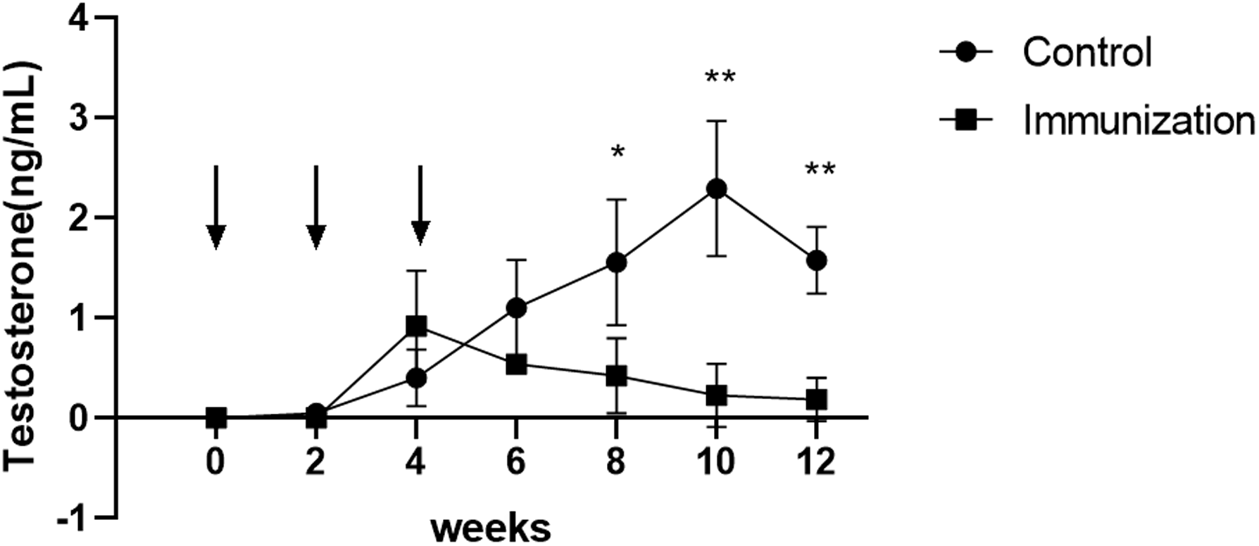
The serum testosterone levels of the control group and the immunized group were monitored as depicted in Fig. 4. Following the second immunization, the testosterone level of immunized rats significantly increased and gradually declined starting from week 4. Testosterone levels in the control group continued to increase from week 4 to reach a peak at week 10 and began to decline at week 10. There was a highly significant difference in serum testosterone levels in the immunized group compared to the control group at weeks 10 and 12 (P < 0.01).
The serum testosterone level of the control and immunization. The abscissa is the age of weeks after first immunization and Arrows indicate the primary vaccination and subsequent boosters. Mean (±SEM) of significantly different from control *P < 0.05; **P < 0.01. Data from null rats were removed.

Body weight
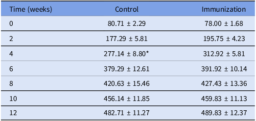
Following the initial immunization, the rats’ body weight was significantly higher than the control group only in week 4 (P<0.05), with no significant differences observed at other time points (Table 2).
Body weight (g) of male rats immunized with GnRH6-kisspeptin protein compared with the control group

Note: *P<0.05.
Weight and size of the testes
The testicular weight was determined after removing all paired testes. The findings in Table 3 indicate that the testes weight was significantly lower (P<0.05) in the immunization group compared to the control group. Furthermore, both the testes length and width were also significantly lower (P<0.05) in the GnRH6-kisspeptin group than in the control group (Table 3). The measurements of transverse and longitudinal circumference of the testis exhibited a very significant decrease (Table 3). It can be visualized in Fig. 5 that the GnRH6 vaccine significantly reduced testicular volume.
Testis weight and size of control and immunization

Note: **P<0.01, *P<0.05.
Representative testicular photographs of the control and immunization.

Histological observation of the testes
Fig. 6A and B display that the testes of the control group exhibited normal seminiferous tubules with spermatogenic cells present at diverse developmental stages, characterized by distinct layers and abundant sperm. Conversely, testicular tissue samples from immunized rats displayed prominent vacuolar degeneration and a considerable reduction in seminiferous tubule diameter (Fig. 6C and D). Additionally, the boundaries of spermatogonia, spermatocytes, and sperm cells were blurred and no sperm was produced in the immunization group.
Histopathological observation of testes samples from the control (A, B), and the immunization (C, D) at 7 weeks after first immunization. S indicates seminiferous tubules, spermatogonia (short arrows), primary spermatocyte (long arrows), spermatid (ellipse area), and spermatozoa (square) are indicated.

Discussion
Over the past few decades, GnRH vaccines have been widely studied as a recognized method used to control the reproduction and behaviour of animals. GnRH is often coupled to a carrier protein for immunization production. While different types of vaccines have been reported in previous studies (Fang et al., Reference Fang, Yang, Liu, Zhang, Tao, Wang, Pu and Zhang2009; Han et al., Reference Han, Ye, Liu, Jiang, Ijaz, Zhao and Tesema2016; Zhang et al., Reference Zhang, Xu, Zhao, Liu and Wu2007), in this study, we focused on the construction of a GnRH6-kisspeptin recombinant protein by linking 6 copies of GnRH (GnRH6) with kisspeptin. Male rats were immunized to induce high levels of anti-GnRH antibody and anti-kisspeptin antibody production in our study, as previously reported (Fang et al., Reference Fang, Li, Liu, Zhang, Tao, Li, Cao, Wang, Wang and Zhang2010; Tesema et al., Reference Tesema, Zhao, Jiang, Liu, Han and Wassie2019). The production of anti-GnRH antibody and anti-kisspeptin antibody leads to atrophy of testicular tissue and a decrease in testosterone hormone levels.
In the present study, tissue sections of the testes showed atrophy of the testicular seminiferous tubules in rats actively immunized with GnRH6-kisspeptin, characterized by a sparse arrangement of spermatogonia cells, spermatocytes, and sperm cells. The seminiferous tubules in testicular tissue are predominantly composed of spermatogenic cells and supporting cells, where supporting cells play a crucial role in both promoting the production of spermatogenic cells and protecting sperm. Both spermatogenic cells and supporting cells were significantly reduced in number in the immunized rats compared to the control group. This observation aligns with the findings of previous studies on GnRH vaccines (Liu et al., Reference Liu, Tian, Zhao, Jiang, Li, Zhang, Zhang, Li, Zhou and Fang2015; Yao et al., Reference Yao, Si, Tian, Ye, Zhu, Li, Ji, Zheng, Liu and Fang2018). Specifically, the decreased number of supporting cells alongside the altered arrangement of spermatogenic cells in the immunized rats indicate an impaired spermatogenesis process, further highlighting the impact of GnRH6-kisspeptin immunization on testicular function.
The testes of the vaccinated rats in our study exhibited atrophy and a severely wrinkled appearance, resulting in a significant decrease in weight and size compared to the control group. Previous study has confirmed that there is a significant correlation between the thickness of the spermatogenic epithelium, cytoplasmic content of testicular stromal cells, and testicular weight (Rottner and Claus, Reference Rottner and Claus2009). Consequently, the reduction or loss of testicular tissue cells in the immunized rats led to a decrease in both testicular weight and volume. This finding aligns with earlier reports on the effects of GnRH immunization in various animal species, such as rats, boars, and male dogs (Fang et al., Reference Fang, Li, Liu, Zhang, Tao, Li, Cao, Wang, Wang and Zhang2010; Liu et al., Reference Liu, Tian, Zhao, Jiang, Li, Zhang, Zhang, Li, Zhou and Fang2015; Yao et al., Reference Yao, Si, Tian, Ye, Zhu, Li, Ji, Zheng, Liu and Fang2018).
The results obtained from the immunization group demonstrated a notable decrease in the length and width of testes compared to those from the control group. This decrease can be attributed to a reduction in spermatogonia within the testicular tissue and the atrophy of the germinal tubules, aligning with previous research findings (Doroteu et al., Reference Doroteu, Viana, Ferreira Junior, Macedo, Oliveira and Pedroso2021; Ferro et al., Reference Ferro, Khan, McAdam, Colston, Aughey, Mullen, Waterston and Harvey2004). The difference between the mean transverse and longitudinal circumferences of the testes in the two groups was found to be highly significant, which is consistent with studies involving GnRH castration vaccine effects on bull testes(Cook et al., Reference Cook, Popp, Kastelic, Robbins and Harland2000; Oatley et al., Reference Oatley, Tibary, de Avila, Wheaton, McLean and Reeves2005).
Immunological castration induces a suppression of LH and FSH production by influencing the hypothalamic axis. This disruption in hormone production leads to a subsequent decrease in testosterone levels due to the reliance of testosterone synthesis on LH stimulation. In this study, the testosterone levels of the immunized rats decreased progressively starting from week 4. Moreover, analogous findings have been reported in various other animal studies (Fang et al., Reference Fang, Li, Liu, Zhang, Tao, Li, Cao, Wang, Wang and Zhang2010; Oonk et al., Reference Oonk, Turkstra, Schaaper, Erkens, Weerd, Nes, Verheijden and Meloen1998).
Active immunization with the GnRH vaccine induces the production of antibodies in the body that specifically target and neutralize GnRH. This action disrupts the biological activity of GnRH in animals, consequently impacting the balance of the endocrine system and influencing the HPGA. As a result, there is a reduction in the synthesis and release of reproductive hormones, leading to aberrant development of the animals’ reproductive system. Notably, this immunization process is considered reversible. With time, the levels of GnRH antibodies in the immunized animals gradually decrease, allowing the relevant reproductive hormone levels to increase. This gradual reversal triggers a restoration of normal reproductive function in the animals’ reproductive organs. Some studies have revealed consistent findings (Junco et al., Reference Junco, Fuentes and Millar2021; Oatley et al., Reference Oatley, Tibary, de Avila, Wheaton, McLean and Reeves2005). Nonetheless, other studies have indicated that active immunization against GnRH may result in prolonged or permanent suppression of reproductive function (D’Occhio et al., Reference D’Occhio, Aspden and Trigg2001; Gökdal et al., Reference Gökdal, Atay, Ülker, Yaralı, Helva, DeAvila and Reeves2008). Consequently, additional research is essential to ascertain whether the GnRH6-kisspeptin fusion protein vaccine assessed in this study elicits a lasting immune response in rats.
Our findings indicate that the GnRH6-kisspeptin vaccine induces the production of GnRH antibodies that can neutralize endogenous GnRH. Consequently, this process leads to the inhibition of gonadal development in rats, resulting in testicular hypoplasia and atrophy, along with the manifestation of morphological and histological alterations in the testes. Further studies are required to determine the actual castration effect of this vaccine in rats, due to various volatile factors including species, vaccine dose, time intervals between doses, and type and dose of adjuvant utilized.
Conclusion
The above results showed that GnRH6-kisspeptin immunization significantly increased the serum antibody level of rats, thereby inhibiting the development of rat testis and eventually atrophy. These findings demonstrates that the engineered GnRH6-kisspeptin exhibits strong immunogenicity.
Acknowledgements
The authors thank the financial support from all aspects, including the Innovation Fund Designated for Graduate Students of Anhui Agricultural University (grant numbers X202210364181, X202210364145, X202310364037).
Competing interests
The authors declare no conflicts of interest with regard to this study.
Ethics standards
This study received approval from the Animal Protection Committee of Anhui Agricultural University and all experimental procedures involving the rats adhered to the Regulations on the Management of Experimental Animals (Ministry of Science and Technology, China; revised in June 2004).