Introduction
Morphology and host plant association are significant sources of aphids classification. Heie (Reference Heie1986) asserted that, identification using the morphological characters and host–plant affiliation are more reliable than those based on morphological data alone. However, identification could be restricted by substantial intraspecific variation (Margaritopoulos et al., Reference Margaritopoulos, Tsitsipis, Zintzaras and Blackman2000). An intimate relationship with host plants based on their unique lifecycle can give raise to high morphological plasticity, such as seasonal polyphenisms and host-specialized biotypes, which can cause misidentification. In particular, those species that are exemplified from some of the polyphagous aphids in the family Aphididae such as Acyrthosiphon pisum (Harris, 1776) (Via, Reference Via1991; Via & Hawthorne, Reference Via and Hawthorne2002; Peccoud et al., Reference Peccoud, Ollivier, Plantegenest and Simon2009), Aphis fabae Scopoli, 1763 (Mackenzie, Reference Mackenzie1996), Cryptomyzus galeopsidis (Kaltenbach, 1843) (Guldemond, Reference Guldemond1990), Aphis gossypii Glover, 1877 (Margaritopoulos et al., Reference Margaritopoulos, Tzortzi, Zarpas, Tsitsipis and Blackman2006; Carletto et al., Reference Carletto, Lombaert, Chavigny, Brevault, Lapchin and Vanlerberghe-Masutti2009b ), Myzus persicae (Sulzer, 1776) (Blackman, Reference Blackman1987; Margaritopoulos et al., Reference Margaritopoulos, Tsitsipis, Zintzaras and Blackman2000; Nikolakakis et al., Reference Nikolakakis, Margaritopoulos and Tsitsipis2003), Rhopalosiphum maidis (Fitch, 1856) (Brown & Blackman, Reference Brown and Blackman1988), and Sitobion avenae (Fabricius, 1775) (Barro et al., Reference Barro, Sherratt, David and Maclean1995; Lushai et al., Reference Lushai, Markovitch and Loxdale2002), consist of two or more subspecies or host plant associated biotypes. Therefore, molecular tools are more widely used nowadays. Using molecular tools could provide supplementary evidences particularly for closely related species, sharing the host plant. By taking that into consideration, the use of integrating morphological, molecular and host–plant information could be the solution to the difficult problems for indistinguishable taxa.
The genus Aphis Linnaeus is the most species rich genus among Aphididae, with approximately 560 species known in the world (Favret, Reference Favret2005) and 39 species within the Korean Peninsula (Lee & Kim, Reference Lee and Kim2006). Compared to other genera, the species in the genus Aphis have remarkable diversity in their host plant utilization (Heie, Reference Heie1986; Von Dohlen & Teulon, Reference Von Dohlen and Teulon2003) and display various types of lifecycles; monoecy on woody or herbal plants and heteroecy (Kim et al., Reference Kim, Hoelmer, Lee, Kwon and Lee2010a ). However, due to their rapid radiation, most of the congeneric species are often cryptic in their morphology (Kim et al., Reference Kim, Lee and Lee2010b ).
The cotton-melon aphid, A. gossypii Glover; the soybean aphid, Aphis glycines Matsumura; and Aphis rhamnicola sp. nov. that were found on the buckthorn, Rhamnus spp. (primary host), showed the same problem in our previous study (Kim et al., Reference Kim, Hoelmer, Lee, Kwon and Lee2010a ). Those three species show morphological similarity with only few different characters in spite of the clear genetic differences (Kim et al., Reference Kim, Hoelmer, Lee, Kwon and Lee2010a ). According to a recent molecular phylogenetic study, these species are closely related and belong to the gossypii group, which is one of the four species groups in Aphis (Kim & Lee, Reference Kim and Lee2008; Kim et al., Reference Kim, Lee and Jang2011). The apterous viviparous females of this group are characterized as follows: smaller body size, shorter appendages than the other species groups (fabae, craccivora, and spiraecola groups) of Aphis, length of Antennal segment IV (Ant.IV) and hind femur (HFM), and the number of setae on mandibular lamina (ML) and cauda, setae on body and appendages relatively short, abdominal tergites (AbdT.) I and VII only with four marginal tubercles (MTu), and abdominal tergite VIII always with two setae (Kim et al., Reference Kim, Lee and Lee2010b ). Although the Aphis rhamnicola sp. nov. is morphologically indistinguishable to both A. gossypii and A. glycines, it has only recently been recognized as a distinct species based on molecular identification (Kim et al., Reference Kim, Hoelmer, Lee, Kwon and Lee2010a ).
In aphids, detecting a life cycle and different host plant associations of cryptic species is sufficient to provide conclusive evidence of a new species (Heie, Reference Heie1986; Thieme, Reference Thieme1987). For example, in Europe, two cryptic species, Aphis frangulae Kaltenbach and A. gossypii can be distinguished on the basis of their ecological differences such as life cycle and host plant association (Carletto et al., Reference Carletto, Blin and Vanlerberghe-Masutti2009a ). In another case, several subspecies belonging to the A. fabae complex, exhibit host alternation between shared primary host, spindle tree (Euonymus europaeus) and various secondary hosts which are distinctively preferred for these subspecies (Raymond et al., Reference Raymond, Searle and Douglas2001). As is the case of the A. fabae, in general, heteroecious aphids show a broader range of secondary hosts than primary hosts (Moran, Reference Moran1992).
Host shift and following specialization between intra specific populations might lead to reproductive isolation and change of life cycle as a consequence of different host preference (Moran, Reference Moran1992; Peccoud et al., Reference Peccoud, Simon, von Dohlen, Coeur d'acier, Plantegenest, Vanlerberghe-Masutti and Jousselin2010). Thus, detecting different host plant affiliation between closely related taxa could be the decisive evidence for discriminating them and describing new species. Ecologically, all three of these species have a typical heteroecious holocyclic life cycle and colonize together on the Rhamnus species; Rhamnus davurica Pall and Rhamnus japonica Maximowicz serve as primary hosts during the overwintering season: fall, winter, and spring in Korea (Blackman & Eastop, 1994; Blackman & Eastop, Reference Blackman and Eastop2007). Similar to the A. fabae complex, even though A. gossypii and A. glycines share the same primary host, they exhibit quite different associations with secondary host plants (Blackman & Eastop, Reference Blackman and Eastop2007). The polyphagous species, A. gossypii, is found on 912 species of plants belonging to 116 families (Inaizumi, Reference Inaizumi1980), whereas A. glycines feeds on the soybean, Glycine max (L.) Merr., as its only secondary host (Blackman & Eastop, Reference Blackman and Eastop2007). For this reason, we made an attempt to find secondary hosts of Aphis rhamnicola sp. nov. based on field research and molecular identification in the past several years.
In this study, we first compared the three species: A. gossypii, A. glycines, and Aphis rhamnicola sp. nov. based on morphometric analysis in order to confirm their morphological relationships. Second, molecular identification using the cytochrome c oxidase subunit I (COI) mitochondrial barcode region was performed on the aphid samples collected from the primary host, R. davurica, and several of the secondary host plants. Based on these multidisciplinary results, we report the cryptic new species, A. rhamnicola sp. nov. We not only provided a description, illustration, and biometric data of a new species, A. rhamnicola sp. nov., but also presented identification key to the species sharing the primary and secondary hosts with other congeneric species such as A. gossypii, A. glycines, and A. frangulae.
Materials and methods
Sample collection
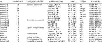
For this study, aphid samples were collected on the primary host R. davurica Pall (n=77) and various herbaceous plants including Capsella bursapastoris Medicus (n=11), Commelina communis Green (n=16), Lysimachia coreana Nakai (n=3), Rubia akane Nakai (n=5), Stellaria media Vill (n=3), and Veronica nakaiana Ohwi (n=3) in the Korean Peninsula between 2003 and 2012 (table 1). In each location, aphid samples from each plant species were sampled in separated colonies. In the overwintering season of the fall, winter, and spring, different morphs of females: fundatrices, viviparae, oviparae, and males were collected on the primary hosts.
Sample list of the Aphis species.

P, primary host; S, secondary host; GG, Gyeonggi-do; GW, Gangwon-do; CN, Chungcheongnam-do; GN, Gyeongsangnam-do; GB, Gyeongsangbuk-do; JB, Jeollabuk-do; KR, Korea; apt., Apterous viviparous female; ma. Male; ovi., Ovipara; fu., Fundatrix.
Morphological analysis
The voucher specimens used for DNA extraction were mounted in Canada balsam, following methods developed by (Blackman & Eastop, Reference Blackman and Eastop2000) and (Martin, Reference Martin1983). Digital images for illustrations were taken using a digital camera (Leica 400B; Leica Microsystems; Germany) at a resolution of 600 dpi. Measurements were performed from the digital images of each specimen using the software, i-Solution (IMT iSolutions, Inc., Chicago, IL, USA). The abbreviations for the descriptions are as follows: al., alate viviparous female; apt., apterous viviparous female; Ant., antennae; Ant.I, Ant.II, Ant.III, Ant.IV, Ant.V, Ant.VI, and Ant.VIb for antennal segments I, II, III, IV, V, VI, and base of VI, respectively; BDAnt.III, basal diameter of antennal segment III; AbdT.I, AbdT.II, AbdT.I II, AbdT.IV, AbdT.V, AbdT.VI, AbdT.VII, and AbdT.VIII, abdominal tergites I–VIII; MTu, marginal tubercle(s); BL, body length; GP, genital plate; 2HT, second segment of hind tarsus; HFM, hind femur; ML, mandibular lamina; PT, processus terminalis; SIPH, siphunculus; and URS, ultimate rostral segment. The holotype and paratype samples were deposited in the College of Agriculture and Life Sciences, Seoul National University (CALS SNU), Seoul, Korea.
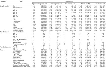
The means as well as the minimum and maximum values were calculated for each morphological characteristic (table 2). Multivariate, one-way analysis of variance test (ANOVA) for 39 morphological characteristics was conducted in order to determine whether there were significant morphological differences between the 56 viviparous females of the following three species: A. gossypii (n=20), Aphis glycines (n=16), and Aphis rhamnicola sp. nov. (n=20). All specimens which included in the morphometric analysis were collected from primary host R. davurica to reduce unexpected error such as phenotypic plasticity according to different host plants. In addition, the post hoc multiple comparisons based on the Tukey-HSD was performed in order to detect pairwise differences in each of the morphological characteristics between the taxa. On the basis of those results, we determined the informative morphological characteristics. Principal component analysis (PCA) was also performed in order to identify the structure in the distribution of the morphological variation among the individuals of those species. In this step, three invariable characteristics: setae on URS, AbdT.VIII, and median of GP were removed. All of the statistical analyzes were conducted using SPSS statistics ver. 20 (IBM, Chicago, IL, USA).
Biometrics of A. rhamnicola sp. nov.

1 Fourth and fifth segments are fused. Abbreviations are explained in ‘Materials and Methods’.
DNA extraction and sequencing
A total of 118 aphid samples were used for the molecular analysis. Whole genomic DNA was extracted from each specimen using a DNeasy Blood & Tissue kit (QIAGEN, Inc., Dusseldorf, Germany) following the manufacturer's protocol. In order to make voucher specimens from the DNA-extracted samples, nondestructive DNA-extraction was performed with a slightly modified method from that developed by Favret (Reference Favret2005). A tissue lysis buffer with protease K solution was used to treat the entire bodies of the aphids at 57°C for 24 h. Primers LEP-F1 5-ATTCAACCAATCATAAAGATAT-3 and LEP-R1, 5-TAAACTTCTGGATGTCCAAAAA-3 were used to amplify the 658 base pairs (bp) fragment of the mitochondrial COI gene.
The DNA fragments were amplified using AccuPowerH PCR PreMix (BIONEER, Corp., Daejeon) in 20 μl reaction mixtures containing 0.4 μM of each primer, 20 μM of the dNTPs, 20 μM of the MgCl2, and 0.05 μg of the genomic DNA template. Polymerase chain reaction (PCR) was performed using a GS1 thermo-cycler (Gene Technologies, Ltd, Essex) according to the following procedure: initial denaturation at 95°C for 5 min, followed by 34 cycles at 95°C for 30 s; an annealing temperature of 43°C for 30–50 s; an extension at 72°C for 30–60 s and a final extension at 72°C for 5 min. The PCR products were visualized by electrophoresis on a 1.5% agarose gel. A single band was observed, purified using a QIAquick PCR purification kit (QIAGEN, Inc.), and then sequenced directly using an automated sequencer (ABI Prism 3730 XL DNA Analyzer) at © MACROGEN LIC. The sequences we obtained in this study were all deposited in the GenBank (KJ195541–KJ195658).
Molecular analysis
In order to identify species, we retrieved 105 COI sequences of the known Aphis species from Rhamnus spp. and other herbaceous species such as Rubia spp. and Capsella spp. from the GenBank database (Supplementary Table 1).
All of the COI sequences to be analyzed were initially examined to merge forward and reverse strands using SeqMan Pro ver. 7.1.0 (DNASTAR, Inc., Madison, WI). After the examination, the DNA sequences were aligned using MEGA software ver. 5.0 (Tamura et al., Reference Tamura, Peterson, Peterson, Stecher, Nei and Kumar2011). For the aligned COI data set, a neighbor-joining (NJ) tree was generated using MEGA based on the Kimura-2-Parameter (K2P) model (Kimura, Reference Kimura1980), which is the best model for species level analysis particularly in low distances (Foottit et al., Reference Foottit, Maw, Von Dohlen and Hebert2008). The COI haplotypes of each species were analyzed using DnaSP ver. 5 (Rozas et al., Reference Rozas, Sánchez-DelBarrio, Messeguer and Rozas2003).
Results
Morphological comparison
The results of the multivariate ANOVA determined that there is significant morphological variation between the following three species: A. rhamnicola sp. nov., A. gossypii, and A. glycines (Wilk's λ=0.000022, hypothesis d.f.=74, error d.f.=32, F=91.55, P<0.0001). However, in the results of post hoc analysis by Tukey, only eight of the characteristics, such as length of HFM, HTB, and BDAnt.III; ratio of SIPH/BL, SIPH/HFM, and SIPH/Cauda; setae on Ant.III/BDAnt.III; and setae on AbdT.V/ BDAnt.III were significantly distinguished between all three of the species. The rest of the characteristics were not classified in accordance with the species level, but instead were overlapped across the species (Supplementary Table 2). Therefore, at least eight of the characteristics could be useful in terms of morphological identification of the three species. In particular, in the results of the pairwise comparison, 25 of the characteristics of the A. rhamnicola sp. nov. were not significantly differentiated from the A. gossypii, and this amount of indistinguishable characteristics was much more than the 10 characteristics that were different between A. glycines and A. gossypii, as well as the 14 characteristics between A. rhamnicola sp. nov. and A. glycines (Supplementary Table 2). This result indicates that A. rhamnicola sp. nov. is more morphologically similar to A. gossypii in many parts of the body than to A. glycines.
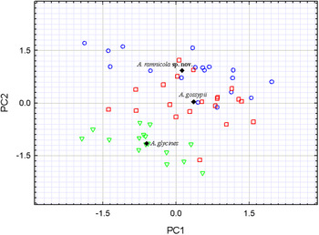
Overall, the results of the PCA were consistent with the result of the ANOVA. In the plot of the principal components analysis with the first two principal components: principal component 1 (PC1) and principal component 2 (PC2), PC1 and PC2, accounted for 32.1 and 15.9% of the total variance, respectively. There was high degree of distributional range overlap between A. rhamnicola sp. nov. and A. gossypii (fig. 1). On average, A. gossypii located on the right side of the middle part and to the center of this, the mean score of A. rhamnicola sp. nov. and A. glycines were distributed on the upper side and lower side, respectively. Unlike A. gossypii, the specimens of A. glycines were clearly separated from A. rhamnicola sp. nov. and they were aggregated on the left side of the lower part. Individuals in the same species were largely scattered in the PC1, which was separated primarily due to the length of the SIPH, Ant.III, and Ant.II. Such large intraspecific variation may be caused by the lack of dominant values among species. While, in the PC2, the length of the HFM and HTB contributed substantially to the separation of the species with a relatively low level of interspecific variation. As a consequence of the PCA, the informative morphological characteristics that contributed to the species separation almost corresponded with the results of the ANOVA test.
Scatter plot of the first two principal components based on 39 the morphological characteristics for the viviparous females of three species: Aphis rhamnicola sp. nov. (blue circles), Aphis gossypii (red rectangles), and Aphis glycines (green triangles). The black diamond symbols represent the mean scores of the principal components on each population level.

Molecular analysis
In total, 223 individuals of the 32 species were included for the COI data set. After the sequence examination, 739 of the sites were aligned, but only 658 bp were used for the analyzes. In the definitive data set, the number of variable sites was 88 and parsimony informative sites were 68. The K2P sequence divergences between taxa ranged from 0.33 to 11.3%, of which the gossypii group ranged from 0.33 to 6.1% (Supplementary Table 3). The overall mean divergences for the total data set and the gossypii group were 4.8 and 2.7%, respectively. Among individuals in the same species, sequence divergence ranged from 0 to 0.61%.
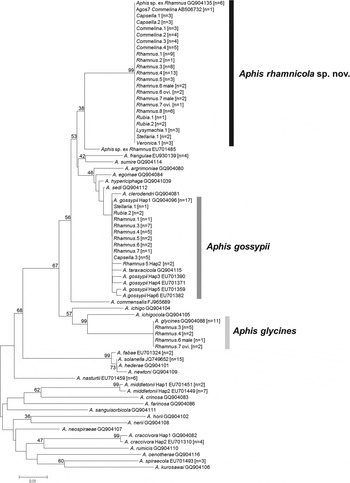
The NJ tree inferred from the COI gene showed that multiple aphid samples were divided into three clades and were identified as the following species: A. gossypii, A. glycines, and A. rhamnicola sp. nov. (fig. 2). The clades were clearly separated from each other, indicating that using the COI barcode region was an effective method for identifying these closely related species. Many of the species such as Aphis commensalism and Aphis sp. ex Rhamnus (EU701485) (Foottit et al., Reference Foottit, Maw, Von Dohlen and Hebert2008), which also occur on Rhamnus spp., intervened between the two clades of A. gossypii and A. rhamnicola sp. nov. (fig. 2). The COI sequences of A. rhamnicola sp. nov. did not match any of the known species from Rhamnus, Rubia, Stellaria, and Capsella except the reference sequences of the same species generated by a former study (Kim et al., Reference Kim, Hoelmer, Lee, Kwon and Lee2010a ). Interestingly, regardless of the host plant differences, the COI sequences of the 77 samples of A. rhamnicola sp. nov. were identical. Besides the R. davurica, A. rhamnicola sp. nov. was found co-existing with A. gossypii on several host plants such as Lysimachia, Capsella, Stellaria, and Rubia. Therefore, this new species seems to be polyphagous. All of the individuals collected from the C. communis and V. nakaiana were identified as A. rhamnicola sp. nov. and completely identical with the Agos7 (AB506732), which was also collected from C. communis (Komazaki et al., Reference Komazaki, Shigehara and Toda2010). Except for A. nasturtii, the entire Rhamnus associated species including A. rhamnicola sp. nov. belong to the clade of the gossypii group.
Neighbor-joining tree of the COI data set of a total 223 individuals containing 77 individuals from R. davurica and 41 individuals from the following secondary hosts: C. communis (n=16), C. bursapastoris (n=11), R. akane (n=5), S. media (n=3), V. nakaiana (n=3), and L. coreana (n=3) (Table 1), and 105 individuals retrieved from GenBank (Supplementary Table 2). The number in brackets indicates the number of duplicate sequences. Bootstrap values are given in percentages for 1000 replications.

Discussion
From the results of the morphological comparison based on 39 characteristics, A. rhamnicola sp. nov., A. gossypii, and A. glycines were morphologically very similar. In particular, the A. rhamnicola sp. nov. was almost indistinguishable from A. gossypii, due to the fact that only some of the meristic characteristics were significantly different. In addition, it makes a difficult situation worse when it is taken into consideration that A. gossypii is phenotypically a very plastic species in response to a variety of environmental factors according to host plant diversity, localities, and abiotic conditions (Rosenheim et al., Reference Rosenheim, Wilhoit and Colfer1994; Wool & Hales, Reference Wool and Hales1997; Nevo & Coll, Reference Nevo and Coll2001; Margaritopoulos et al., Reference Margaritopoulos, Tzortzi, Zarpas, Tsitsipis and Blackman2006; Agarwala & Choudhury, Reference Agarwala and Choudhury2013). Phenotypic plasticity induced by the interaction of environmental and genetic components has been well documented (Gorur et al., Reference Gorur, Lomonaco and Mackenzie2005). This result suggests that so far, A. rhamnicola sp. nov. has been misidentified to A. gossypii mainly due to the overlapping host plant range as well as the absence of conspicuous morphological characteristics. Aphis frangulae also is known to possibly alternate Rhamnus and other secondary hosts. Although we did not include the samples of A. frangulae in morphometric analysis because the species is not distributed in our region at least in East Asia, we luckily compared the species with A. rhamnicola and other Aphis species using the GenBank sequences in DNA analysis.
Unlike the morphological similarity, the results of our molecular analysis based on the COI region revealed that the aphid samples used in this study were divided into three distinct clades. In terms of species identification, the use of the COI region enabled rapid and effective identification between the three species. Obviously, the clade of A. rhamnicola sp. nov. was clearly distinct from those of A. gossypii and A. glycines with a high divergence level. In comparison to the closely related species of A. gossypii, the divergence level between A. gossypii and A. rhamnicola sp. nov. was much greater. Except for A. gossypii, there was little or no intraspecific variation in the COI region. Six different haplotypes were identified in the A. gossypii, which is considered to be the only species that has many unrelated primary hosts for the sexual phase in the gossypii group. Several studies showed host-associated genetic differences of A. gossypii (Margaritopoulos et al., Reference Margaritopoulos, Tzortzi, Zarpas, Tsitsipis and Blackman2006; Carletto et al., Reference Carletto, Blin and Vanlerberghe-Masutti2009a , Reference Carletto, Lombaert, Chavigny, Brevault, Lapchin and Vanlerberghe-Masutti b ; Komazaki et al., Reference Komazaki, Shigehara and Toda2010). Therefore, this level of haplotype diversity may be due to its amazing diversity of host plant associations. Similar to A. glycines, all of the specimens of A. rhamnicola sp. nov. shared an identical haplotype despite their host plant diversity.
In this study, the complex host plant associations of A. rhamnicola sp. nov. were revealed by comparing the COI region of the aphid samples from various host plants, which is one of the critical factors for species identification in aphids. Aphis rhamnicola sp. nov. has distinct host plant associations even though several hosts were shared with A. gossypii. So far, the specimens of A. rhamnicola sp. nov. were found on seven different host plants, which are known as the host plant of A. gossypii. Interestingly, the entire sample of aphids collected from C. communis (n=16) and V. nakaiana (n=3) were identified as A. rhamnicola sp. nov. based on the COI region. However, because of few specimens, it is cautious to assure that V. nakaiana (n=3) is the only host plant used by A. rhamnicola sp. nov.
Inaizumi (Reference Inaizumi1980) found that some aphids on C. communis had a unique host plant association and used Rhamnus spp. as their primary host. It was demonstrated experimentally that the aphids on the C. communis also survived on R. japonica, Veronica persica, C. bursapastoris, and Cayratia japonica (Inaizumi, Reference Inaizumi1981). Those results are consistent with our findings in regards to host plant association. In particular, aphids that hibernated on Hibiscus, one of the primary hosts for A. gossypii, could not survive on C. communis, suggesting that C. communis may be the only secondary host for A. rhamnicola sp. nov. (Inaizumi, Reference Inaizumi1981). Similar to this host association, every subspecies of A. fabae have one exclusive secondary host, though they share many hosts (Müller, Reference Müller1986; Thieme, Reference Thieme1987). Therefore, it is possible that A. rhamnicola sp. nov. may be diverged by a change in their host plant association. Host plant specialization has been known to be a crucial evolutionary force in many herbivore insects including aphids (Via, Reference Via1991; Via et al., Reference Via, Bouck and Skillman2000; Drès & Mallet, Reference Drès and Mallet2002; Via & Hawthorne, Reference Via and Hawthorne2002; Simon et al., Reference Simon, Carre, Boutin, Prunier-Leterme, Sabater–Muñoz, Latorre and Bournoville2003). Besides the most well known example of A. pisum, several aphid species showed host associated genetic differentiation (Peccoud et al., Reference Peccoud, Ollivier, Plantegenest and Simon2009).
More recently, Komazaki et al. (Reference Komazaki, Shigehara and Toda2010) suggested that the aphids collected on C. communis were thought to be a subspecies of A. gossypii based on its distinct genetic divergence level. However, A. rhamnicola sp. nov. with Agos7 obviously formed a distinct clade from A. gossypii with an approximately 3.4% genetic divergence. In addition, such a level of genetic divergence was high in comparison to the mean divergence of 0.5% between A. gossypii and the other closely related species such as Aphis taraxacicola, Aphis clerodendri, and Aphis sedi. Those gossypii group species showed an approximately 3% genetic divergence with A. frangulae and this value was slightly lower than the 3.3% divergence level of A. rhamnicola sp. nov. In the case of A. fabae, there was only a 0.4% genetic divergence compared to its sibling species A. solanella, which had been considered as subspecies of A. fabae. Therefore, the high level of differentiation at the COI region as well as host plant restriction strongly supports the fact that A. rhamnicola sp. nov. is a distinct species rather than a subspecies.
Despite the opportunity for hybridization between the three species by sexual reproducing on the same primary hosts, genetic divergence seems to be well maintained. Mate recognition and reproductive isolation have been demonstrated in several aphid species that also share the same primary host during the mating season. For example, A. fabae and Aphis solanella copulate preferentially based on a species specific mate-recognition system (Thieme & Dixon, Reference Thieme and Dixon1996). On the contrary, in the case of Cryptomyzus, interspecific mating and post-zygotic isolation primarily occurred by the indiscriminate courtship behavior of the males (Adriaan Guldemond et al., Reference Guldemond, Dixon and Tigges1994). In both of the studies, hybrids between the species exhibited reduced fitness in terms of both survival and reproductive performance. In addition, hybrids may suffer a disadvantage by inhibiting the optimal use of the parental secondary host plants. Considering the hybrid inferiority, assortative mating could be favored between the three species and this might contribute to the maintenance of the high genetic divergence level.
Most of the studies on the ecological speciation of aphids predict that the host races may have originated from a sympatric population in the face of gene flow and the drawbacks of hybridization (Mackenzie & Guldemond, Reference Mackenzie, Guldemond, Leather, Wait, Mills and Walters1994). The results obtained in this study are in agreement with those of previous studies. Species in the gossypii group such as A. gossypii, A. glycines, A. commensalism, A. frangulae, and A. rhamnicola sp. nov. alternate their hosts with a common primary host plant, Rhamnus sp., which is a unique trait within the genus Aphis. Even though they share their primary host, each of the species showed different associations with their secondary host plants, indicating that those species may be diverged from a Rhamnus associated common ancestor by a change in their secondary host preference followed by specialization. This might eventually lead to speciation by enhancing the reduced gene flow between secondary host specialized races (Mackenzie & Guldemond, Reference Mackenzie, Guldemond, Leather, Wait, Mills and Walters1994; Via et al., Reference Via, Bouck and Skillman2000; Raymond et al., Reference Raymond, Searle and Douglas2001).
In conclusion, our results demonstrated that A. rhamnicola sp. nov. should be regarded as a distinct species and not a subspecies of A. gossypii. Aphis rhamnicola sp. nov. seems to be a typical heteroecious species that alternates between one restricted primary host, Rhamnus spp, and various secondary hosts. Due to the shift in the secondary host association, it might be diverged from a common ancestor of the gossypii group. Based on the high level of genetic divergence and the different secondary host association, A. rhamnicola sp. nov. is a new species clearly distinguishable from A. gossypii.
Systematic accounts Aphis rhamnicola Lee, Lee, and Kim, sp. nov.
Type materials. Holotype: apt., Coll#120503-YR1-i, Donam-ri, Banpo-myeon, Gongju- si, Chungcheongnam-do, Korea, 3-V-2012, on R. davurica.
Paratypes: 5 apt., Coll#120503-YR1-ii–vi, same data with holotype; 1 apt., Coll#110526-HJ5-i, SNU Arboretum, Seodun-dong, Gwonseon-gu, Suwon-si, Gyeonggi-do, Korea, 28-IV-2011, on R. davurica., 5 apt., Coll#110428YR3-i-v, Mulhyanggi Arboretum, Geumam-dong, Osan-si, Gyeonggi-do, Korea, 28-IV-2011, on R. davurica., leg. Yerim Lee., 6 apt. and 4 al., Coll#120825-SH2-i-x. Daewon-ri, Okseong-myeon, Gumi-si, Gyeongsangbuk-do, Korea, 25-VIII-2012, on C. communis. 3 al., Coll#080820-HJ1-i-iii, Jisimdo, Jisepo- ri, Irun-myeon, Geoje-si, Gyeongsangnam-do, Korea.
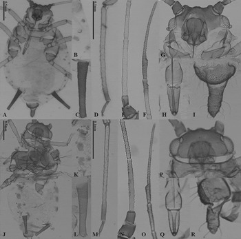
Description. Apterous viviparous female. Color in life. Head blackish green including Ant.I, and Ant.II; Ant.III, IV pale; Ant.VI and distal 1/3 of Ant.V dusky. Rostrum gradually dusky from rostal segment III to URS. Thorax and abdomen entirely blackish green, green mottled dark green, or green, lightly dusted with wax. Legs pale; coxae, 1/6 of tibia, and tarsi dusky. SIPH black, distal 1/3 of SIPH little bit brighter than base part. Cauda concolorous with body. Morphology. Body oval, 1.31–1.86 mm from antennal tubercle to end of cauda (fig. 3). Head: smooth dorsally and ventrally, three pairs of acuminate setae on dorsum, longest seta slightly shorter than BDAnt.III; median tubercle on frons not developed, frontal line between antennal tubercles almost straight with 4 setae on vertex (fig. 3). Antennae 0.3–0.76 times as long as body length without secondary rhinaria; Ant.III and Ant.IV sometimes fused; Ant.I smooth with 3–5 setae; Ant.II smooth with 3–5 setae; Ant.III imbricate with 4–6 setae; Ant.IV-V imbricate; Ant.VI strongly imbricate with 2–3 setae on Ant.VIb, 3–4 apical setae on PT, PT 2.13–3.09 times as long as Ant.VIb (fig. 3). Rostrum generally reaching to hind coxae; clypeus with 4 setae; mandibular laminae with 1 setae on each side; URS 0.87–1.27 times as long as 2HT, 0.88–1.16 times as long as Ant.VIb, bearing 2 very long accessory setae. Thorax: pronotum smooth with 2 spinal setae, 1 spino-pleural setae and 1 marginal setae close to marginal tubercle on each side; polygonal reticulations of marginal sclerites distinct on meso- and metathorax. Hind coxae smooth with approximately 5 setae; posterior seta on hind trochanter 0.7 times as long as width of trochantro-femoral joint; hind femur smooth, bearing slightly long setae, longest of posterior setae 0.68 times as long as middle width of segment; the longest seta of tibiae 0.83 times as long as middle width of segment; first tarsal chaetotaxy 3, 2, 2; 2HT imbricate with 3–5 setae entirely. Abdomen: dorsum smooth, membranous with 6 setae on each segment of AbdT.I-VII; AbdT.I, VII with MTu, at least 4 additional marginal tubercles on AbdT.II-VI; marginal seta on AbdT.I 0.66–1.03 times as long as height of marginal tubercle of the same segment (fig. 3); the longest seta of tergite V 0.38–0.99 times as long as BDAnt.III; AbdT.VI with 2 setae between SIPH; AbdT.VIII with 2 setae; genital plate with 2 median setae and 9–14 setae on posterior margin. SIPH cylindrical, strongly imbricate, much narrower at apex than at base, 1.83–2.5 times as long as cauda; apical flange weakly developed (fig. 3). Cauda tongue-shaped, constricted, with 5–9 setae; dorsal spinules denser than ventral ones (fig. 3).
Apterous viviparous female (A–I) and Alate viviparous female (J–R) of Aphis rhamnicola sp. nov.: (A) body; (B) marginal tubercle on abdominal tergite I; (C) siphunculus; (D) hind tibia and tarsus; (E) antennal segments III–IV; (F) antennal segments V–VI; (G) head; (H) ultimate segment of rostrum; (I) cauda; (J) body; (K) marginal tubercle on abdominal tergite I; (L) siphunculus; (M) hind tibia and tarsus; (N) antennal segments III–IV; (O) antennal segments V–VI; (P) head; (Q) ultimate segment of rostrum; and (R) cauda.

Alate viviparous female. Color in life. Head blackish green including Ant.I, and Ant.II; the rest of the Ant. dusky. Rostrum gradually dusky from rostral segment III to URS. Thorax and abdomen entirely blackish green to black. Legs pale; coxae, 1/6 of tibia, and tarsi dusky. Forewings pale with veins bordered by narrow dark pigmentation. SIPH black, distal 1/3 of SIPH little bit brighter than base part. Cauda concolorous with body. Others like apterous viviparous female. Morphology. Ant. with 5–6 secondary rhinaria lined up on Ant.III; 1 secondary rhinaria on Ant.IV or excluded; PT very long 2.84–3.86 as long as Ant.VIb. SIPH strongly imbricate, 1.78–2.05 times as long as cauda. Cauda tongue-shaped, constricted, with 5–6 setae. Others like apterous viviparous female.
Fundatrix. Morphology. Ant. about 0.68–0.86 mm length without secondary rhinaria; Ant.III and Ant.IV fused. Whole Ant. 0.44–0.49 times as long as BL. BL 1.52–1.75 mm, larger than apterous viviparous female with 0.79 times shorter legs and 0.58 times shorter SIPH. Others like apterous viviparous female.
Gynopara. Morphology. Ant. with 6–13 secondary rhinaria lined up on Ant.III; 1–5 secondary rhinaria on Ant.IV; PT very long 1.96–3.31 as long as Ant.VIb. Cauda 1.1–1.2 times longer and SIPH 0.81–1.1 times shorter than viviparous alate female. Others like alate viviparous female.
Ovipara. Morphology. Antenna about 0.63–0.65 mm length without secondary rhinaria; Ant.III and Ant.IV fused. Whole Ant. 0.39–0.56 times as long as BL. BL 1.22–1.39 mm, shorter than apterous viviparous female. HFM and HTB almost half length of apterous viviparous female with rounded scent glands on enlarged HTB. SIPH 0.32–0.36 times shorter and cauda 0.67–0.8 times shorter than apterous viviparous female. Posterior margin of GP bears 12–19 hairs. Others like apterous viviparous female.
Diagnosis. Aphis rhamnicola sp. nov. is morphologically similar to other aphids of the A. gossypii group (A. gossypii Glover, 1877, A. glycines Matsumura, 1917, Aphis sumire Takahashi, 1961, A. sedi Kaltenbach, 1843, etc.). Aphis rhamnicola sp. nov. can be distinguished from A. gossypii by having at least 4 MTu on AbdT.II-VI, whereas A. gossypii has no MTu on AbdT.II-VI.
Host plants and distribution. Collected on R. davurica in Gongju-si, Chungcheongnam-do, Suwon-si Gyeonggi-do, Osan-si Gyeonggi-do, C. communis in Jisimdo, Gyeongsangnam-do, Icheon-si, Gyeonggi-do, and Gumi-si, Gyeongsangbuk-do, C. bursapastoris in Jeju-si, Jeju Island, Taean-gun, Chungcheongnam-do, and Namhae- gun, Gyeongsangnam-do, R. akane Yangyang-gun, Gangwon-do, Suwon-si, Gyeonggi-do, S. media Namhae-gun, Gyeongsangnam-do, Veronika nakaiana Andong- si, Gyeongsangbuk-do, L. coreana Pyeongchang-gun, Gangwon-do in Korea.
Biology. Fundatrices of this species were observed on R. davurica in the early spring; March to early April (fig. 4A). In the middle of March, small colonies were found on the tip of buds and under the leaves (fig. 4B). From the late summer to autumn, viviparous females were observed on various secondary hosts; C. bursapastoris C. communis, R. akane, S. media, L. coreana and V. nakaiana (fig. 4C). Sexual morphs were observed from September to November (fig. 4D). It seems to take holocyclic heteroecious life cycle between primary host davurica and secondary host C. bursapastoris C. communis, R. akane, S. media, L. coreana and V. nakaiana in Korea.
Pictures of Aphis rhamnicola sp. nov. (A–D): (A) fundatrix nymphs on the shoot of R. davurica in the early spring; (B) apterous viviparous female on the stem of R. davurica in the spring; (C) apterous viviparous female on the stem of the secondary host, C. communis, in the summer; (D) gynoparae, oviparous nymphs and male on the leaf of R. davurica in the late autumn.

Etymology. The species name, rhamnicola, is derived from the genus name of primary host plant (Rhamnus) and the Latin suffix, -cola (dweller, inhabitant).
A modified key of identification for apterous viviparous female to the species of the genus Aphis on Rhamnus spp. (Blackman & Eastop, Reference Blackman and Eastop1994)
-
1. Stridulatory apparatus present on AbdT V and VI, and a row of peg like hairs on HTB...............................................2
-
- No stridulatory apparatus.................................................3
-
-
2. SIPH very short, truncated conical shape, 0.4–0.9 times as long as cauda. PT 2.3–3.4 times as long as Ant.VIb. Length of setae on Ant.III 2.6–3.5 times as long as BDAnt.III............................................................Aphis odinae*
-
- SIPH usually longer than cauda (1.2–1.99 times). PT 3.0–5.14 times as long as Ant.VIb. Length of setae on Ant.III 1.0–1.5 times as long as BDAnt.III....................................................................Aphis aurantii*
-
-
3. Pronotum and AbdT.I-IV and VII with extremely large, hemispherical marginal tubercles.............Aphis mammulata
-
- Pronotum and AbdT.II and VII with normal sized marginal tubercles..............................................................4
-
-
4. SIPH 0.05–0.08 times as long as BL. Rostrum relatively short, reaching barely middle coxa......................................5
-
- SIPH 0.09–0.25 times as long as BL. Rostrum reaching to middle of hind coxa............................................................6
-
-
5. Antennae usually 5 segmented, PT 1.6–2.2 times as long as Ant.VIb, and 0.79–0.91 times as long as Ant.III..............................................................Aphis commensalism
-
- Antennae usually 6 segmented PT 3 times as long as Ant.VIb, and 1.4 times as long as Ant.III..........................................................................Aphis utilis
-
-
6. Femora dark at least distally. AbdT.VII and VIII with dark cross bands, and more anterior tergites with some dark segmental and intersegmental markings..........................................................................Aphis fabae
-
- Femora pale or dusky. AbdT.VII and VIII without or with only faint cross bands, and more anterior tergites without any dark markings...............................................7
-
-
7. Cauda with 11–19 hairs.........................................................8
-
- Cauda with 4–9 hairs........................................................10
-
-
8. AbdT.VIII with 3–7 hairs. MTu usually only on prothorax and AbdT.I and VII.......................................Aphis spiraecola
-
- AbdT.VIII with 2 hairs. MTu frequently exist on AbdT.II and IV...................................................................................9
-
-
9. Length of setae on Ant.III 0.9–1.2 times as long as BDAnt.III.........................................................Aphis zamorana
-
- Length of setae on Ant.III 0.4–0.7 times as long as BDAnt.III.......................................................Aphis nasturtii
-
-
10. Dorsal abdomen almost wholly covered by an extensive solid black sclerite..........................................Aphis craccivora
-
- Dorsal abdomen mainly membranous...........................11
-
-
11. Dorsal cephalic hairs finely pointed, 35–45 μm long. HFM with some ventral hairs more than 45 μm long, exceeding diameter of trochantro femoral suture. Cauda almost as dark as SIPH, with 6 hairs...........................Aphis rhamnifila
-
- Dorsal cephalic hairs blunt, less than 30 μm long. Hairs on hind femur all not exceeding diameter of trochantro-femoral suture. Cauda usually paler than SIPH, with 4–9 hairs.....................................................................................12
-
-
12. Head and Ant.I-V pale, only Ant.VI dusky. Cauda much paler than SIPH, often with a midpoint constriction, and bearing 6–9 hairs. Posterior marginal GP usually with 5–9 hairs ...................................................................Aphis glycines
-
- Head and at least Ant.I,II and last segment (V or VI) dusky or dark. Cauda often dusky not contrasting with SIPH. Posterior marginal GP usually with 6–15 hairs.....................................................................................13
-
-
13. 1–4 MTu frequently on AbdT.II – IV.................................14
-
- Usually no MTu on AbdT.II – IV.....................................15
-
-
14. SIPH 0.8–1.8 times as long as cauda. Cauda tongue shaped without constriction.......................................Aphis frangulae
-
- SIPH 1.8–2.5 times as long as cauda (normally 1.9). Cauda tongue shaped with constriction...............................................Aphis rhamnicola sp. nov.
-
-
15. Length of setae on Ant.III 0.7–1.5 times as long as BDAnt.III. Cauda triangular with 4–6 hairs. PT 1.0–1.9 times as long as Ant.VIb...............................................Aphis iranensis
-
- Length of setae on Ant.III 0.41–0.78 times as long as BFAnt.III. Cauda tongue shaped with 6–8 hairs. PT 1.73–3.24 times as long as Ant.VIb.......................Aphis gossypii
-
*Species of the genus Toxoptera are currently merged into the genus Aphis (Favret, Reference Favret2014).
The supplementary material for this article can be found at http://www.journals.cambridge.org/BER
Acknowledgements
We are grateful for the helpful comments of subject editor, Ryan Garrick, and two anonymous reviewers. This research was supported by the Basic Science Research Program through the National Research Foundation of Korea (NRF) funded by the Ministry of Education, Science, and Technology (Project Code No. 2011-0012300), a fund (Z-1541745-2013-15-01) by Research of Animal and Plant Quarantine Agency, South Korea, a grant from the National Institute of Biological Resources (NIBR), funded by the Ministry of Environment (MOE) of Republic of Korea (NIBR 201401203), and Research Institute for Agriculture and Life Sciences.