Introduction
Canadian amber was called an ‘entomological treasure-chest’ by McAlpine and Martin (Reference McAlpine and Martin1969) for a good reason. This mostly transparent, reasonably preparation-propitious fossil resin is readily available in several large collections and provides a taphonomical window to the Late Cretaceous Era. Mites (Acari), ubiquitous and diverse as they are, were notably among the first fossils reported from this source (Carpenter et al. Reference Carpenter, Folsom, Essig, Kinsey, Brues, Boesel and Ewing1937). Both mites were Prostigmata: formally described as Bdella vetusta Ewing, 1937 (Trombidiformes: Bdellidae) and an unidentified Erythraeidae (Trombidiformes) larva. The latter was subsequently described by Vercammen-Grandjean (Reference Vercammen-Grandjean1973) under the name Proterythraeus southcotti Vercammen-Grandjean, Reference Vercammen-Grandjean1973 in the new family Proterythraeidae. These remain the only mites among the more than 100 arthropod species formally described from Canadian amber (for review see McKellar et al. Reference McKellar and Wolfe2008; McKellar and Wolfe Reference McKellar, Wolfe, Tappert and Muehlenbachs2010).
The overall richness of mite fossils in this material is significant. Skidmore (Reference Skidmore1999) summarised the label data from the 1281 inclusions housed in the Canadian National Collection of Insects, Arachnids, and Nematodes (Ottawa, Ontario, Canada), and listed 141 specimens under Acarina, among which 11 families were identified, and Sidorchuk (in Rasnitsyn et al. Reference Rasnitsyn, Bashkuev, Kopylov, Lukashevich, Ponomarenko and Popov2016) expanded this to 20 mite families, more than half of which (12) belong to Oribatida. This is not surprising; oribatids, or moss mites, are both among the oldest known mites (Late Devonian: Norton et al. Reference Norton, Bonamo, Grierson and Shear1988; Subías and Arillo Reference Subías and Arillo2002) and among the commonest forest animals (Krivolutsky et al. Reference Krivolutsky, Lebrun, Kunst, Akimov, Bayartogtokh and Vasiliu1995; Norton and Behan-Pelletier Reference Norton and Behan-Pelletier2009). Along with prostigmatid mites, they comprise the majority of mites found in fossil resins worldwide (E.S., unpublished database).
The main reason why these numerous and diverse fossils are still undescribed may be the technical difficulty of their observation. To see a mite, whose size is normally under a millimetre, with all taxonomically important detail, one has to obtain a preparation suitable for compound light microscopy, and suitable methods are quite recent (Sidorchuk Reference Sidorchuk2011, Reference Sidorchuk2013). The second difficulty is to obtain a high-quality image of the mite inclusion. When amber is transparent and inclusions contain air or water, this is relatively easy and requires only normal compound microscopy with transmitted (brightfield or differential interference contrast) and incident illumination (see for example Sidorchuk and Norton (Reference Sidorchuk and Norton2010, Reference Sidorchuk and Norton2011a, Reference Sidorchuk and Norton2011b). In Canadian amber, many inclusions are filled with the fossil resin, and only fragments of air films remain on the surface, hampering rather than helping observation. Confocal microscopy, successfully used in descriptions of insect and fungal amber fossils (Speranza et al. Reference Speranza, Wierzchos, Alonso, Bettucci, Martín-González and Ascaso2010; Kirejtshuk et al. Reference Kirejtshuk, Chetverikov and Azar2015), has the potential for better viewing resin-filled specimens, so we tried this technique on the oribatid mite.
The aims of this contribution are to: describe and name the first oribatid mite species from Canadian Cretaceous amber, Megeremaeus cretaceous new species (Acari: Oribatida: Megeremaeidae), provide a key to described species, and discuss advantages that confocal microscopy provides for the study of dark, resin-filled, miniature amber inclusions.
Material and methods
Provenance of fossil resin
This study is based on two Canadian amber inclusions (CAS 668 and CAS 806) housed in the Canadian National Collection of Insects, Arachnids, and Nematodes, Ottawa, Ontario, Canada. Originally, this fossil resin commonly called ‘Canadian amber’ is found in Alberta and Manitoba, Canada. It belongs to the Taber Coal Zone, and was deposited in the upper Foremost Formation, in beds of coal and of shale saturated with organic matter. The age of the amber is upper Campanian (~76–72 million years ago), and the source of the original resin is, predominantly, a plant genus Parataxodium Arnold and Lowther (Cupressaceae) (McKellar and Wolfe Reference McKellar, Wolfe, Tappert and Muehlenbachs2010). The two pieces with mites for our study were collected near Medicine Hat, Alberta (see Occurrence).
Preparations
Each mite was originally fossilised in a small piece of red fossil resin. Tools and techniques applied during preparation are those described by Sidorchuk (Reference Sidorchuk2013) and Sidorchuk and Vorontsov (Reference Sidorchuk and Vorontsov2016). Specimen CAS 806 was mounted in a dorso-ventral preparation between two coverslips. As preserved, the specimen is dark and compressed dorsoventrally, not translucent. Most setae were preserved but those of the venter were hard to discern and easy to confuse with filamentous material (probably fungal) that is abundant on the ventral side; the dorsal side is partly obscured by a white aggregation of minute bubbles. Specimen CAS 668 was accidentally split during preparation, roughly along the sagittal plane, with the legs partly lost. The two parts, CAS 668a and 668b, are each mounted in a lateral preparation between two coverslips. As preserved, the body imprint is translucent, with cuticle crumbled and displaced. The legs are life-like in appearance, dark, partly folded, with tarsi and long solenidia partly hidden in the optically irregular layering of amber.
Light microscopy and imaging
All observations were made with a Nikon Eclipse 800 compound microscope (Nikon Corporation, Tochigi, Japan), mostly with a 60× water immersion lens, in brightfield or Nomarski differential interference contrast transmitted illumination and incident light from a halogen source (Chiu Technical Corporation, New York, New York, United States of America). Stacks of images were obtained with a Nikon DS-Fi1 digital camera, colour-enhanced, noise-enhanced, and sharpness-enhanced with Adobe Photoshop Lightroom 5.7.1. (Adobe Systems Incorporated, San Jose, California, United States of America) Stacks were combined with the aid of Helicon Focus Pro 6.5.2 (Helicon Soft, Kharkov, Ukraine), algorithm A, with subsequent manual transfer of significant details taken from the individual images, as necessary.
Drawings were prepared with the aid of InkScape (v. 0.48 r9654) (InkScape) and VLC media player (v. 0.9.8a) (VideoLAN, Paris, France), using Wacom Intuos 4 drawing tablet (Wacom Technology Corporation, Portland, Oregon, United States of America) and live capture from the digital camera AmScope MU 900 (AmScope, Irvine, California, United States of America) installed on the microscope (Sidorchuk and Vorontsov Reference Sidorchuk and Vorontsov2014).
Confocal microscopy and imaging
Confocal microscopy was performed by Denise Chabot, at the Microscopy Unit, Agriculture and Agri-Food Canada (Ottawa, Ontario, Canada). A Zeiss LSM 510 Duo confocal microscope with objective EC Plan-Neofluar 10X/0.30 M27 and electronic zoom 0.9 (Carl Zeiss, Oberkochen, Germany) was applied to receive the image stacks. Three settings were used, and respective images were colour-coded: (1) in red, excitation with laser He/Ne 633 nm, emission long pass 650 nm; (2) in green, excitation with Argon/2 Laser line 488 nm, emission long pass 505 nm; (3) in mixed red and green, (1) and (2) combined. Pixel dwell time was 1.97, and stacks of images were taken with 10 μm intervals. Procedure for colour correction and combining was the same as for the light micrographs.
Terminology
Morphological terminology is adapted from F. Grandjean (see Travé and Vachon Reference Travé and Vachon1975 for references and Travé et al. Reference Travé, André, Taberly and Bernini1996 or Norton and Behan-Pelletier Reference Norton and Behan-Pelletier2009 for overview); most terms are translated from French (van der Hammen Reference van der Hammen1980). Paired structures are described in the singular, unless noted.
Measurements
All measurements are in micrometres (μm), rounded to the nearest 5, if beyond 50. They were taken in non-ideal orientations, so should be treated as minimal estimates.
Taxonomic and ecological background
The monogeneric oribatid family Megeremaeidae and genus Megeremaeus include seven extant species. Five species are Nearctic: Megeremaeus montanus Higgins and Woolley, Reference Higgins and Woolley1965; M. ditrichosus Woolley and Higgins, Reference Woolley and Higgins1968; M. hylaius Behan-Pelletier, Reference Behan-Pelletier1990; M. keewatin Behan-Pelletier, Reference Behan-Pelletier1990; and M. kootenai Behan-Pelletier, Reference Behan-Pelletier1990. In addition, Megeremaeus expansus Aoki and Fujikawa, Reference Aoki and Fujikawa1971 is known from Japan (Aoki and Fujikawa Reference Aoki and Fujikawa1971), South Korea (Choi Reference Choi1987), the Kuril Islands (Golosova Reference Golosova1980), and Primorskii krai and Khabarovskii krai in the Russian Far East (Golosova Reference Golosova1989; Ryabinin Reference Ryabinin2015), and M. spinosus Aoki and Yamamoto, Reference Aoki and Yamamoto2000 from China (Wang et al. Reference Wang, Wen and Chen2002). Megeremaeus rameus Wen and Zhao, 1993 described from Hainan in China, is incorrectly assigned to Megeremaeus; it is clearly a member of the Ameroidea (it expresses ventral neotrichy), but genus and family placement would require re-examination of the type specimens.
Species of Megeremaeus are found in moist temperate to subarctic habitats, associated with litter and moss in primarily coniferous but also mixed forests (Behan-Pelletier Reference Behan-Pelletier1990). Megeremaeus montanus is found in canopy habitats in the Pacific Northwest of North America (Winchester et al. Reference Winchester, Lindo and Behan-Pelletier1999, Reference Winchester, Behan-Pelletier and Ring2008), and is also known as ~10 000-year-old subfossils from Queen Charlotte Islands, Cape Ball, British Columbia, Canada. Megeremaeus hylaius is most commonly found on conifer tree trunks in northeastern North America. Megeremaeus keewatin is known only from northern Yukon and Northwest Territories, Canada, and as a subfossil record somewhat older than two million years (Matthews and Ovenden Reference Matthews and Ovenden1990; Matthews and Telka Reference Matthews and Telka1997), from Meighen Island, Queen Elizabeth Islands, Nunavut, Canada (Behan-Pelletier Reference Behan-Pelletier1997).
Systematic palaeontology
Megeremaeus cretaceous Sidorchuk and Behan-Pelletier, new species
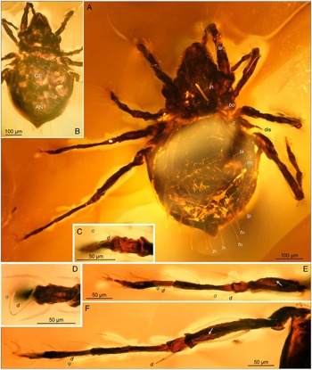
Megeremaeus cretaceous new species holotype CAS 806, combined incident and transmitted brightfield illumination (A, B) and brightfield light micrographs. A, dorsal view; B, partial ventral view, compare to confocal images in Fig. 3; C–F, leg details, note proportions of solenidia and coupled setae d: C, genu I; D, genu II; E, leg III; F, leg IV; note reticulation visible on femora (arrows). Each image is layered from 40 to 70 focal planes. AN, anal opening (barely visible); dis, discidium; GE, genital opening.

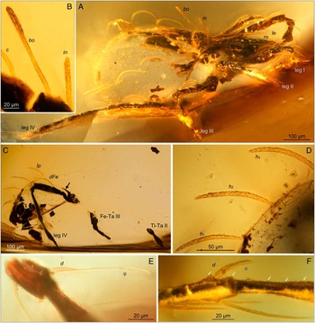
Megeremaeus cretaceous new species paratype CAS 668a, b, combined incident and transmitted brightfield illumination (A, F, E) and brightfield light micrographs, lateral views. A, CAS 668a overview, note that posterior is broken away; B, detail of posterior prodorsum and anterior notogaster; C–F, CAS 668b: C, overview, arrow points to dorsal spine of trochanter IV; D, posterior notogastral setae; E, tibia II, F, tibia IV, note numerous cerotegumental granules (arrows). Each image layered from 30 to 70 focal planes. Fe, femur; Ta, tarsus; Ti, tibia; Tr, trochanter.

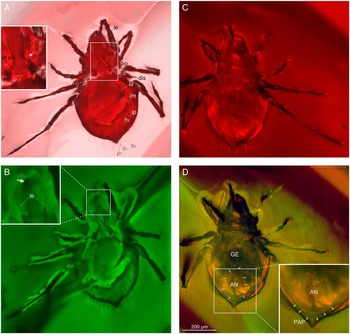
Megeremaeus cretaceous new species confocal images, all to same scale. A, dorsal aspect, excitation with laser He/Ne 633 nm, emission long pass 650 nm, pixel dwell time 1.97 us, arrow points to subtriangular tubercle opposing tutorium; B, same as A, but excitation with Argon/2 laser line 488 nm, emission LP 505 nm, arrow points to rounded projection on rostrum; C, ventral aspect with same parameters as A; D, same as C but two laser lines (He/Ne 633 nm and Argon/2 laser line 488) combined. Each image is layered from 10 to 20 focal planes. AN, anal opening, outlined with arrows; in alv, alveolus of missing seta in; GE, genital opening; PAP, postanal process, outlined with arrowheads; tu, tutorium.

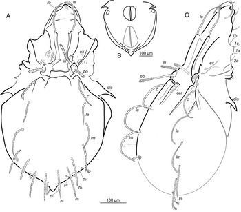
Megeremaeus cretaceous new species body. A, holotype CAS 806, dorsal aspect, legs not shown; B, same, partial ventral view, schematic (setae and legs not shown); C, paratype CAS 668a, lateral aspect, partial (posterior broken off), legs and gnathosoma not shown. Grey lines used for poorly discerned structures.

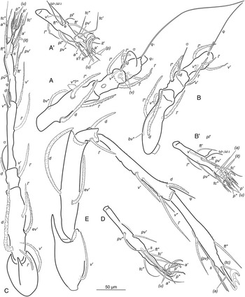
Megeremaeus cretaceous new species legs. A–D, CAS 806. A, femur-tibia I, dorsal aspect; A′, tarsus I, ventral aspect of the specimen, note that seta v″ Fe is probably aberrant; B, femur-tibia II, dorsal aspect; B′, tarsus II, ventral aspect of the specimen; C, leg III, dorsal aspect; D, tarsus IV, ventral aspect of the specimen; E, CAS 668a, leg IV in lateral abaxial aspect. A, A′, B, B′, D foreshortened due to orientation. Grey lines used for poorly discerned structures.

Classification. Arachnida: Acari: Acariformes: Oribatida: Megeremaeidae.
Type material. Canadian amber inclusions holotype CAS 806 and paratype CAS 668 (two parts, CAS 668a and CAS 668b), each mounted between two circular, 1-cm coverslips with Buehler’s EpoThin epoxy resin. Both holotype and paratype housed in the Canadian National Collection of Insects, Arachnids, and Nematodes.
Occurrence. In Canadian Cretaceous amber. Holotype CAS 806 collected near Medicine Hat, Alberta, 1973 by P. Boston; paratype CAS 668 collected near Medicine Hat, Alberta, May 1973 by J. F. McAlpine and H. J. Teskey.
Diagnosis. Total length 710–845 μm, maximum notogastral width 385 μm. Lamella length 100 μm, tutorium length 150 μm, minimal mutual distances 55 and 100 μm, respectively. Translamella not evident. Prodorsal setae coarsely serrate; setae in and bo thick, weakly clavate. Maximum measured lengths: ro 100 μm, le 150 μm, in 95 μm, bo 140 μm, ex 70 μm; mutual distances ro–ro 65 μm, le–le 72 μm, in–in 80 μm, bo–bo 120 μm, ex–ex 130 μm; proportion of lengths bo:in between 1.3 and 1.5. Notogaster length to width ratio 1.2, with conspicuous posterior projection. Notogastral setae c, la, lm, lp, and h 3 overlapping insertion of next most posterior seta. Discidium large, length 65 μm, sharply pointed. Postanal process present. Dorsal spine on trochanters III and IV about 2/3 length of segment. Solenidion φ1 of tibia I ~240 μm, tarsus I solenidion ω1 ~80 μm. Seta d on tibiae II–IV and genua I–III robust, coarsely serrate, comparable in length to coupled solenidion.
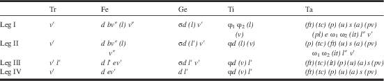
Description. Measurements. CAS 806 total length in dorsal aspect 710 μm, maximum (notogastral) width 385 μm. CAS 668 length as preserved (broken posteriorly) 745 μm; including broken part ~845 μm; height 315 μm. Colour as preserved dark brown. Integument. Legs with net-like pattern of ridges (Fig. 1E–F). Cerotegument partly detached in both specimens; on legs, notogastral and prodorsal ridges visible as granules 1–3 long (Fig. 2F). Prodorsum. Slightly longer than wide (width 260 μm, length 280–310 μm), rostrum with rounded projection anteriorly (Fig. 3B, arrow). Bothridium diameter 30 μm, with evident posterior tubercle (Figs. 2B, 3A, 4A, 4C). Lamella length 100 μm, tutorium length 150 μm, minimal mutual distances 55 and 100 μm, respectively (Figs. 2A, 3A–B, 4A, 4C). Translamella not evident; transverse fold present at mid-distance between lamellar tip and rostrum (Fig. 4A). Lamellar cusp rounded, bearing seta le (Figs. 2A, 4A, 4C). Lamella fused to bothridium posteriorly (Figs. 3A–B, 4A, 4C). Seta in situated on medial margin of lamella (Figs. 3A, 4A). Tutorium posteriorly opposed to subtriangular tubercle; latter connected to bothridium by short ridge (Fig. 2A, 4A, 4C). Prodorsal setae coarsely serrate, especially thick, weakly clavate setae in and bo (Figs. 2B, 4A, 4C). Maximum measured lengths: ro 100 μm, le 150 μm, in 95 μm, bo 140 μm, ex 70 μm; mutual distances ro–ro 65 μm, le–le 72 μm, in–in 80 μm, bo–bo 120 μm, ex–ex 130 μm; length bo:in between 1.3 and 1.5 μm. Notogaster. Slightly elongate: length 455 μm, length to width ratio 1.2 in CAS 806. With conspicuous posterior projection (extent of projection possibly artefact of desiccation in amber). Anterior tubercles rounded; lateral pair well separated from notogastral border anteriorly; medial pair short, almost merging to notogastral border (Figs. 1A, 2B, 3A–B, 4A, 4C). Setae blunt, robust, coarsely serrate (Fig. 2A, 2D): c, la, and lm curved, 170, 200, and 210 μm; lp and h-series almost straight, 115–125 μm; p-series finer, curved, p 1 80 μm, p 2 55 μm, p 3 30 μm. Notogastral setae c, la, lm, lp, and h 3 overlapping insertion of next most posterior seta (Figs. 1A, 2A, 2D, 4A, 4C). Lyrifissures not visible. Lateral region. Pedotecta I and II well developed. Discidium large, length 65 μm, sharply pointed (Figs. 1A, 4A). Ventral region. Poorly preserved in both specimens: coxisternal integument, most setae, and all lyrifissures not discernible (Figs. 1B, 3C–D). General appearance, as far as visible, similar to modern Megeremaeus species (Behan-Pelletier Reference Behan-Pelletier1990). Genital opening oval, slightly longer than wide, 115×90 μm; anal opening distanced from it by 40 μm, pear shaped (Fig. 3D, arrows), length 155 μm, maximal width 110 μm (Fig. 4B). Postanal process wide, visible in CAS 806 (Fig. 3D insert, PAP, arrowheads). Coxisternal setae 1a–1c and 2a in CAS 668a 30–34 long, fine, smooth, tapered (Fig. 4C). Gnathosoma. Poorly discernible (Figs. 1B, 2A, 3C–D). Subcapitular setae in CAS 668a 32–34 μm long, fine, smooth. Legs. Slender, with net-like pattern of cuticular ridges at least on femora and genua (Fig. 1E–F arrows). Claws lightly serrate dorsally, strong, lateral finer than median. Setae robust, long, and coarsely serrate on femora, genua, and tibiae; finer, tapered and barbed on trochanters and tarsi (Figs. 1C–F, 2C, 2E–F, 5). Porose area present at least on femur I ventrally. Dorsal spine on trochanter III, IV about 2/3 length of respective segment (Fig. 2C arrow, Fig. 5E). Leg IV (635 μm) longest, leg II (430 μm) shortest, leg I and III measuring 460 μm and 520 μm in CAS 806. In CAS 668 only leg IV measurable, 635 μm. Presumed setal homologies shown in Table 1. Solenidion φ1 of tibia I extremely long, 240 μm (Fig. 5A); tarsus I solenidia much shorter, ~80 μm (Fig 5A′). Setae d on tibiae II–IV and genua I–III robust, coarsely serrate (Figs. 1C–F, 2E–F, 5A–C, 5E): smallest of setae d, 20, d genu I about 1/4 respective solenidion σ (85 μm), d tibia IV as long as respective solenidion φ (50 μm); d genu IV (42) as long as respective leg segment, as for femoral setae d. Famulus – tiny spine (Fig. 5A′); eupathidia could not be confirmed, on tarsus I most fundamental setae appearing glabrous. Seta v″ poorly visible on both femora I in CAS 806, appearing short and truncate (Fig. 5A); in CAS 668, formed as normal large seta similar to bv″.
Setiform organs in adult Megeremaeus cretaceous based on CAS 806 (holotype) and CAS 668 (paratype).

Notes: Setae (Roman) and solenidia (Greek) are indicated; parentheses indicate pairs of setae. Seta d coupled to solenidion where present together on article.
Fe, femur; Ge, genu; Ta, tarsus; Ti, tibia; Tr, trochanter.
Etymology. Specific epithet is a noun in apposition, referring to the geological age of the described fossils.
Comparison with other Megeremaeus species
Adults of Megeremaeus cretaceous can be distinguished from congeners by the following combination of character states: (a) notogaster pointed posteriorly; (b) body length ~708–845 μm; (c) translamellar ridge not evident, possibly absent; (d) bothridial seta isodiametric along most of length, slightly thickened distally; (e) length ratio of bo:in between 1.3 and 1.5; (f) notogastral setae c, la, lm, lp, and h 3 relatively long, overlapping insertion of next most posterior seta; (g) postanal process well developed; (h) setae d on tibiae II–IV and genua I–III robust, coarsely serrate.
Megeremaeus cretaceous is among the few species with the notogaster pointed posteriorly. It shares this character state with M. hylaius and M. expansus, but it is more pronounced in M. cretaceous (Figs. 1A–B, 3, 4A–B). Megeremeus cretaceous has the shortest body of described Megeremaeus species. The dorsodistal spine on trochanter IV of M. cretaceous, at 0.3 the length of trochanter in lateral view, is longer than in any species other than M. ditrichosus, at least among those species for which this character has been described.
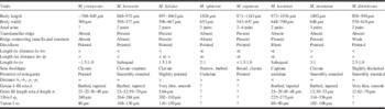
Solenidion φ1 of tibia I of Megeremaeus cretaceous is similar in length to that of other species of Megeremaeus for which we have comparable data (Table 2). In these species φ1 of tibia I is ~3× the length of tarsus I solenidion ω1 (Table 2), and in M. cretaceous solenidion φ1 is slightly more than half the length of leg I.
Character states of Megeremaeus adults.

Note: Data after: Higgins and Woolley Reference Higgins and Woolley1965; Woolley and Higgins Reference Woolley and Higgins1968; Behan-Pelletier Reference Behan-Pelletier1990; Aoki and Yamamoto Reference Aoki and Yamamoto2000.
An extant undescribed species of Megeremaeus from the Russian Far East shows a number of similarities to M. cretaceous; Nikolay Ryabinin has kindly shown to us his drawings and five specimens of this species, which he is describing. Most strikingly, the Cretaceous and Far Eastern species share the size and robustness of setae d on leg tibiae II–IV and genua I–III. Seta d is also barbed and tapered in M. montanus, M. keewatin, and M. kootenai, but is not as long or robust (Table 2). The Far Eastern species also shares the posterior notogastral projection with M. cretaceous, though it is less prominent in Far Eastern specimens, and could be exaggerated due to some lateral compression with fossilisation in M. cretaceous. However, the Far Eastern specimens are larger, and their notogastral setae and discidium differ from those of M. cretaceous. Megeremaeus cretaceous is clearly distinct from the Far Eastern species; still, it is remarkable that such similarity exists between these mite species separated by 80 million years.
Discussion
Megeremaeus cretaceous is ~80 million years old, yet we have no hesitation including it in this genus. It shares with extant species of Megeremaeus the two pairs of tubercles on the anterior margin of notogaster; a costate lamella with short cusp; a costate tutorium with its posterior margin opposing a small, anteriorly directed tubercle on the prodorsum; 10 pairs of notogastral setae; a well-developed discidium; trochanters III, IV with a pointed dorsodistal spine; and femora without a ventral carina. We outline differences in character states among described species of Megeremaeus in Table 2, recognising that data are incomplete for M. expansus, M. spinosus, and M. cretaceous. Differences in the shape of coupled seta d on genua I–III and tibiae II–IV are striking, as noted above, with d reduced to a thin, smooth seta in M. hylaius and M. ditrichosus (Behan-Pelletier Reference Behan-Pelletier1990). None of the character states expressed in M. cretaceous required modification of the generic diagnosis of Behan-Pelletier (Reference Behan-Pelletier1990).
This is another case of remarkably long evolutionary stasis in oribatid mites, examples of which are not uncommon in their fossil record. The oldest indubitable Oribatida fossils are Devonian (Norton et al. Reference Norton, Bonamo, Grierson and Shear1988; Subías and Arillo Reference Subías and Arillo2002), and five oribatid families with modern members are known since the Lower Carboniferous (over 359 million years ago) (Subías and Arillo Reference Subías and Arillo2002). Oribatid genera that include fossil and extant species are known from the Mesozoic (252–66 million years ago): Cultroribula Berlese, 1908 (Astegistidae) and Hydrozetes Berlese, 1902 (Hydrozetidae) from the Jurassic (150–200 million years ago) (Krivolutsky and Krasilov Reference Krivolutsky and Krasilov1977; Krivolutsky and Druk Reference Krivolutsky and Druk1986); and Trhypochthonius Berlese, 1904 (Trhypochthoniidae), Ametroproctus Higgins and Woolley, 1968 (Cymbaeremaeidae), Eupterotegaeus Berlese, 1916, and Ommatocepheus Berlese, 1913 (both Compactozetidae) from the Lower Cretaceous amber of Spain (Arillo and Subías Reference Arillo and Subías2002; Arillo et al. Reference Arillo, Subías and Shtanchaeva2008, Reference Arillo, Subías and Shtanchaeva2009, Reference Arillo, Subías and Shtanchaeva2012). As Caenozoic fossils, at least 44 extant genera are known from Paleogene Baltic amber (Labandeira et al. Reference Labandeira, Phillips and Norton1997 (their appendix 1)), and 17 genera from Oligocene-Miocene Dominican and Mexican ambers (Woolley Reference Woolley1971; Norton and Poinar Reference Norton and Poinar1993; Heethoff et al. Reference Heethoff, Helfen and Norton2009). Compared with the list of 10–12 genera described based on Tertiary fossils (i.e., presumed to be extinct) (Dunlop et al. Reference Dunlop, Penney and Jekel2016), 67 oribatid genera are known to have survived from the Mesozoic or early Caenozoic (i.e., from ~113 to 38 million years ago), to the present. Of these survivors, Megeremaeus is among the oldest, with Canadian amber being Campanian (~84–72 million years old).
Confocal microscopy was very helpful in this study. We used the reflected laser mode because we could not detect autoexcitation from darkened, resin-filled M. cretaceous specimens while the amber was ‘glowing’, especially in the red part of the spectrum. This allowed for the visibility of surface features comparable with what could be obtained under a compound microscope from bright (air-filled) inclusions with ideal incident illumination (see e.g., figs. 14–16 in Sidorchuk and Norton Reference Sidorchuk and Norton2011a). Structures only hardly discernible with compound microscopy, but clearly visible on confocal images of CAS 806 were the spine on trochanter III, prodorsal ridges, and an alveolus of seta in. Also, the transverse ridge connecting the lamella and tutorium is absent in M. cretaceous as it is in M. keewatin (Behan-Pelletier Reference Behan-Pelletier1990). With compound microscopy it was impossible to determine presence or absence of this transverse ridge (Fig. 1A), but with confocal microscopy its absence is evident (Fig. 3B). Anal and genital openings were almost untraceable with compound microscopy (Fig. 1B), but could be vaguely discerned in the confocal images (Fig. 3C, 3D). On the other hand, compound microscopy allowed for higher magnification than the confocal imagery, owing to the longer working distances of the Nikon objectives, especially the water immersion lenses. This allowed for study of leg setation, cerotegument and cuticle ornamentation, and the structure of particular setae (compare Figs. 3 and 1D–F, 2B, 2D–F). The combination of the two microscopy systems allowed interpretation of the morphology of these ~80-million-year-old fossils with detail sufficient for direct comparison with extant congeners.

Acknowledgements
The authors are grateful to Professor Roy A. Norton (State University of New York College of Environmental Science and Forestry, Syracuse, New York, United States of America) for valuable suggestions and improvements to this manuscript. They are cordially grateful to Denise Chabot (Microscopy Unit, Agriculture and Agri-Food Canada) for the confocal imaging, to Drs. Fred Beaulieu and Evert Lindquist (Canadian National Collection of Insects, Arachnids, and Nematodes, Ottawa) for organising E.S.’s collection study visit, and to Dr. Nikolay Ryabinin (Institute of Water and Ecological Problems Far Eastern Branch, Russian Academy of Sciences, Khabarovsk, Russia) for sharing information and specimens of an undescribed Megeremaeus species from the Russian Far East.
Financial Support
E.S. was supported by CanaColl Collection Study Grant (Canadian National Collection of Insects, Arachnids, and Nematodes) and by the Russian Foundation for Basic Research (project 12-04-01177).