Introduction
Myofibromas are rare soft tissue tumours which account for up to 1 per cent of benign tumours.Reference Kayes, Bancroft, Tennyson and O'Connor1 First described by Stout in 1954 as a ‘congenital generalized fibromatosis’, these tumours usually present in young children as a dermal, subcutaneous or, more rarely, intramuscular nodule.Reference Stout2
Further classification by Chung and Enzinger in 1981 differentiated between solitary and multicentric myofibromas, the former being twice as common as the latter. These authors renamed the tumour infantile myofibromatosis due to its age of onset (being predominantly diagnosed in newborns and infants, with 90 per cent identified during the first two years of life) and after recognition of its myofibroblastic nature.Reference Chung and Enzinger3
In 1996, the term myopericytoma was first proposed by Requena et al. as an alternative designation for the solitary myofibroma.Reference Requena, Kutzner, Hügel, Rütten and Furio4 In 1998, this term was adopted to describe a spectrum of tumours with striking concentric, perivascular proliferation of spindle cells (perivascular myoid cells). This spectrum also includes infantile-type myofibromatosis, solitary myofibroma, benign myopericytoma, infantile haemangiopericytoma and glomangiopericytoma.
Since 1981, sporadic adult cases of myofibroma have been reported, most frequently as solitary tumours affecting the head and neck.Reference Chung and Enzinger3 Lesions occurring in adults and infants are clinically and histopathologically similar. The commonest sites in adults are the scalp, forehead and oral cavity, but cases involving the nose, external auditory canal and cheek have also been reported.Reference Chung and Enzinger3, Reference Lingen, Mostofi and Solt5–Reference Gozeler, Suoglu, Enoz, Colhan and Demiryont11
The presented patient represents only the second reported case of myofibroma affecting the pinna of the ear, and the first case of myofibroma of the pinna developing after a history of trauma.Reference Balakrishnan, Pujary, Shah and Hazarika12
Case report
A 29-year-old Caucasian man presented to our ENT department with a 12-month history of an exquisitely painful lump over the anti-helix of his left ear. He had suffered repeated trauma whilst playing rugby, and had also been wearing a helmet as an infantryman whilst on a recent tour of duty.
On examination, the lump was 6 mm in diameter, very tender and remarkable in that it involved the skin.
The lump was excised via an anterior approach using an elliptical incision. The underlying cartilage was also removed within a narrow, 1 mm margin. Care was taken to preserve the outline of the outer helix.
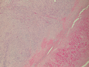
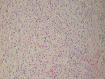
Histopathological examination revealed myofibroblastic features extending from below the dermis to the level of the cartilage, with no evidence of necrosis (Figures 1 and 2). The patient's history of trauma raised the possibility of a florid cellular repair process.
Photomicrograph showing fascicular myofibroblastic proliferation abutting cartilage. (H&E; original magnification ×50)

Higher power photomicrograph showing bland spindle cells. Note the lack of mitotic activity and atypia. (H&E; original magnification ×200)

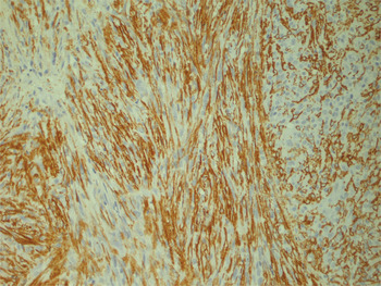
Immunohistochemical analysis showed negative reactions to desmin, S-100 and cytokeratin. The cells focally expressed α-smooth muscle actin (Figure 3). A diagnosis of myofibroma was made.
Photomicrograph showing immunohistochemical staining for α-smooth muscle actin. (Original magnification ×200)

The patient made a good recovery and his ear appearance returned to a normal. He was monitored on a six-monthly basis; at his first follow-up appointment (at the time of writing) there was no recurrence of disease.
Discussion
Our case differs from Balakrishnan's 1999 report of a pinnal myofibroma in two main respects: a history of repeated trauma as a causative agent was present, and immunochemistry was used alongside histopathology to formulate the diagnosis.Reference Balakrishnan, Pujary, Shah and Hazarika12
In the presented case, the main differential diagnosis was chondrodermatitis nodularis chronica helices, given the history of trauma and the tenderness of the lesion. Clinically, myofibromas usually present as painless, firm nodules.Reference Balakrishnan, Pujary, Shah and Hazarika12 Histologically, they are distinct from chondrodermatitis nodularis chronica helicis and other differential diagnoses in terms of their biphasic pattern, with zones of multinodular proliferation. At the periphery, there are mature, spindle-shaped cells in fascicles and bundles, with more immature ‘haemangiopericytoma-like’ areas in the centre.Reference Lingen, Mostofi and Solt5 This pattern was consistent with our patient's histology report of spindle cell lesions with a fascicular growth pattern. However, a myofibroma may be misdiagnosed histologically as a neural tumour, leiomyoma, leiomyosarcoma, myofibroblastic sarcoma, nodular fasciitis, infantile fibromatosis, fibrous histiocytoma or haemangiopericytoma.Reference Chung and Enzinger3, Reference Balakrishnan, Pujary, Shah and Hazarika12, Reference Hartig, Koopmann and Esclamado13 These other neoplasms have several overlapping and subtle histological changes which sometimes make diagnosis difficult.Reference Azevedo, Pires, Della Coletta, De Almeida, Kowalski and Lopes14
Hence, in our case, immunohistochemistry was used to support the diagnosis of myofibroma. Positive staining for vimentin and α-smooth muscle actin, along with negativity for S100, cluster of differentiation 34 glycoprotein and desmin, is highly suggestive of myofibroma, and our patient's results were consistent with this pattern.Reference Azevedo, Pires, Della Coletta, De Almeida, Kowalski and Lopes14 Whilst not pathognomonic of myofibroma, positive immunochemistry is still an important tool in aiding the diagnosis, as it is supportive of myofibroblastic cell proliferation.
Regarding the aetiology of our patient's myofibroma, we postulate that repetitive pinnal injury from rugby and helmet use was causative. The lesion may have resulted as a reactive process secondary to this trauma.Reference Kassenoff, Tabaee and Kacker9 Myofibroblasts are thought to be important in wound healing, as they develop gradually from granulation tissue fibroblasts.Reference Darby, Skalli and Gabbiani15 There have been sporadic reports of trauma at the site of myofibromas prior to presentation.Reference Kassenoff, Tabaee and Kacker9 Azevedo et al. have described trauma as accounting for some of the findings of myofibromas within the oral cavity.Reference Azevedo, Pires, Della Coletta, De Almeida, Kowalski and Lopes14
• Myofibromas of the pinna are rare; they are mesenchymal, spindle cell tumours which usually present in infancy but have been reported sporadically in adults
• Pinnal trauma is a potential cause, resulting in myofibroblastic proliferation
• Histological diagnosis can be difficult; immunohistochemistry may be helpful in excluding other diagnoses
• The treatment is surgical excision
Conclusion
Myofibroma of the pinna is rare. Its aetiology is unclear, but repeated trauma may be a potential trigger for development. Immunohistochemistry is recommended in conjunction with histology in cases with an ambivalent diagnosis.