Introduction
While studying the family Hysteriaceae (Dothideomycetes) from southern South America (Lorenzo & Messuti Reference Lorenzo and Messuti1998; Messuti & Lorenzo Reference Messuti and Lorenzo2009), an interesting specimen, the type of Hysterium graphideum Speg. was examined.
The Hysteriaceae is characterized by carbonaceaous hysterothecioid ascomata (lirellae) with longitudinal slits, which are immersed to erumpent or entirely superficial, solitary or gregarious, ellipsoid to elongated, simple or branched, with or without a subiculum. The hamathecium is composed of persistent cellular pseudoparaphyses forming an epithecium. The asci are bitunicate, clavate to cylindrical, and 8-spored. The ascospores are hyaline to dark brown, obovoid, clavate, ellipsoid or fusoid, didymospores (1-septum), phragmospores (2- or more transverse septa) or dictyospores (submuriform or muriform), at times surrounded by a gel coating, often showing bipolar asymmetry (Boehm et al. Reference Boehm, Schoch and Spatafora2009). Some authors (Zogg Reference Zogg1962; Purvis et al. Reference Purvis, Coppins, Hawksworth, James and Moore1992; Brodo et al. Reference Brodo, Sharnoff and Sharnoff2001; Ertz & Diederich Reference Ertz and Diederich2007) pointed out that the Hysteriaceae could be morphologically confused with members of the lichenized family Opegraphaceae (Arthoniomycetes), but there is only a superficial resemblance of the ascomata.
Most members of the Hysteriaceae are saprophytic, mainly lignicolous or corticolous. There are, however, scattered reports of species with a very casual association with algae. Spegazzini (Reference Spegazzini1909) suggests the presence of algae associated with the fungus (“Species habitu valde lichenino sed thallo omnimo destituta…”) in the case of Hysterium graphideum, while Lohman (Reference Lohman1933) recorded Hysterium angustatum Alb. & Schw. in which hysterothecia grow superficially upon a crust with algal cells. In more recent years, Kantvilas & Coppins (Reference Kantvilas and Coppins1997) cited a terricolous-saxicolous Glonium species, G. circumserpens Nyl., which has an inapparent ‘thallus’ sometimes occurring among a discontinuous greenish to blackish film associated with a green alga. The occurrence of a facultative or poorly developed lichenization within the same genus or species, is not unique to the Hysteriaceae. There are various fungal-algal associations with intimate or irregular connections between the bionts that are generally not considered lichens or studied by lichenologists (Hawksworth Reference Hawksworth1988; Grube & Hawksworth Reference Grube and Hawksworth2007). Some facultative lichenizations have been suggested to occur in other Ascomycota such as the Pyrenulaceae and Requienellaceae (Aptroot Reference Aptroot1991). The case of ‘optional lichenization’ where species are lichenized or saprotrophic depending on their substratum, was described in the Stictis-Conotrema complex in the mainly saprotrophic Stictidaceae (Wedin et al. Reference Wedin, Döring and Gilenstam2004). Several additional genera in the Stictidaceae include closely related lichenized and saprotrophic species, as in the genus Cryptodiscus (Baloch et al. Reference Baloch, Gilestam and Wedin2009). Additional examples of facultative associations with algae in Basidiomycota are Trametes and Resinicium which incorporate algae into their basidiomes (Zavada & Simoes Reference Zavada and Simoes2001; Grube & Hawksworth Reference Grube and Hawksworth2007).
Materials and Methods
Specimens from LPS and S were studied. Hand-cut sections were examined in tapwater, 5% KOH, Lugol's reagent, without (I) or with KOH pre-treatment (KI). Macroscopic photographs were taken on an Olympus SZH10 stereoscopic microscope, with a Sony SSC-DC50 digital camera and Image Pro-Plus 4.0 Media Cybernetics software. Microscopic photographs were prepared with an Olympus BX 40 optical microscope, and the same digital camera.
The Species
Gloniella graphidoidea Rehm
Hedwigia 42: 290 (1903); type: Tiches dessechèes de Pteris aquil. Sous des rochers proche le mer. Boci de la Chaise. Côte de Normantier Vendèe. leg. Dr Viand Marais Ex. herb. Arnold. (exsiccata H. Rehm, Ascomyceten nr. 1460; S reg. nr. F 11143!—lectotype, designated here).
Hysterium graphideum Speg., Anal. Mus. Nac. Bs. As. (ser. 3, v. 12) 19: 438 (1909); type: Ad truncos cariosos vetustos Corallodendri cristaegalli, Isla Santiago, prope La Plata, ix 1906, C. Spegazzini (LPS 28171!—holotype).
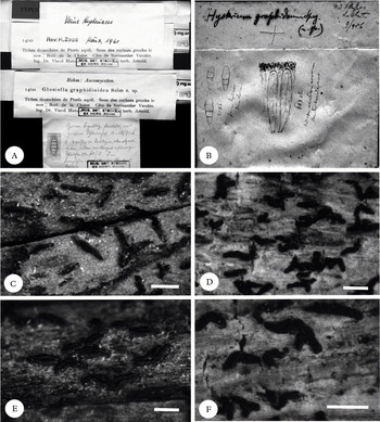
Gloniella graphidoidea (S reg. nr. F 11143, lectotype); A, herbarium packet; C, general view of the ascomata; E, detail of hysterothecia. Hysterium graphideum (LPS 28171, holotype); B, herbarium packet; D, general view of the ascomata; F, detail of hysterothecia. Scales = 1 mm.

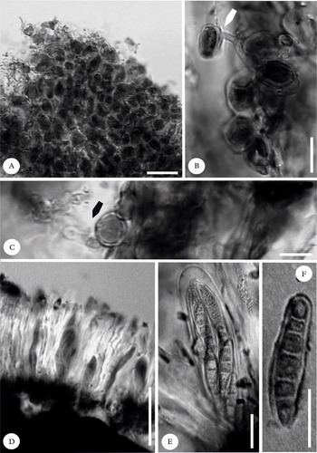
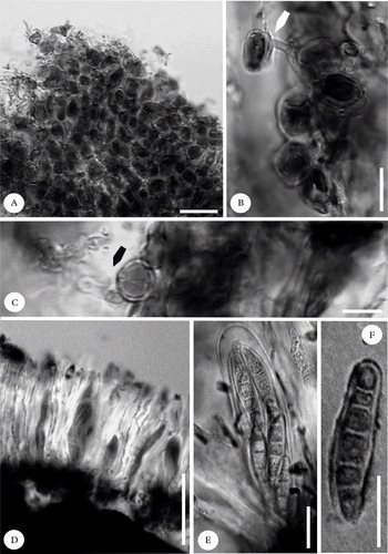
Gloniella graphidoidea, anatomy (S reg. nr. F 11143, lectotype). A, section of the thallus; B & C, association between alga and hypha (arrows); D, hymenium; E, ascus; F, ascospore. Scales: A = 20 μm; B, C, E & F = 10 μm; D = 40 μm.
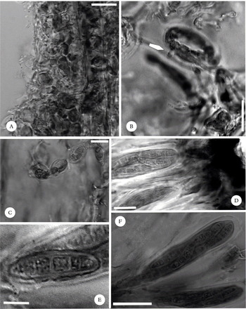
Hysterium graphideum, anatomy (LPS 28171, holotype). A, section of the thallus; B & C, association between alga and hypha (arrow); D, epithecium; E, ascospore; F, ascus. Scales: A = 20 μm; B, C, D & E = 10 μm; F = 20 μm.

Lichenized thallus inapparent, endosubstratal. Vegetative hyphae hyaline, interwoven with unicellular green algae 8–12 (–20) μm diam.
Ascomata lirelliform, carbonaceous, brittle, 0·47–1·43 (–2) × 0·1–0·2 mm, superficial to semiimmersed over the substratum, straight to flexuous, occasionally branched, scattered or crowded in groups of 2–5 hysterothecia, with a remarkable slit.
Hymenium colourless, I−, I/K+ blue. Asci 8-spored, claviform, (44–) 60–64 × 12–14 μm, tholus I/K+ blue. Pseudoparaphyses hyaline, septate, branched and anastomosing, up to 1 μm thick. Epithecium reddish brown, up to 22 μm thick. Ascospores hyaline, rarely pale yellow (fide Spegazzini Reference Spegazzini1909), bi- to triseriate, ellipsoid to claviform, slightly curved, upper part broader than the lower one, (3–) 4–5-transverse septate, 16–20 ×4–6 μm.
Habitat and distribution. A very uncommon species, but most probably overlooked. The type material of Gloniella graphidoidea was collected on the stalks of Pteris aquilina L. in France. In Argentina this species was described by Spegazzini (Reference Spegazzini1909) as Hysterium graphideum growing on twigs of Erythrina crista-galli L. in La Plata, Isla Santiago, Buenos Aires.
Remarks. The genus Gloniella Sacc. includes hysteriaceous fungi that possess hyaline, 3–9-septate phragmospores, while the genus Hysterium Pers. is characterized by pigmented, 3- or more transverse-septate, asymmetric phragmospores, with the cells often constricted, especially at the median septum. Most authors have considered the two genera to be closely related (Zogg Reference Zogg1962). Our morphological and anatomical examination of the type of H. graphideum (LPS 28171) revealed that the species does not belong to Hysterium, but it is better placed in Gloniella (Hysteriaceae). The holotype of H. graphideum is almost identical in many respects to the European G. graphidoidea. Both species have lirelliform carbonaceous ascomata (hysterothecia) that are more or less open, up to 2 mm long, 0·1–0·2 mm wide, and (3–) 4–5-septate, hyaline ascospores, 16–20 × 4–6 μm (Figs 1, 2 & 3). We have no hesitation in considering H. graphideum as a synonym of the previously described G. graphidoidea, rather than a distinct species. The samples studied were clearly lichenized, having a rudimentary thallus with green algal cells distributed homogeneously throughout the substratum (Fig. 2A). The presence of haustoria or connections with the colourless hyphae was also observed (Figs 2B & C, 3B & C). Therefore, it can be stated that this species is another example of a lichenized species included within the family Hysteriaceae which is mostly represented by non-lichenized members.
Our sincere thanks to Gintaras Kantvilas, Tasmania, for his critical comments and suggestions on the manuscript, and to the curators of LPS and S for the loan of the type material. Funds for this research were provided by the Secretaría de Investigación (UNComahue) and CONICET.