

Introduction
The ultimate goal of rehabilitation after stroke is to promote return to productive daily activities that occur in real-world environments, not just in the clinic or laboratory. For people whose upper-limb is affected by stroke, this refers to use of the upper-limb in everyday tasks that were possible pre-stroke, such as doing the laundry, preparing meals or playing golf. Currently, there is an assumption that training provided in the clinic or laboratory setting will lead to a corresponding improvement in use of the upper-limb outside these environments. Using the International Classification of Functioning, Disability and Health (World Health Organisation, 2001) definition of activity limitations, this implies that if training can change an individual's capacity for movement during the execution of tasks or actions (what they can do), they will also experience a change in their performance of everyday tasks (what they actually do). The concept of learned non-use (Taub, Uswatte, Mark, & Morris, Reference Taub, Uswatte, Mark and Morris2006) after stroke however, suggests that the opposite is likely; people often do not use their paretic upper-limb in everyday tasks to the full extent of their capability (de Niet, Bussmann, Ribbers, & Stam, Reference de Niet, Bussmann, Ribbers and Stam2007). Indeed, stroke survivors can have significant improvements in capacity (e.g., Action Research upper-limb Test or Box and Block Test), without any change in performance (e.g., upper-limb activity count) (Rand & Eng, Reference Rand and Eng2012, Reference Rand and Eng2015). Thus, being able to objectively and accurately measure use of the paretic upper-limb in real-world environments is critical.
The primary method to measure performance or real-world upper-limb use has been through self-report questionnaires, such as the Motor Activity Log (Uswatte, Taub, Morris, Light, & Thompson, Reference Uswatte, Taub, Morris, Light and Thompson2006) or Rating of Everyday Upper-Limb Use (REACH) (Simpson, Eng, Backman, & Miller, Reference Simpson, Eng, Backman and Miller2013). While these measures allow us to explore the relationship between capacity and performance, they do have inherent weaknesses as they rely on self-report. These include errors in recall due to memory, cognitive or perceptual difficulties and social desirability to report the behaviour of interest, all of which may be heightened in stroke populations, where cognitive and perceptual impairments are common (Dobkin & Dorsch, Reference Dobkin and Dorsch2011). Therefore, being able to gain an objective measure of upper-limb performance after stroke is important.
Non-invasive, wearable sensors such as accelerometers have gained acceptance as a way to objectively index upper-limb use in real-world environments (Uswatte et al., Reference Uswatte, Miltner, Foo, Varma, Moran and Taub2000). The current paper aims to explore the feasibility of accelerometers to measure upper-limb use after stroke and discuss the translation of this measurement tool into clinical practice. Here, we present a discussion of: (1) how accelerometers determine amount of upper-limb use; (2) the reliability, validity and sensitivity to change of accelerometry data; (3) how the accelerometer signal can be turned into clinically meaningful data and (4) the practicalities of developing an accelerometer protocol. We conclude by summarising the facilitators and barriers to the clinical uptake of accelerometers and future directions for research using accelerometers to capture upper-limb use.
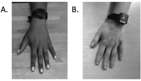
A search of the PubMed database was completed to identify relevant papers investigating the use of accelerometers to measure real-world upper-limb use after stroke. The search was performed up to March 31, 2015. Search terms included ‘stroke’, ‘accelerometer’ and ‘upper-limb’. Relevant papers were those that included a cohort of stroke survivors at any stage of recovery who had their upper-limb real-world arm use measured by an accelerometer. This paper discusses the commercially available Actigraph (Actigraph, Pensacola FL, Figure 1A) and Actical (Mini Mitter Co, Bend OR, Figure 1B) units as they are the most routinely cited in upper-limb stroke literature (Bailey, Klaesner, & Lang, Reference Bailey and Lang2014; Bailey & Lang, Reference Bailey and Lang2014; Lang, Wagner, Edwards, & Dromerick, Reference Lang, Wagner, Edwards and Dromerick2007; Rand & Eng, Reference Rand and Eng2010, Reference Rand and Eng2012, Reference Rand and Eng2015; Urbin, Hong, Lang, & Carter, Reference Urbin, Hong, Lang and Carter2014; Urbin, Waddell, & Lang, Reference Urbin, Bailey and Lang2015). While we anticipate that the commercially available options for accelerometers will expand rapidly; we note that technology will advance but the underlying concepts of accelerometry will likely remain the same. Finally, the reference lists of relevant original research and review papers in the field were also hand searched to identify any additional papers.
Accelerometer devices commercially available. A: Actigraph, Actigraph, Pensacola FL. B: Actical, Mini Mitter Co, Bend OR.

Accelerometers: Measurement, Metrics and Accuracy
Accelerometers were initially developed to monitor sleep cycles, where arm movement was considered a proxy for sleep time. Subsequently, they were translated to index upper-limb use in people with stroke and other neurological conditions, as well as in healthy individuals. At the present time, both the Actigraph and Actical devices resemble a wrist watch, are small and lightweight (Actigraph: 4.6 cm × 3.3 cm × 1.5 cm, 43 grams; Actical: 2.8 cm × 2.7 cm × 1.0 cm, 17 grams) and are able to measure movement of the upper-limb across multiple axes.
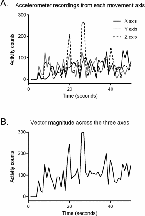
Accelerometers measure movement of the upper-limb through acceleration. Acceleration is the change in speed with respect to time. While acceleration is typically measured in gravitational acceleration units (g; 1 g = 9.8 m/s2), commercially available accelerometers often measure it in arbitrary units called activity counts. Sensors contained within the device convert mechanical motion (movement) into electrical signals. The electrical signals are then converted into a digital signal, which is stored as an activity count. Different brands of accelerometers have different processes for integrating the signal to produce activity counts, which are not publically available. This inherently makes it difficult to directly compare activity counts provided by different accelerometer brands. Depending on the unit used, an activity count may be produced for one, two or three axes of movement. With respect to the upper-limb, everyday functional tasks typically require movement to occur throughout multiple axes simultaneously, not a single axis. As such, data from two or three axes can be combined into a vector magnitude, a single value to quantify how much acceleration occurred at an instant in time, independent of direction. Instances in time are integrated over a pre-specified period called an epoch. The duration of the epoch can be set by the user, and may be as short as a fraction of a second or as long as a few minutes. Data are stored internally and can be downloaded via a USB or wireless connection to a computer for further evaluation. Figure 2 shows an example of 50 seconds of upper-limb accelerometry data from the left upper-limb using the Actigraph.
Example of accelerometry data from an Actigraph, which was worn on the left upper-limb of a neurologically intact community dwelling adult while completing a bilateral sorting task (from Bailey et al., Reference Bailey and Lang2014). The participant was asked to sort objects into a tackle box. A: Data from each axes (X, Y and Z). B: Vector magnitude data reflecting combined acceleration across all three axes.
The summed data from each epoch are used to compute a range of metrics to describe upper-limb use. The most common metrics quantify the amount of upper-limb use with respect to magnitude and/or duration. Magnitude typically refers to the total activity count across all observed epochs, with a higher number indicating greater upper-limb use. Duration refers to the period of time the upper-limb is used and is the sum of all the epochs when the upper-limb was moving (i.e., when a minimum threshold of activity counts were recorded). Calculations of duration of upper-limb activity simply reflect whether or not the upper-limb was moving, irrespective of the magnitude of the movement. When determining duration of movement, studies to date have used thresholds of one or two activity counts to categorise epochs into movement versus no movement (Bailey & Lang, Reference Bailey and Lang2014; Uswatte et al., Reference Uswatte, Miltner, Foo, Varma, Moran and Taub2000). In this way, epochs where the upper-limb was used can be summed to determine the total duration of upper-limb use during a given time period, such as hours in a day or days of the week. A greater duration indicates greater upper-limb use. A limitation of these metrics is that they do not give any indication about how movement of the paretic upper-limb occurred in relation to the non-paretic upper-limb. If an accelerometer device is applied to each upper-limb, the ratio of activity between both upper-limbs can be used to provide an indication of movement characteristics of the paretic upper-limb normalised to the non-paretic upper-limb (Uswatte et al., Reference Uswatte, Miltner, Foo, Varma, Moran and Taub2000). If the activity ratio is nearing 1.0, it indicates that the paretic and non-paretic upper-limb are used for the same magnitude or duration; however, if the activity ratio is less than 1.0, it indicates that the non-paretic upper-limb is used for a greater magnitude or duration than the paretic upper-limb. Because these are summary statistics over specified time periods, a ratio nearing 1.0 means that the limbs moved the same duration over the time period, but not necessarily that they were being moved simultaneously. Interestingly, the activity ratio appears to be relatively constant in non-disabled, neurologically intact, community-dwelling adults, averaging 0.95 (SD = 0.06), regardless of the person's activity levels, i.e., very sedentary or very active (Bailey & Lang, Reference Bailey and Lang2014). This stable value suggests that both upper-limbs are critical to function in everyday life and that this ratio is useful for distinguishing loss of upper-limb activity from normal. While metrics of magnitude, duration and the activity ratio are frequently used to indicate summed activity across a day, it is possible to examine only portions of the data to explore activity at specific time-points during the day, such as in the morning or during a meal.
The psychometric properties of metrics of accelerometry-derived activity counts are an important consideration to research and clinical uptake. Accelerometry-derived metrics of upper-limb use have test–retest reliability. Strong correlations between activity counts at two time-points (3-days duration separated by 2-weeks) have been identified for upper-limb magnitude metrics derived from the paretic (r = 0.87) and non-paretic (r = 0.81) upper-limb, as well as the ratio of activity (r = 0.90) (Uswatte et al., Reference Uswatte, Taub, Morris, Light and Thompson2006).
Accelerometry-derived activity counts of upper-limb use has been found to have a high level of agreement with human-observed purposeful repetitions during group (r = 0.58 to 0.92, (Rand, Givon, Weingarden, Nota, & Zeilig, Reference Rand, Givon, Weingarden, Nota and Zeilig2014)) and individual therapy sessions (r = 0.93, (Uswatte et al., Reference Uswatte, Miltner, Foo, Varma, Moran and Taub2000); r = 0.627, (Connell, McMahon, Simpson, Watkins, & Eng, Reference Connell, McMahon, Simpson, Watkins and Eng2014)). However, there was no correlation between the accelerometry-derived activity count and all human-observed repetitions, that is, purposeful plus non-purposeful repetitions (Connell et al., Reference Connell, McMahon, Simpson, Watkins and Eng2014). Accelerometry-derived duration of use has been found to differ from human-observed duration of use. During a single therapy session, accelerometry data indicated the duration of use was 35.9 minutes (or 75.7% of the session), while the human-observed reported duration of use was 31.2 minutes (or 63.8% of the session) (Connell et al., Reference Connell, McMahon, Simpson, Watkins and Eng2014). Discrepancies between accelerometry metrics and human-observed repetitions are likely underpinned by differences between accelerometry and human-made recordings. Accelerometers record all accelerations that occur and a threshold of one or two activity counts (which does not discriminate based on purpose) is often applied to categorise epochs into movement versus no movement. This may lead to some discrepancy with human-observed movement, as observers may discount or not detect small accelerations or non-purposeful movements that occurred. Thus, if more accelerations are detected by accelerometers, a greater duration of activity will be defined compared to human-observed movement. In addition, for duration, the selected epoch may have been insensitive to inactive periods. For example, if it takes a patient 8-seconds to complete the task of reaching to pick up a cup, the observer would record duration of use as 8-seconds; but if the accelerometer defined epoch is 15-seconds, it would record duration of use as 15-seconds. Extrapolating this difference in duration out across a 1-hour therapy session or a 24-hour day serves to highlight how accelerometry data may overestimate upper-limb activity. This highlights the need for small (e.g., 1-second or less) user-selected epoch duration.
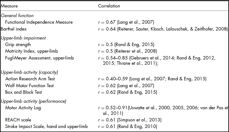
There is agreement between accelerometry activity counts and standardised measures of upper-limb function, which demonstrates convergent validity. Accelerometry data have been found to correlate to general measures of disability such as the Functional Independence Measure, upper-limb impairment measures such as Fugl-Meyer Assessment of Upper-Limb function, upper-limb capacity measures such as Action Research Arm Test and self-reported upper-limb performance measures such as the REACH Scale (Table 1). More recently, a reduced upper-limb activity count has been found to be associated with increased sedentary time (r = −0.36, (Bailey et al., Reference Bailey, Klaesner and Lang2014)).
Relationships Between Accelerometer Metrics and Measures of Function According to the ICF
Generally accelerometers have good discriminative validity. They can accurately distinguish upper-limb use between people with and without stroke (de Niet et al., Reference de Niet, Bussmann, Ribbers and Stam2007; Lang et al., Reference Lang, Wagner, Edwards and Dromerick2007), and between use of the paretic and non-paretic upper-limb (de Niet et al., Reference de Niet, Bussmann, Ribbers and Stam2007; Vega-Gonzalez & Granat, Reference Vega-Gonzalez and Granat2005). There is however, conflicting evidence surrounding their sensitivity to change during upper-limb training interventions. An increase in accelerometer activity counts has been demonstrated after therapeutic interventions of task-specific training (Urbin et al., Reference Urbin, Bailey and Lang2015; Waddell, Birkenmeier, Moore, Hornby, & Lang, Reference Bailey and Lang2014) and constraint induced movement therapy (Taub et al., Reference Taub, Uswatte, Bowman, Mark, Delgado, Bryson and Bishop-McKay2013), but not after robotic therapy (Lemmens et al., Reference Lemmens, Timmermans, Janssen-Potten, Pulles, Geers, Bakx and Seelen2014) or mental practice (Timmermans et al., Reference Timmermans, Verbunt, van Woerden, Moennekens, Pernot and Seelen2014). The conflicting findings to date may occur for several reasons. It could be proposed that it relates to the training undertaken, which includes reasons such as: (1) how the upper-limb is used within therapy is not consistent with how the upper-limb is used outside of therapy (Lang et al., Reference Lang, Wagner, Edwards and Dromerick2007); (2) therapeutic interventions are targeted to achieve a change in a stroke survivor's capacity, but not their performance (Gebruers, Vanroy, Truijen, Engelborghs, & De Deyn, Reference Gebruers, Vanroy, Truijen, Engelborghs and De Deyn2010); and (3) there is a minimal threshold of capacity relative to use that is required to achieve a change in performance as a result of training (Kokotilo, Eng, McKeown, & Boyd, Reference Kokotilo, Eng, McKeown and Boyd2010). Also, potential for a change in activity counts may be impacted on by factors related to the stroke survivor (e.g., how well they feel that day), their environment (e.g., presence of social support) and the period of monitoring (e.g., weekend vs. weekday, 1-day vs. 7-days). Given the relatively few studies in this area, there is a pressing need to unpack these findings to determine the drivers of change in accelerometry-derived activity counts during training interventions in future studies.
Most recently, relationships between accelerometry data and areas of the brain activated during a magnetic resonance imaging (MRI) performed with a functional task (functional MRI, fMRI) and at rest (resting state-MRI, rs-MRI) have been investigated in individuals with stroke. When completing a functional task during MRI, people with stroke with a lower accelerometry count, that is, rarely used their paretic upper-limb, were found to have significantly greater activity in secondary motor areas (e.g., contralesional areas including the pre-motor cortex r = −0.648, primary motor cortex r = −0.721) (Kokotilo et al., Reference Kokotilo, Eng, McKeown and Boyd2010) compared with an age-matched control group. Similarly, when at rest in the scanner, a lower accelerometry count was associated with an increase in activity between non-related regions of the ipsilesional and contralesional hemispheres (Urbin et al., Reference Urbin, Hong, Lang and Carter2014). Whereas, a higher accelerometry count, that is, used their paretic upper-limb a lot, was associated with an increase in activity between related regions of the ipsilesional and contralesional hemispheres (Urbin et al., Reference Urbin, Hong, Lang and Carter2014). These early findings suggest that accelerometry counts and brain recovery are related, but much work is needed to understand these likely complex relationships.
Turning the Accelerometer Signal into Clinically Meaningful Information
Despite growing evidence of their accuracy as a tool to measure upper-limb use in real-world environments, there are several challenges to facilitating widespread clinical use of accelerometers. The first challenge relates to the fact that the devices record all movement, without respect to purpose or quality. As mentioned above, the devices register counts of activity whenever the body part is moving. Thus, wrist-worn accelerometers cannot distinguish between different types of upper-limb movements, such as purposeful actions to complete a task with the upper-limb versus upper-limb swing during gait. As accelerometers do not capture the context within which movement occurred it is increasingly difficult to differentiate types of movements. Quantifying upper-limb use as a ratio, as described above, partially but not completely eliminates this challenge. The inability to distinguish purposeful versus non-purposeful movements has generated a great deal of debate amongst rehabilitation researchers. On one side of the debate is the wish to quantify only purposeful movements that are part of a focused upper-limb related task (Gebruers et al., Reference Gebruers, Vanroy, Truijen, Engelborghs and De Deyn2010; Uswatte et al., Reference Uswatte, Foo, Olmstead, Lopez, Holand and Simms2005; Wade, Chen, & Winstein, Reference Wade, Chen and Winstein2014). On the other side of the debate, there are plausible reasons to quantify all motion including: (1) interest in upper-limb swing as a ‘purposeful’ action; (2) knowledge that time spent walking in many patient populations is quite small (Roos, Rudolph, & Reisman, Reference Roos, Rudolph and Reisman2012); and (3) the desire to increase any movement, not just upper-limb movement in inactive patient populations (Billinger, Coughenour, Mackay-Lyons, & Ivey, Reference Billinger, Coughenour, Mackay-Lyons and Ivey2012; Billinger et al., Reference Billinger, Arena, Bernhardt, Eng, Franklin, Johnson and Tang2014). Algorithms can be developed to remove upper-limb swing during gait from the accelerometer recordings, but it does require people wear an additional accelerometer on the leg (hip, thigh or ankle) at the same time as wearing wrist accelerometers. In addition, there has been some research to investigate the role of aligning snapshot activity mapping (e.g., Measuring Activity Recall for Adults and Children [MARCA] (English, Coates, Olds, Healy, & Bernhardt, Reference English, Coates, Olds, Healy and Bernhardt2014)) to accelerometry data to capture the context of activity. From a research perspective, exploration of these avenues will likely be ongoing. From a clinical perspective however, these approaches may have less clinical utility because they may impact on patient experiences (e.g., comfort, aesthetics, workload) and provider and service resources (e.g., cost of unit procurement and amount and cost of data processing). At the present time, there is therefore a strong clinical rationale for including all voluntary actions, purposeful or not, in accelerometry recordings as it will be substantially easier to implement this technology at an individual patient level within a service.
A second challenge is that wrist-worn accelerometers are not able to measure movement quality. That is, they cannot distinguish between a reaching movement with appropriate shoulder and elbow biomechanics versus a reaching movement where the shoulder and elbow biomechanics are altered. The inability to readily determine movement quality has also generated a great deal of debate, with this second debate engaging both rehabilitation clinicians and researchers. On one side of the debate clinicians and researchers, consider the goal of rehabilitation to be retraining normal movement patterns (Krakauer, Carmichael, Corbett, & Wittenberg, Reference Krakauer, Carmichael, Corbett and Wittenberg2012; Levin, Kleim, & Wolf, Reference Levin, Kleim and Wolf2009). For these individuals, the premise is that allowing practice of abnormal movements will prevent restoration of optimal movement. Others argue that the goal of rehabilitation is to restore function by whatever means possible (Lang, Bland, Bailey, Schaefer, & Birkenmeijer, Reference Lang, Bland, Bailey, Schaefer and Birkenmeijer2013; Levin et al., Reference Levin, Kleim and Wolf2009). It is likely however, that recovery of upper-limb function is often a result of both more normalised movement patterns for some movements and more compensatory movement patterns for others depending on the deficits of the patient (DeJong, Birkenmeier, & Lang, Reference Lang, Guccione, Wong and Avers2012). At present, there is no definitive answer to the debate. There is accumulating data on the ineffectiveness of therapeutic approaches focused on movement quality (for recent review, see Veerbeek et al., Reference Veerbeek, van Wegen, van Peppen, van der Wees, Hendriks, Rietberg and Kwakkel2014) and the limited amount of time that patients actually use their arm during inpatient rehabilitation, both inside and outside of therapy (Bernhardt, Chan, Nicola, & Collier, Reference Bernhardt, Chan, Nicola and Collier2007; Hayward, Barker, Wiseman, & Brauer, Reference Hayward, Barker, Wiseman and Brauer2013; Hayward & Brauer, Reference Hayward and Brauer2015; Lang et al., Reference Lang, Wagner, Edwards and Dromerick2007; Rand & Eng, Reference Rand and Eng2012). Taken together, it would suggest that erring on the side of quantifying any voluntary movement may be most beneficial after stroke at this point in time.
A third challenge is to turn the data recorded by the accelerometer (i.e., activity counts quantifying accelerations) into information that has clinical relevance. A number of different approaches have tried to identify the performance of specific tasks/actions (e.g., feeding, bathing, reaching) from the accelerometer signals. One method has been to identify known ‘wave-forms’ of specific movements in the data (e.g., time and/or frequency series of accelerometer values during reaching), and then to use the known wave-form to identify other instances where and when the same specific movement occurred (e.g., search through multiple hours of recording to try to identify the same reach pattern) (Jain, Duin, & Mao, Reference Jain, Duin and Mao2000; Lemmens et al., Reference Lemmens, Seelen, Timmermans, Schnackers, Eerden, Smeets and Janssen-Potten2015; Wade et al., Reference Wade, Chen and Winstein2014). A second method has been to use machine-learning algorithms (Bao & Intille, Reference Bao, Intille, Ferscha and Mattern2004; Del Din, Patel, Cobelli, & Bonato, Reference Del Din, Patel, Cobelli and Bonato2011; Mannini & Sabatini, Reference Mannini and Sabatini2011; Preece et al., Reference Preece, Goulermas, Kenney, Howard, Meijer and Crompton2009). In this method, specific known movements are used as the ‘teacher’ to train the algorithm and then unknown movements can be identified and categorised based on one or more extracted characteristics that are shared with the ‘teacher’ movements. The machine-learning algorithm can be created for use across people (inter-subject) or created uniquely for each person (intra-subject). The inter-subject approach is computationally appealing, since the ‘teacher’ movements and algorithm could be generated only once, but the intra-subject approach is more realistic for use in patient populations with heterogeneous movement patterns. Nonetheless, the eventual success of these methods may be challenging for three reasons. The primary reason is that human movement, within and across people, is highly variable (Bailey et al., Reference Bailey, Klaesner and Lang2014; Stergiou & Decker, Reference Stergiou and Decker2011). Movements that may be viewed as relatively invariant, such as reaching, are actually highly variable across the day, as people reach for many different objects, in many locations, for many purposes. The second reason is that humans perform an enormous number of movements and actions throughout the day with their upper-limbs (Lang, Reference Lang, Guccione, Wong and Avers2012). Therefore, even if waveform recognition or machine-learning approaches could accurately identify 10–20 movements, it is unlikely that they could identify and categorise all movements (i.e., all data points in the recordings). If they could, this would provide a substantial amount of information, which may be redundant or not necessary. For example, does a therapist need to know that the stroke survivor opened their hand to pick up a coffee cup compared to a toothbrush or do they just need to simply know that the hand opened and closed? The third reason these options are challenging is related to classification and the fact that there are not ‘pure’ categories of upper-limb movements in daily life. One can readily identify actions based on a number of factors, such as the goal of the movement or whether it is unilateral or bilateral. Very quickly, however, these factors start to become less distinct. For instance, is a reach to pick up an object on the counter during food preparation the same ‘category’ as a reach to bring the hand to the mouth during feeding? How would one classify the action when the food preparer sneaks a bite of food during preparation activities? Likewise, actions can be bilateral and symmetric, such as carrying a large laundry basket, or bilateral and asymmetric, such as stabilising with one hand and cutting with the other hand. Following this line of reasoning, it quickly becomes apparent that a ‘perfect’ categorisation solution would always be complicated.
A newer method for turning accelerometer data into clinically relevant information involves second-by-second quantification of two key aspects of upper-limb movement: the intensity of activity (i.e., magnitude of accelerations) from both limbs; and the relative contribution of each upper-limb to activity (Bailey et al., Reference Bailey, Klaesner and Lang2014). Both of these aspects, quantified as the Bilateral Magnitude and the Magnitude Ratio respectively, are critical for successful performance in daily life where the majority of activities require the interaction of both limbs (Kilbreath & Heard, Reference Kilbreath and Heard2005; McCombe Waller & Whitall, Reference McCombe Waller and Whitall2008). Lang and colleagues recently evaluated these two parameters in a sample of community-dwelling, neurologically intact adults (Bailey, Klaesner, & Lang, in review). Figure 3 shows example data from three people, in two-dimensional density plots. The intensity of activity (y-axis, Bilateral Magnitude) and the contribution of each limb (x-axis, Magnitude Ratio; zero indicating that both limbs contribute the same amount) are calculated for every second of data during the 25–26 hour wear period. The colour represents the frequency that each combination of values occurred, with cooler colours representing lower frequencies (less time) and warmer colours representing higher frequencies (more time). The two bars on either side show the amount of isolated dominant and non-dominant limb use. The shape in the middle shows the large amount of time when the two limbs are used together. The plots are symmetrical, indicating that, contrary to what one might imagine, the dominant and non-dominant upper-limbs are used about the same amount of time and mostly together. The plots are wider on the bottom, indicating that most upper-limb movements are low intensity. The ‘rims’ of the bowl-like shape occur during activities where one limb is accelerating while the other is relatively still, such as placing objects in a container with one hand and holding the container with the other (Bailey et al., Reference Bailey, Klaesner and Lang2014). The ‘warm glow’ in the middle occurs during lower intensity movements where both limbs are working together, such as cutting food with a knife and fork (Bailey et al., Reference Bailey, Klaesner and Lang2014). The top peak occurs with higher intensity movements involving both limbs, such as stacking boxes on a shelf (Bailey et al., Reference Bailey, Klaesner and Lang2014). Figure 3A shows a person who moved the upper-limbs an average amount in the 24-hour period. Figures 3B and 3C show persons who were more sedentary (3B), and more active (3C), respectively. Despite the difference in activity level, the plots are remarkably consistent in shape and symmetry. This consistency held across a sample of more than 70 people evaluated and suggests relatively constant features of upper-limb use during daily life.
Example data from three community-dwelling, neurologically intact adults, showing the importance of both upper-limbs for daily activity. Data are quantified on a second-by-second basis over the 24+ hour wearing period. The y-axis represents the intensity of movement in both limbs (in activity counts, 1 count = 0.001664 g) and the x-axis represents the contribution from each limb (0 = equal contributions from the limbs). There is remarkable consistency in the shape and symmetry across individuals, despite differing levels of overall activity. A: Moderate activity, 11.7 hrs of upper-limb activity. B: Lower activity, 8.5 hrs. C: Higher activity, 13.6 hrs.
The consistent shape and symmetry of the figures means that this accelerometry method may be ideal for distinguishing typical from atypical upper-limb activity in real-world environments. Figure 4 shows example data from people with stroke. Figure 4A has data from three people, one each from persons with low, moderate and high upper-limb function. These plots are less symmetrical and have lower peaks than the plots in Figure 3. Interestingly, the plots of the low and moderate functioning individuals look remarkably similar, suggesting that a higher level of functional capacity may be needed to restore activity in real-world environments (Bailey et al., in review; Rand & Eng, Reference Rand and Eng2012). The slight asymmetry in the plot from the high-functioning person suggests that this person has almost, but not quite returned to normal upper-limb activity in daily life. Figure 4B shows data from one person near the beginning and then at the end of an intensive upper-limb training programme during an inpatient rehabilitation stay (Waddell et al., Reference Waddell, Birkenmeier, Moore, Hornby and Lang2014). The plots in Figure 4B suggest that these figures might be used to assess the need for and progress during rehabilitation services.
Example data from people with stroke. A: Data from three persons with chronic stroke. Note that upper-limb activity is quite similar in the lower and moderate functioning individuals, despite differences in functional capacity (ARAT scores). The higher functioning individual has asymmetrical upper-limb activity. Data from Bailey et al. in review. B: Data from one person 10 days and then 33 days post stroke, showing clear restoration of upper-limb activity after intensive therapy. Data from Waddell et al. (Reference Waddell, Birkenmeier, Moore, Hornby and Lang2014). ARAT: Action Research Arm Test, range = 0 to 57, with higher scores = better. IRF = inpatient rehabilitation facility.
The value of quantifying daily upper-limb activity with this approach extends beyond people with stroke. Figure 5A shows an example of an older adult receiving rehabilitation in a skilled nursing facility. As might be expected from an older adult with significant disability and complex medical conditions, the plot is symmetrical, but overall movement is less intense. Figure 5B and 5C show examples of a typically developing child (age 8 years) and a child with mild hemiparesis due to brain injury (age 8 years). The height of the plot and the narrow width in Figure 5B suggest more intensive and more bilateral activity in children compared to adults (Figure 3A–C). In comparison to Figure 5B, the plot in figure 5C is not as high (although still more active than some adults) and slightly asymmetrical. The peak of the plot sits a bit to the left, suggesting that the dominant hand contributes a bit more to the higher intensity activities. [Please see www.accelerometry.wustl.edu for the opportunity to learn about and process acceleromtry as shown in the example figures.]
Example data showing that accelerometer quantification of upper-limb activity can be used for many populations. A: An older adult with complex medical conditions (not stroke) undergoing post-acute rehabilitation in a skilled nursing facility. The activity is symmetrical but far less intense than in community-dwelling adults. B: An 8 yr old typically developing child, showing higher intensity and more activity where the upper-limbs are moving at similar intensities. C: An 8 yr old child with mild hemiparesis due to brain injury, showing intensities more similar to adults, but an asymmetrical use of the limbs. Note the range of the y-axis scale for B and C is different from A and from the y-axes in Figures 2 and 3.
Collectively, the plots in Figures 3–5 suggest that, despite the challenges of capturing unintentional movement and inability to quantify movement quality, accelerometry data can provide clinically meaningful information that has the potential to be informative for clinicians, patients, family and care givers. Anecdotally, occupational and physical therapists and stroke survivors who have viewed these pictures (showing data from their own patients or themselves) indicate that the information is very helpful in understanding the status of the recovery and how much more improvement is needed. The pictures have also been useful in encouraging participants to engage their upper-limb more outside of therapy.
Practicalities of Developing An Accelerometer Protocol
Considerable evidence has been presented to demonstrate that accelerometers are simple to use, produce reliable (Uswatte et al., Reference Uswatte, Taub, Morris, Light and Thompson2006) and valid (Gebruers, Truijen, Engelborghs, & De Deyn, Reference Gebruers, Truijen, Engelborghs and De Deyn2014; Rand & Eng, Reference Rand and Eng2012, Reference Rand and Eng2015; Thrane, Emaus, Askim, & Anke, Reference Thrane, Emaus, Askim and Anke2011; Uswatte et al., Reference Uswatte, Miltner, Foo, Varma, Moran and Taub2000, Reference Uswatte, Foo, Olmstead, Lopez, Holand and Simms2005, Reference Uswatte, Taub, Morris, Light and Thompson2006; van der Pas, Verbunt, Breukelaar, van Woerden, & Seelen, Reference van der Pas, Verbunt, Breukelaar, van Woerden and Seelen2011) metrics of upper-limb use, and can provide clinically meaningful information. Yet, accelerometers are not routinely adopted in research protocols that explore upper-limb recovery after stroke. If the goal of rehabilitation is to improve real-world upper-limb use, then there is a strong rational for including an objective measure of upper-limb use outside of the laboratory setting across all research domains, whether the studies are asking questions about the neurobiology of behaviour or the efficacy of clinical interventions. Further, this would build a large collection of data, which would be available to then understand the threshold level of functional capacity required to achieve a change in performance, and thus restore activity in real-world environments. As such, there is a clear need to collect repeated, objective measures of upper-limb performance alongside measures of capacity (Dobkin & Dorsch, Reference Dobkin and Dorsch2011). To facilitate widespread adoption of accelerometers however, we need to consider how best to collect data to facilitate pooling across studies. To increase consistency in data collection in stroke research, we propose the following arguments and implications for research.
Selecting a Device
Accelerometers are generally described as uniaxial or multiaxial (bi- or tri-axial) depending on their sensitivity to register acceleration that occurs across one, two or three axes of movement. Early studies suggested that uniaxial accelerometers could be used to provide the same information as multiaxial accelerometers about whether or not the limb was moving (Redmond & Hegge, Reference Redmond and Hegge1985), likely because upper-limb movements nearly always involve joint rotations in multiple axes of movement, and not in a single axis (Lang et al., Reference Lang, Wagner, Edwards and Dromerick2007). Although there is evidence to suggest that uniaxial and multiaxial units are correlated, correlation coefficients reported for multiaxial devices are much higher (Trost, McIver, & Pate, Reference Trost, McIver and Pate2005). This suggests that collecting accelerations across multiple axes to produce a vector summation is ideal. Implications for practice: Multiaxial devices should be used where possible.
Selecting the Length of the Epoch
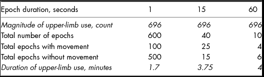
As described, sampling frequency is the rate at which data are collected and epochs are the time period in which individual points of data are viewed. Data may be sampled at fast frequencies (e.g., 30 Hz, 1 Hz) but then entered into analyses with the same (0.03s) or longer epochs (1 second out to 60 seconds). Regardless of sampling frequency, the epoch length needs to reflect the underlying rate at which human movement occurs. Shorter epochs, i.e., fractions of a second, can quantify milliseconds of movement and are ideal for unpacking unilateral (i.e., raise right arm then raise left arm) from bilateral movements (i.e., right and left arm pick up a box together). In capturing short epochs, the unit must have sufficient storage capacity for the high number of data collection time-points. Longer epochs, i.e. 10–60 seconds, allow for data to be collected over longer durations (e.g., weeks or months instead of a days), but sacrifice sensitivity in accelerometry metrics. Longer epochs also increase the risk that periods of inactivity will be classified as periods of activity, which may in turn overestimate total duration of activity. The impact of epoch selection (1-second versus 15-seconds versus 60-seconds) on duration of upper-limb use is demonstrated in Table 2. Current reports in the literature of accelerometry for the upper-limb chunk data into 1-second (Bailey & Lang, Reference Bailey and Lang2014; Bailey et al., Reference Bailey, Klaesner and Lang2014; Urbin et al., Reference Urbin, Bailey and Lang2015) or 15-second (Rand & Eng, Reference Rand and Eng2010, Reference Rand and Eng2012, Reference Rand and Eng2015) epochs. We have included 60-seconds for demonstration purposes. Here, you see that while magnitude remains consistent, the duration of use varies across the 10-minute interval explored. Given this is just a snapshot of data from a device worn for 3-days, the longer epoch durations could lead to considerable errors in per day duration of use estimations. Implications for practice: Short epochs should be chosen to support accurate representations of upper-limb activity. A middle value of 1-second epochs may be optimal for accounting for time-varying upper-limb activities that can be sampled over reasonable hours of wearing time. As units advance, i.e., longer battery life and greater storage capacity, then fraction of a second epochs can be adopted.
Influence of Different Epoch Durations (1-second, 15-second, 60-second) Over a Selected 10-minute Period of Observation (Lakhani et al., Reference Lakhani, Borich, Jackson, Wadden, Peters, Villamayor, MacKay, Vavasour, Rauscher and Boyd2015)

Selecting the Duration of Time to Monitor Activity
The duration of wear depends on the reason for capturing upper-limb use. Shorter durations (e.g., 1-hour) are generally used in clinic or laboratory settings, where the goal is to characterise activity during a therapy session or during performance of a specific series of tasks (Bailey et al., Reference Bailey, Klaesner and Lang2014; Urbin, Bailey, & Lang, Reference Urbin, Bailey and Lang2015). In comparison, longer durations (e.g., 1-, 3- or 7-days) are generally used when the goal is to characterise activity outside the clinic or laboratory setting, including real-world environments such as the home (Lang et al., Reference Lang, Wagner, Edwards and Dromerick2007; Rand & Eng, Reference Rand and Eng2012, Reference Rand and Eng2015; Rand et al., Reference Rand, Givon, Weingarden, Nota and Zeilig2014). The rationale for choice of duration of wear is infrequently reported in the literature. Longer durations of wear (7-days as compared to 1- or 3-days) enable exploration of the variability in daily upper-limb use (e.g., weekdays compared to weekends or mornings to afternoons; presence of carers/visitors; fatigue and environmental factors such as weather etc.). Given that use may vary across days, it provides support for multiple days of collection that is counterbalanced between people. Indeed during lower-limb monitoring, higher reliability is evident over a longer period of monitoring; 3-days is preferred over 1-day (Mudge & Stott, Reference Mudge and Stott2008) and 7-days preferred over 3-days (Hale, Pal, & Becker, Reference Hale, Pal and Becker2008). The challenge is however, that longer durations of wear can reduce compliance (Barak, Wu, Dai, Duncan, & Behrman, Reference Barak, Wu, Dai, Duncan and Behrman2014). For the upper-limb, wearing periods of 1-day have been suggested to be a practical compromise between sufficient wear time and participant willingness to wear the accelerometers (Bailey & Lang, Reference Bailey and Lang2014; Lang et al., Reference Lang, Wagner, Edwards and Dromerick2007), particularly when participants are asked to wear accelerometers repeatedly e.g., in longitudinal studies or clinical trials with follow-up. This challenge highlights the need for further investigations to determine the most appropriate period of time to record. Unfortunately, it is likely that the appropriate duration may vary across patient populations or subpopulations (e.g., people with stroke who have returned to work versus those who have not). Implications for practice: Where possible, more days of monitoring are ideal and days of monitoring should cover a weekday and weekend day, if the participant is employed or reports different activities on weekends.
Selecting the Number of Accelerometers and Body Position
One, two or three accelerometers can be used. If a single accelerometer is used, it is applied to the stroke affected upper-limb. Given the large proportion of upper-limb tasks that involve use of both upper-limbs (Bailey et al., Reference Bailey, Klaesner and Lang2014), this is less than ideal. Further, a single device could serve as an external cue to the participant to use the monitored limb more during the wearing period than is typical. Application of an accelerometer to each upper-limb at the wrist overcomes these limitations and supports the calculation of ratio metrics, which can mitigate some upper-limb use that occurs during walking. The addition of a third device to the leg would allow thorough exploration of the contribution of walking (e.g., upper-limb swing) and/or body position (e.g., sitting, standing) to upper-limb use. Implications for practice: An accelerometer should be applied to both the paretic and non-paretic upper-limb. If possible, application of a device to the leg is beneficial to control for upper-limb use during walking and lower-limb activities.
Selecting the Metrics for Analysis
Standard metrics to quantify the magnitude, duration and ratio of paretic to non-paretic upper-limb use should be produced to indicate use per day. The majority of studies completed to date provide such information. If there are indications that use differs throughout the day then the day can be broken down (e.g., morning, afternoon, evening). Alternatively, if use differs across days, e.g., due to employment or carers present, then individual days should be evaluated. The main challenge at present exists as to whether to include or exclude upper-limb swing in calculations of magnitude or duration. There are benefits gained from either approach. Algorithms can be developed to remove upper-limb swing during gait from the accelerometer recordings if people wear accelerometers on the leg (hip, thigh, ankle) at the same time as wearing wrist accelerometers. It is relatively simple to construct accurate algorithms to remove periods of gait from recordings in healthy, neurologically intact participants. For example, gait can be detected> 95% of the time during periods of known walking (unpublished data; Bailey et al., in review). Using a similar mathematical approach with lower thresholds, gait could be accurately detected only 50% of the time during periods of known walking in people with stroke using the Actigraph (unpublished data; Bailey et al., in review). Some studies have applied rules, such as when five consecutive steps occur in an epoch (15-seconds) upper-limb count is considered consistent with upper-limb swing (non-purposeful) and therefore, excluded (Rand & Eng, Reference Rand and Eng2010). The psychometric properties of the unit are likely to be important in the capacity to accurately detect gait. The Actical and Actigraph appear to have more difficulty in detecting gait in patient populations with abnormal gait (e.g., stroke) compared to other units, such as the Step Activity Monitor (SAM) (Fini, Holland, Keating, Simek, & Bernhardt, Reference Fini, Holland, Keating, Simek and Bernhardt2015) and FitBit (Klassen et al., Reference Klassen, Eng, Chan, Hassall, Lim, Louie and Zbogar2014), which was accurate at gait speeds as slow as 0.3 m/s. Despite this, as it is impossible to know the context of the activity that is taking place during gait, removing all upper-limb movements that occur may lead to exclusion of periods of purposeful upper-limb movement, such as carrying a cup of tea or the newspaper, or putting on shoes. Another consideration is inclusion or exclusion of upper-limb movements during periods of sleep. Deciding what constitutes non-functional movement (e.g., a tick or jerk) during quiescent periods is subjective. Movement during a nap or night time may be associated with functional movements such as an unconscious scratch or reaching for a glass of water and would be lost if upper-limb movements during sleep were removed (Bailey et al., Reference Bailey, Klaesner and Lang2014). Implications for practice: Frequency and duration metrics should be reported across all studies that include accelerometry measurement. Given the heterogeneity of walking movements within and across people with stroke (Meijer et al., Reference Meijer, Plotnik, Zwaaftink, van Lummel, Ainsworth, Martina and Hausdorff2011; Patterson et al., Reference Patterson, Mansfield, Biasin, Brunton, Inness and McIlroy2015; Reisman, Wityk, Silver, & Bastian, Reference Reisman, Wityk, Silver and Bastian2007; Roos et al., Reference Roos, Rudolph and Reisman2012) and variable sensitivity of units to capture gait, the ratio of activity of one upper-limb to the other is currently the simplest and most efficient way to remove the effect of gait from upper-limb activity counts. As movement during sleep is likely to be minimal, it is plausible for it to be included if patients are comfortable wearing the unit while sleeping.
By highlighting the decisions required to develop an accelerometer data collection protocol, the challenges of integrating results across studies emerge. At present, there is limited ability to pool accelerometry data due to the variety of devices used, which have different specifications and options e.g., sampling rates, epoch durations, internal memory storage and algorithms to collate data internally in the accelerometer and externally after downloading data to a computer, all of which impact activity count metrics. The lack of conversion factor to compare activity counts reported using different accelerometers compounds the issue of data pooling. A conversion factor would enable people to use the accelerometer device they have access to and subsequently afford comparison to previously completed studies using a different accelerometer. This has been developed for accelerometer derived step counts (Paul, Kramer, Moshfegh, Baer, & Rumpler, Reference Paul, Kramer, Moshfegh, Baer and Rumpler2007; Straker & Campbell, Reference Straker and Campbell2012). It would appear timely to do the same for the upper-limb. Finally, it is important to highlight that the challenges described above in developing an accelerometry protocol are not isolated to measurement of upper-limb activity after stroke, but are evident when monitoring physical activity after stroke (Fini et al., Reference Fini, Holland, Keating, Simek and Bernhardt2015). From a clinical perspective, where monitoring of both upper- and lower-limb activity may occur concurrently, there is scope to consider the development of an overall streamlined approach to effectively monitor both upper-limb use and physical activity.
Barriers and Facilitators to Moving Accelerometers Into Clinical Practice
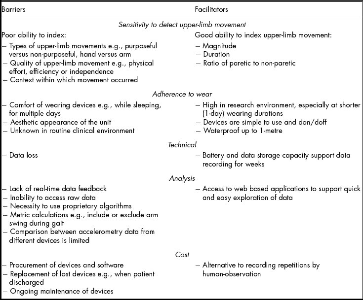
With ongoing advancements in wearable sensors, such as accelerometers, it is possible that they will replace the need for time-consuming and sometimes imprecise clinical tests and questionnaires in the future. From the discussion presented above, several barriers and facilitators to the deployment of accelerometers in research and clinical practice have been highlighted. These are summarised in Table 3 and relate to sensitivity to detect upper-limb activity, adherence to wear, technical aspects, data processing aspects and cost.
Summary of Identified Barriers and Facilitators to Clinical Implementation of Accelerometers to Measure Upper-Limb Activity After Stroke
There is ongoing research in this field that may serve to overcome some identified barriers to the measurement of real-world upper-limb use. The key barrier that causes great discussion in research and clinical professions is the inability of devices to capture the contribution of wrist and hand movements to use. One tool currently under investigation to overcome this barrier uses force sensor resistors, which extract forces produced by muscles of the wrist and hand during upper-limb tasks. Preliminary data with healthy participants indicates this approach is reliable and valid during performance of reaching for a cup (Xiao & Menon, Reference Xiao and Menon2014). Another tool for monitoring hand and wrist movement is a magnetic ring worn on the patient's finger, which sends signals to a data acquisition device (similar to current accelerometers) worn on the wrist (Friedman, Rowe, Reinkensmeyer, & Bachman, Reference Friedman, Rowe, Reinkensmeyer and Bachman2014). This device can detect movements of the wrist and hand, e.g., wrist flexion/extension, wrist deviation, finger flexion/extension, as well as gross upper-limb movements. Ongoing evaluations will confirm the contribution of such applications in the clinical environment.
The other key challenge emerging is to determine how to use accelerometry data as a tool to motivate patients to increase use of their upper-limb in their real-world environments. This may help to reduce the development and impact of learned non-use after stroke. While there is some work underway to explore the role of accelerometers as ‘persuasive technology’, current systems appear to be limited to real-time feedback of duration of paretic upper-limb use only (Markopoulos, Timmermans, Beursgens, van Donselaar, & Seelen, Reference Markopoulos, Timmermans, Beursgens, van Donselaar and Seelen2011). Additional metrics to integrate into the device could include magnitude of paretic upper-limb use, ratio of paretic to non-paretic upper-limb use and tracking of use throughout the day to identify, in real time, periods of greatest inactivity or activity. Nonetheless, it must be recognised that simply giving a stroke survivor the capacity to self-monitor and gain feedback on performance is unlikely to change performance. In trials of activity monitoring of physical activity after stroke, several studies have demonstrated no improvement in the number of steps taken between people who did and people who did not receive performance-related feedback (Dorsch et al., Reference Dorsch, Thomas, Xu, Kaiser and Dobkin2015; Mansfield et al., Reference Mansfield, Wong, Bryce, Brunton, Inness, Knorr and McIlroy2015). As such, this highlights the need to embed accelerometers within a broader overall upper-limb self-management programme that focuses on behaviour change and includes goal setting and action planning (Connell, McMahon, Redfern, Watkins, & Eng, 2015; Jones, Mandy, & Partridge, Reference Jones, Mandy and Partridge2009; Jones & Riazi, Reference Jones and Riazi2009).
Conclusion
Accelerometers offer a feasible option to index upper-limb use after stroke. There is growing evidence to indicate that accelerometers are a reliable and valid tool, but the sensitivity to change over time remains uncertain. Considerable work is underway to overcome challenges to widespread use and turn accelerometry data into clinically meaningful information. This provides the impetus for widespread uptake in research and clinical settings. As uptake increases, there is a greater need for consistency in how accelerometry data are collected to ensure that the most meaningful representation of use is captured and pooling across studies is possible. In building the evidence base, it will help to build a deeper understanding of what stroke survivors actually do in their everyday lives. This information has the potential to positively impact on the ability of researchers and clinicians to objectively evaluate function, set benchmarks for treatment as well as develop and adapt rehabilitation protocols on an individual basis.
Financial Support
KSH was supported by the Heart and Stroke Foundation of Canada; Michael Smith Foundation for Health Research British Columbia; and the National Health and Medical Research Council of Australia (NHMRC 1088449); BL was supported by the Heart and Stroke Foundation of Canada and the Michael Smith Foundation for Health Research British Columbia Canada; LAB was supported by Canada Research Chairs (950-203318) and JB was supported by the National Health and Medical Research Council of Australia (NHMRC 1058635).This work was partially supported by grants from the Canadian Institutes of Health Research (JJE,MOP-137053; LAB,MOP-130269) and the US National Institutes of Health (CEL,R01 HD068290).
Conflict of Interest
None.
Acknowledgements
We have benefited greatly from the input of Dr. Ryan Bailey OTR/L, PhD, whose dissertation made a significant contribution to our understanding of accelerometry.