
Introduction
The protozoan parasite Trypanosoma cruzi is the aetiological agent of Chagas disease (CD), considered as a neglected disease. CD is an important parasitic disease in America, nevertheless the disease has been reported in non-endemic countries among immigrants coming from endemic Latin America country (Porrás et al., Reference Porrás, Yadon, Altcheh, Britto, Chaves, Flevaud, Martins-Filho, Ribeiro, Schijman, Shikanai-Yasuda, Sosa-Estani, Stobbaerts and Zicker2015).
The parasite develops as an intracellular parasite in the host, and it relies on an arsenal of virulence factors and strategies to face the hostile immune environment and invade host cells (Osorio et al., Reference Osorio, Ríos, Gutiérrez and González2012). The parasite also affects metabolic pathways of the host such as glucose uptake, nucleotide metabolism, fatty acid and phospholipids synthesis (Gironés et al., Reference Gironés, Carbajosa, Guerrero, Poveda, Chillón-Marinas and Fresno2014). The changes in metabolic pathways during parasite infection may be used to identify potential biomarkers for the progression of CD cardiomyopathy and response to treatment (Sherbuk et al., Reference Sherbuk, Okamoto, Marks, Fortuny, Clark, Galdos-Cardenas, Vasquez-Villar, Fernandez, Crawford, Do, Flores-Franco, Colanzi, Gilman and Bern2015; Pinho et al., Reference Pinho, Waghabi, Cardillo, Mengel and Antas2016).
The treatment for CD is limited to nifurtimox and benznidazole. These drugs induce adverse reactions such as hypersensitivity, cutaneous toxicity, hepatic damage, mucocutaneous and digestive symptoms, arthralgia, dyspnoea and sensitive neuropathy among others (Molina et al., Reference Molina, Salvador, Sánchez-Montalvá, Treviño, Serre, Sao Avilés and Almirante2015; Jackson et al., Reference Jackson, Wyssa and Chappuis2020). Consequently, some patients interrupt the therapy (Molina et al., Reference Molina, Salvador, Sánchez-Montalvá, Treviño, Serre, Sao Avilés and Almirante2015; Jackson et al., Reference Jackson, Wyssa and Chappuis2020). Combination therapies with benznidazole and vaccine candidates are a promising alternative to reduce drug doses and consequently to reduce adverse reactions while maintaining therapeutic efficacy (Dumonteil et al., Reference Dumonteil, Bottazzi, Zhan, Heffernan, Jones, Valenzuela, Kamhawi, Ortega, de Leon Rosales, Lee, Bacon, Fleischer, Slingsby, Cravioto, Tapia-Conyer and Hotez2012; Jones et al., Reference Jones, Versteeg, Damania, Keegan, Kendricks, Pollet, Cruz-Chan, Gusovsky, Hotez and Bottazzi2018). Nonetheless, multiple recent clinical trials have evidenced the limitation of current end-points to evaluate treatment efficacy in Chagasic patients. Indeed, seronegativization, which may be considered the best indicator of therapeutic cure, takes several years to be observed (Cancado, Reference Cancado2002; Sguassero et al., Reference Sguassero, Roberts, Harvey, Comandé, Ciapponi, Cuesta, Aguiar, Castro, Danesi, de Andrade, de Lana, Escribà, Fabbro, Fernandes, Flores-Chávez, Hasslocher-Moreno, Jackson, Lacunza, Machado-de-Assis, Maldonado, Meira, Molina, Monje-Rumi, Muñoz-San Martín, Murcia, Nery de Castro, Sánchez Negrette, Segovia, Silveira, Solari, Steindel, Streiger, Vera de Bilbao, Zulantay and and Sosa-Estani2018). While T. cruzi detection in the blood by polymerase chain reaction (PCR) is a good indicator of treatment failure, it is less useful for the evaluation of treatment efficacy and may not always correlate with cardiac tissue damage and cardiac alterations (Britto, Reference Britto2009; Pinazo et al., Reference Pinazo, Thomas, Bustamante, de Almeida, Lopez and Gascon2015; Sulleiro et al., Reference Sulleiro, Muñoz-Calderon and Schijman2019). Novel host/parasite biomarkers to monitor disease progression as well as the therapeutic response in chronic CD is thus critically needed.
An ideal target product profile for biomarkers for evaluating therapeutic responses in Chagasic patients has been proposed (Pinazo et al., Reference Pinazo, Thomas, Bua, Perrone, Schijman, Viotti, Ramsey, Ribeiro, Sosa-Estani, López and Gascon2014; Porrás et al., Reference Porrás, Yadon, Altcheh, Britto, Chaves, Flevaud, Martins-Filho, Ribeiro, Schijman, Shikanai-Yasuda, Sosa-Estani, Stobbaerts and Zicker2015). Accordingly, a biomarker must be present at high levels in chronic T. cruzi patients, and gradually return to normal levels following effective drug treatment (Pinazo et al., Reference Pinazo, Thomas, Bua, Perrone, Schijman, Viotti, Ramsey, Ribeiro, Sosa-Estani, López and Gascon2014). In addition, the biomarkers must be easily detected in biological samples such as blood or urine, using a simple and low-cost method (Pinho et al., Reference Pinho, Waghabi, Cardillo, Mengel and Antas2016). Several issues have precluded the identification of the ideal biomarker for CD: (i) the incomplete validation of candidates (Pinho et al., Reference Pinho, Waghabi, Cardillo, Mengel and Antas2016), (ii) the lack of a biomarker for all T. cruzi discrete typing units circulating in the different endemic region and (iii) the diversity of serological patterns in infected patients (Granjon et al., Reference Granjon, Dichtel-Danjoy, Saba, Sabino, Campos de Oliveira and Zrein2016).
Several candidate biomarkers have been nonetheless been proposed (Pinazo et al., Reference Pinazo, Thomas, Bustamante, de Almeida, Lopez and Gascon2015; Pinho et al., Reference Pinho, Waghabi, Cardillo, Mengel and Antas2016). These include: Troponin I, L-ficolin, N-terminal brain natriuretic peptide (NTproBNP), Matrix metalloproteinase 2 (MMP-2) and MMP-9, inhibitor of metalloproteinase 1 (TIMP-1), inhibitor of metalloproteinase 2 (TIMP-2), hypercoagulability factors, a fragment of the apolipoprotein A (APoA1) and a fragment of fibronectin 1 (FN) (Ndao et al., Reference Ndao, Spithill, Caffrey, Li, Podust, Perichon, Santamaria, Ache, Duncan, Powell and Ward2010; Fares et al., Reference Fares, Gomes, Garzoni, Waghabi, Saraiva, Medeiros, Oliveira-Prado, Sangenis, Chambela, de Araújo, Teixeira-Carvalho, Damásio, Valente, Ferreira, Sousa, Rocha and Correa-Oliveira2013; Pinazo et al., Reference Pinazo, Posada, Izquierdo, Tassies, Marques, de Lazzari, Aldasoro, Muñoz, Abras, Tebar, Gallego, de Almeida, Reverter and Gascon2016; Pinho et al., Reference Pinho, Waghabi, Cardillo, Mengel and Antas2016). For example, The APoA1 and FN fragments, identified by mass spectrometry, are increased in the serum of chronic Chagasic patients and their levels return close to normal following treatment nifurtimox (Ndao et al., Reference Ndao, Spithill, Caffrey, Li, Podust, Perichon, Santamaria, Ache, Duncan, Powell and Ward2010; Santamaria et al., Reference Santamaria, Chatelain, Jackson, Miao, Ward, Chappuis and Ndao2014). Fragments of FN and APoA1 present in children with Congenital CD disappeared at the end of benznidazole treatment in approximately 60% of the patients, and the correlation between seroreversion and absence of these fragments was confirmed (Ruiz-Lancheros et al., Reference Ruiz-Lancheros, Rasoolizadeh, Chatelain, Garcia-Bournissen, Moroni, Moscatelli, Altcheh and Ndao2018). FN is a protein from the matrix extracellular involved in several biological processes and it plays a role in adherence to and invasion of host cells by bacteria. FN occurs in both soluble and insoluble forms. The soluble form in the blood is called plasma fibronectin (pFN), it is synthetized by hepatocytes and it circulates in a compact conformation. The insoluble form known as cellular FN is secreted locally by cells (Pankov and Yamada, Reference Pankov and Yamada2002; Maurer et al., Reference Maurer, Ma and Mosher2015; Lemańska-Perek and Adamik, Reference Lemańska-Perek and Adamik2019). Cruzipain, a major T. cruzi cysteine proteinase, degrades fibronectin during infection, and this may play a role in host cell invasion (Maeda et al., Reference Maeda, Cortez, Izidoro, Juliano and Yoshida2014). Nonetheless, it is still unclear how fibronectin degradation changes during the course of T. cruzi infection, and how it may correlate with different aspects of cardiac tissue damage progression. Therefore, in this study, we took advantage of two mouse models of T. cruzi infection to investigate changes in fibronectin levels, and how these are associated with acute parasitaemia as well as with cardiac parasite burden and tissue damage during the infection. We also assessed the usefulness of fibronectin degradation as a biomarker for therapeutic response following drug treatment and immunotherapeutic vaccination.
Materials and methods
Model of experimental Trypanosoma cruzi infection and treatment
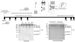
Groups of 6–10 Balb/c and ICR mice were infected with 500 trypomastigotes of H1 strain via intraperitoneal as before (Dumonteil et al., Reference Dumonteil, Escobedo-Ortegon, Reyes-Rodriguez, Arjona-Torres and Ramirez-Sierra2004; de la Cruz et al., Reference de la Cruz, Villanueva-Lizama, Dzul-Huchim, Ramírez-Sierra, Martinez-Vega, Rosado-Vallado, Ortega-Lopez, Flores-Pucheta, Gillespie, Zhan, Bottazzi, Hotez and Dumonteil2018). Parasitemia was measured during 30–50 days post-infection and animals were followed up to 220 days post-infection. A control group received only saline solution. In subsequent experiments, some groups of infected mice were treated with benznidazole and/or an immunotherapeutic vaccine (de la Cruz et al., Reference de la Cruz, Villanueva-Lizama, Dzul-Huchim, Ramírez-Sierra, Martinez-Vega, Rosado-Vallado, Ortega-Lopez, Flores-Pucheta, Gillespie, Zhan, Bottazzi, Hotez and Dumonteil2018). For benznidazole treatment, mice were treated orally with benznidazole at 25 mg kg day−1 during days 7–27 post-infection, and followed for 50 or 200 days post infection. Another group of animals received Benznidazole treatment as above combined with two doses of a therapeutic vaccine candidate at days 7 and 14 post-infection. The therapeutic vaccine consisted in rTSA-1-C4 and rTC24-C4 recombinant antigens formulated with a TLR4 agonist adjuvant (monophosphoryl Lipid A) for the evaluation at 50 days post-infection (de la Cruz et al., Reference de la Cruz, Villanueva-Lizama, Dzul-Huchim, Ramírez-Sierra, Martinez-Vega, Rosado-Vallado, Ortega-Lopez, Flores-Pucheta, Gillespie, Zhan, Bottazzi, Hotez and Dumonteil2018) and rTSA1-C4 formulated with a TLR4 agonist GLA (GLA-s.e., IDRI, Seattle, WA) for the evaluation at 200 days post-infection. Different formulations are assayed in the laboratory in order to select the best candidates to therapeutic vaccine. The protocol was approved by the Institutional Bioethics Committee of the Regional Research Center ‘Dr. Hideyo Noguchi’ of the Autonomous University of Yucatan (CIRB-2018-0008). Animals were sacrificed at days 50 post-infection for the acute phase and at days 195–220 post-infection for the chronic phase.
Plasma and cardiac tissue
Blood samples obtained from mouse-tail were collected in a EDTA containing tube, and the plasma were separated in small aliquots. Laemmli buffer and a cocktail of proteases inhibitors (iodoacetamine 10 mm, n-methylmaleimide 1 mm, benzamidine 1 mm, sodium orthovanadate 1 mm, PMSF 1 mm and Complete®) were added to the plasma and the samples were stored at −20°C. Cardiac tissue was removed to assess parasite burden by real-time qPCR (in a Illumina® EcoTM thermocycle) and tissue inflammation by histopathological examination as described previously (Quijano-Hernández et al., Reference Quijano-Hernández, Castro-Barcena, Vázquez-Chagoyán, Bolio-González, Ortega-López and Dumonteil2013; de la Cruz et al., Reference de la Cruz, Villanueva-Lizama, Dzul-Huchim, Ramírez-Sierra, Martinez-Vega, Rosado-Vallado, Ortega-Lopez, Flores-Pucheta, Gillespie, Zhan, Bottazzi, Hotez and Dumonteil2018).
Quantification of plasma proteins
The plasma proteins were quantified by the Bradford method (Bradford, Reference Bradford1976), using a standard curve with known concentrations of BSA (A2156, Sigma-Aldrich). The absorbance values at 620 nm were obtained in microplate reader (51119000, ThermoFisher Scientific).
Sodium dodecyl sulphate polyacrylamide gel electrophoresis (SDS-PAGE)
The plasma proteins were analysed by SDS-PAGE. A total of 20 μg of protein in Laemmli buffer and with the protease inhibitor cocktail were loaded on a 12% polyacrylamide gel and visualized by staining with PageBlue Protein Staining Solution (24620, ThermoFisher Scientific).
Immunoblotting
The plasma proteins were transferred to a PVDF membranes (PolyVinylidene DiFluoride, IPVH00010, Merck Millipore), and the membrane were stained with 0.2% Ponceau S red solution (1.14275.0010, Merck Millipore). The dye was removed by washing three times with PBS-tween 0.1% solution. The membrane was blocked with 5% milk solution at 4°C overnight, before incubation with a 1:800 dilution of anti-fibronectin antibody (F3648, Sigma Merck, a polyclonal antibody against fibronectin isolated from human plasma) for 1h30 at room temperature. After three washes, the membrane was incubated with a 1:5,000 dilution of an anti-rabbit secondary antibody (SA1200, Thermo Scientific;) for 1h30 at room temperature. The membranes were washed three times, and the immunoreactions were detected with 3,3′,5,5′-Tetramethylbenzidine substrate (T0565, SIGMA). A semi-quantitation of the intensity of the bands corresponding to fibronectin protein (FN) and fibronectin degradation fragments (dFN) was performed by densitometric analysis using AlphaeaseFCTM software. A constitutive and invariable band corresponding to serum albumin was used to normalize the values of the FN signal.
Sequence analysis
Human (accession #NP997647) and mouse (#XP017171337 and NP001263342) fibronectin I protein sequences were aligned and cysteine protease cleavage sites were predicted using PROSPERous (Song et al., Reference Song, Li, Leier, Marquez-Lago, Akutsu, Haffari, Chou, Webb and Pike2017) and PROSPER (Song et al., Reference Song, Tan, Perry, Akutsu, Webb, Whisstock and Pike2012) algorithms.
Cardiac parasite burden and inflammation
Data on cardiac parasite burden and inflammation have been published previously (de la Cruz et al., Reference de la Cruz, Villanueva-Lizama, Dzul-Huchim, Ramírez-Sierra, Martinez-Vega, Rosado-Vallado, Ortega-Lopez, Flores-Pucheta, Gillespie, Zhan, Bottazzi, Hotez and Dumonteil2018). Briefly, parasite burden was assayed by real-time qPCR with an Illumina® EcoTM thermocycler. Cardiac biopsies were embedded in paraffin, to perform tissue sections, which were stained with haematoxylin and eosin. Tissue micrographs were taken at 40 × magnification, and analysed using Multispec to determine the density of inflammatory cells.
Statistical analysis
Numerical data are presented as mean ± standard deviation (s.d.). Student's t test was used to compare the level of FN and dFN in infected vs non-infected mice, and an ANOVA test was used when comparing more than two groups, followed by Tukey post-hoc test for multiple comparisons among group, and tests were performed in GraphPad Prism software. The correlation between dFN levels and the parasitemia [maximum parasitemia and the area under the curve (AUC)], cardiac parasite burden and cardiac tissue inflammation (de la Cruz et al., Reference de la Cruz, Villanueva-Lizama, Dzul-Huchim, Ramírez-Sierra, Martinez-Vega, Rosado-Vallado, Ortega-Lopez, Flores-Pucheta, Gillespie, Zhan, Bottazzi, Hotez and Dumonteil2018) was evaluated, and linear regression was performed (GraphPad Prism).
Results
Changes in pFN levels in mice during the acute phase of T. cruzi infection
We first evaluated how pFN levels are affected during the acute phase of infection with T. cruzi in Balb/c mice. The plasma protein samples collected at 15, 30, 45 and 60 days post-infection were analysed by SDS-PAGE and western blot using a fibronectin-specific antibody (Fig. 1A–C). A clear high molecular weight band (about 260 kDa) was detected in the plasma of non-infected and infected mice, corresponding to intact fibronectin (Fig. 1C). Low molecular weights fibronectin fragments corresponding to its degradation were detected in the plasma of infected mice at 30 and 45 dpi (Fig. 1C). Densitometric analysis of FN levels as well as of its degradation fragments confirmed that there were statistically significant differences in fribronectin level between the plasma samples of non-infected and infected mice although no differences were observed in intact FN levels (Fig. 1D). Together, these results indicated that pFN is degraded during the acute phase of T. cruzi infection. Additionally, we noticed that the high level of parasitemia detected was at 30 dpi (Fig. 1I).
Fibronectin levels in plasma of Balb/c mice during acute T. cruzi infection. (A) Coomassie staining of a SDS-PAGE of plasma protein profile of non-infected (NI) and infected (INF) mice. (B) Transferred proteins to a PDVF membrane stained with Ponceau red dye. (C) Fibronectin western blot to evaluate fibronectin (262 kDa, FN arrow) and dFN levels in plasma of infected and non-infected mice at 15, 30, 45 and 60 days post-infection (dpi). Albumin band enclosed in dotted lines was used for normalization of fibronectin levels. (D) Densitometric analysis of fibronectin and fibronectin degradation fragment levels. The degradation fragments (dFN) of approximately 150, 100, 40 and 30 kDa are indicated with grey arrows. (E, F, G and H) Densitometric analysis of fibronectin degradation fragment levels. The dFN of approximately 150, 100, 40 and 30 kDa were observed more abundant at 30 and 45 dpi. (I) Parasitemia. ****, ***, ** and * indicate P < 0.0001, P < 0.001, P < 0.001 and P < 0.005, respectively. (Student's t test, n = 10).

The animals infected for 30 and 45 days showed several dFN with the main ones of approximately 150, 100, 40 and 30 kDa (Fig. 2A). All these fragments were abundant at 30 and 45 dpi. The fragment of approximately 150 kDa had similar levels during all the acute phase of the infection (Fig. 1E) whereas the fragment of approximately 100 kDa had the higher intensities at 30 and 45 dpi (Fig. 1F). The higher levels of the fragment of approximately 40 kDa was observed at 30 dpi (Fig. 1G). Finally, the fragment of approximately 30 kDa was increased during all the acute phase, and the higher levels were observed at 30 and 45 dpi (Fig. 1H).
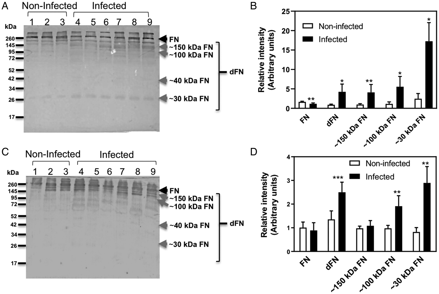
Fibronectin levels in plasma of mice during chronic T. cruzi infection. (A and C) Western blot of fibronectin (262 kDa, FN arrow) and its degradation fragments (dFN) detected in the plasma of Balb/c and ICR mice, respectively, after 200 days of infection. (B and D) Densitometric analysis of fibronectin (FN) and its degradation fragments (dFN) detected in the plasma of Balb/c and ICR mice, respectively, after 200 days of infection. Lanes from 1 to 3 and 4 to 9 correspond to individual mice. ***, ** and * indicate P < 0.001 P < 0.01 and P < 0.05, respectively (Student's t test, n = 6).

Changes in pFN levels in mice during the chronic phase of T. cruzi infection
Next, we analysed how pFN levels are affected by T. cruzi infection during the chronic phase. We used two mouse models, Balb/c mice as above, as well as ICR mice, and measured fibronectin levels in plasma samples collected 195–220 days after infection. Intact fibronectin was detected by western blot analysis in non-infected mice of both strains. In chronically infected Balb/c mice, pFN levels were significantly decreased compared to non-infected mice (Fig. 2A and B), and no difference was observed in chronically infected ICR mice (Fig. 2C and D). In addition, dFN were clearly and significantly increased in chronically infected mice of both strains (Fig. 2B and D). In Balb/c mice, a fibronectin degradation fragment of approximately 30 kDa was increased in the plasma of infected mice compared to uninfected ones (Fig. 2A and B). The fragments of approximately 100 and 150 kDa were significantly increased in infected mice (Fig. 2B).
The 30 kDa FN degradation fragment is comparable in size to that identified in human Chagasic patients (Santamaria et al., Reference Santamaria, Chatelain, Jackson, Miao, Ward, Chappuis and Ndao2014; Ruiz-Lancheros et al., Reference Ruiz-Lancheros, Rasoolizadeh, Chatelain, Garcia-Bournissen, Moroni, Moscatelli, Altcheh and Ndao2018). Comparative analysis of human and mouse fibronectin sequences revealed some sequences differences in the amino-terminus side of the protein, but multiple predicted cysteine protease cleavage sites could be identified in the sequences from both species, as well as some unique to human or mouse sequences, that could generate a proteolytic fragment of 28–31 kDa from this end of the FN (Fig. 3). Thus, digestion of human and murine fibronectin by the parasite cruzipain may yield a similar proteolytic profile.
Predicted cleavage sites of fibronectin by cysteine proteases. Partial sequence alignment of a human and two mouse fibronectin sequences. Arrows indicate predicted cleavage sites by cysteine proteases in the amino-terminus side of the protein that may generate a 28–31 kDa proteolysis fragment. Grey arrows indicate cleavage sites common to human and mouse sequences, red arrows cleavage sites specific for the human fibronectin and blue arrows cleavage sites for the mouse fibronectin only. The black arrow indicates the proposed cleavage site in Chagasic patients and the boxed aminoacid indicate a peptide identified from the analysis of the human 30 kDa fragment by mass spectrometry (From Santamaria et al., Reference Santamaria, Chatelain, Jackson, Miao, Ward, Chappuis and Ndao2014).

pFN degradation is reversed in infected mice treated with benznidazole and a combination therapy
We next assessed how fibronectin degradation during T. cruzi infection may be affected by therapeutic treatment. Infected mice were treated during the acute phase with benznidazole alone or the combination of benznidazole and therapeutic vaccine candidates, which had previously be shown to reduce cardiac inflammation and parasite burden (de la Cruz et al., Reference de la Cruz, Villanueva-Lizama, Dzul-Huchim, Ramírez-Sierra, Martinez-Vega, Rosado-Vallado, Ortega-Lopez, Flores-Pucheta, Gillespie, Zhan, Bottazzi, Hotez and Dumonteil2018). pFN levels were measured at the end of the acute phase (50 dpi) and during the chronic phase (195–200 dpi). As described above, the levels of dFN were strongly increased in infected mice compared to healthy mice (Fig. 4). Importantly, benznidazole treatment was associated with lower levels of degraded fibronectin compared to untreated infected mice, reaching levels identical to that of healthy mice. Similarly, the treatment of mice with a combination of benznidazole and immunotherapeutic vaccine (rTSA-1-C4 + rTC24-C4 + MPLA) also decreased the levels of degraded fibronectin to levels identical to that observed in healthy mice (Fig. 4A). Fibronectin degradation was also decreased following treatment with the MPLA adjuvant alone. Evaluation of fibronectin degradation levels during the chronic phase again indicated a high level of degradation in untreated infected mice (Fig. 4B). Prior treatment with benznidazole significantly decreased the levels of dFN, although this remained higher than that of healthy mice. Prior treatment with a combination of benznidazole and therapeutic vaccine (rTSA-1 + GLA) had no significant effect on fibrobnectin degradation during the chronic phase (Fig. 4B). Together, these results indicate that fibronectin degradation can be reversed by therapeutic treatment in mice during the acute phase and this reversion may be maintained during the chronic phase.
Fibronectin levels in plasma of T. cruzi infected mice and response to treatment. (A) Densitometric analysis of degradation fragments (dFN) detected in the plasma of Balb/c after 50 days of infection. Infected mice were treated with benznidazole, the concomitance benznidazole plus the vaccine candidate rTSA-1-C4 + rTC24-C4 + MPLA and the alone adjuvant MPLA. (B) Infected ICR mice during 194 days were treated with benznidazole as well as the concomitance benznidazole plus a vaccine candidate (rTSA-1 + GLA + BNZ). *** and ** indicate P < 0.01 and P < 0.05 with non-infected group, respectively and # indicate P < 0.05 of treated groups with infected group (Tukey post-hoc test).

Correlation between fibronectin levels and parasitaemia, parasite burden and cardiac tissue inflammation
We then assessed how pFN degradation correlate with parasitological and histological variables in each individual mouse. We observed that the levels of fibronectin degradation fragment positively correlated with parasitemia (Fig. 5A and B). More importantly, the levels of fibronectin degradation fragment positively correlated with cardiac parasite burden as well as with cardiac tissue inflammatory cell density. These data suggest that fibronectin degradation can represent an attractive biomarker for cardiac tissue damage.
Correlation between fibronectin degradation and parasitological variables and cardiac tissue damage. The relationship between fibronectin degradation level and maximum parasitemia (A), overall parasitemia (AUC) (B), cardiac inflammatory cell density (C) and cardiac parasite burden (D) are shown.

Discussion
Monitoring the progression of CD, assessing treatment efficacy and potential cure are still elusive due to the lack of adequate biomarkers. Recently, Ruiz-Lancheros and co-workers proposed a APOA1 and fibronectin fragments as potential biomarkers for parasitological cure in children treated with benznidazole (Ruiz-Lancheros et al., Reference Ruiz-Lancheros, Rasoolizadeh, Chatelain, Garcia-Bournissen, Moroni, Moscatelli, Altcheh and Ndao2018). Fibronectin is a target for cruzipain, a Trypanosoma cruzi cysteine proteinase and fibronectin degradation has been suggested as an important process during host cell invasion (Maeda et al., Reference Maeda, Cortez, Izidoro, Juliano and Yoshida2014). Fibronectin participates in several cell processes such as adhesion, migration, growth and survival (Lemańska-Perek and Adamik, Reference Lemańska-Perek and Adamik2019). pFN also has a role in maintaining the balance between haemostasis and thrombosis (Wang and Ni., Reference Wang and Ni2016). We assessed here in greater detail how pFN levels may be affected by experimental infection in mice, and how this may correlate with disease progression and response to therapeutic treatment.
The semi-quantitation of pFN by western blot clearly indicated that it is initially degraded during the acute phase of infection with T. cruzi, and that dFN accumulated in the plasma during that time. Interestingly, the degradation profile detected appeared similar to that observed by Maeda et al, when FN was incubated in vitro with recombinant cruzipain (Maeda et al., Reference Maeda, Cortez, Izidoro, Juliano and Yoshida2014). However, other parasite proteases may also be involved in fibronectin degradation. Toward the end of the acute phase, the levels of dFN decreased, which may indicate some protein homeostatic response to compensate for fibronectin degradation (Clausen et al., Reference Clausen, Abildgaard, Gersing, Stein, Lindorff-Larsen, Hartmann-Petersen and Donev2019).
During the chronic phase (195–220 dpi) fibronectin degradation fragment levels continued to be elevated, and fragments of smaller molecular weight became detectable, including a 29–30 kDa fragment in Balb/c mice. This fragment is similar to that detected when purified FN was incubated with metacyclic trypomastigotes (Maeda et al., Reference Maeda, Cortez, Izidoro, Juliano and Yoshida2014), as well as to a 29 kDa FN fragment previously identified by proteomic approach in patients with CD (Santamaria et al., Reference Santamaria, Chatelain, Jackson, Miao, Ward, Chappuis and Ndao2014) and children with congenital T. cruzi infection (Ruiz-Lancheros et al., Reference Ruiz-Lancheros, Rasoolizadeh, Chatelain, Garcia-Bournissen, Moroni, Moscatelli, Altcheh and Ndao2018). Further analysis of human and murine fibronectin sequences indicated that several possible cleavage sites were predicted to generate a comparable 28–31 kDa fragment for both species. Together, these observations suggest that a similar proteolytic cleavage of fibronectin may be occurring in T. cruzi infected mice as in human Chagasic patients.
Following benznidazole drug treatment, we observed a reversal of fibronectin degradation in treated mice, with the level of dFN shortly after treatment similar to that of non-infected healthy mice. During the chronic phase, fibronectin degradation remained lower in benznidazole-treated mice, although it did not reach the level of healthy mice. These observations are in agreement with initial studies of nifurtimox treatment in Chagasic patients, which resulted in the 29 kDa fibronectin degradation fragment to return to levels similar as the healthy volunteers (Santamaria et al., Reference Santamaria, Chatelain, Jackson, Miao, Ward, Chappuis and Ndao2014), and benznidazole treatment of children with congenital infection, which lead to the disappearance of this degradation fragment as seronegativization occurred (Ruiz-Lancheros et al., Reference Ruiz-Lancheros, Rasoolizadeh, Chatelain, Garcia-Bournissen, Moroni, Moscatelli, Altcheh and Ndao2018).
Combination therapies are a promising perspective to reduce drug doses and the concomitant adverse reactions (Urbina, Reference Urbina2015; Jones et al., Reference Jones, Versteeg, Damania, Keegan, Kendricks, Pollet, Cruz-Chan, Gusovsky, Hotez and Bottazzi2018), and therapeutic vaccination may complement drug treatment. Our preliminary testing of different vaccine formulations in combination with a low-dose benznidazole treatment suggest that such combination therapy can also reduce fibronectin degradation, as observed at 50 dpi. The limited long-term efficacy of the combined therapy may be due to the low efficacy of the therapeutic vaccine based on a single parasite antigen, and strengthen the need for a multi-antigen vaccine able to stimulate a broader immune response.
While fibronectin fragments had previously been proposed as biomarkers of cure following drug treatment, there was no data on the correlation of this biomarkers with specific variable of the infection and cardiac tissue damage. Our model of experimental infection allowed to evaluate parasitemia as well as cardiac parasitic burden and histological damage (Jelicks and Tanowitz, Reference Jelicks and Tanowitz2011). Importantly, we observed significant positive correlations between the levels of dFN and each one of these variables. This observation strongly supports fibronectin degradation as an important biomarker candidate for T. cruzi infection and CD progression, as it may represent a valuable proxy for cardiac parasite burden and tissue damage.
These results warrant the development of easier and more quantitative tests to measure fibronectin degradation, including as ELISA or immunochromatographic rapid tests for the point of care monitoring of disease progression and response to treatment in patients. Fibronectin is present in blood at a relatively high concentration of about 0.6 mg mL−1 in mice and 0.3 mg mL−1 in humans (Mosesson and Umfleet, Reference Mosesson and Umfleet1970; Zardi et al., Reference Zardi, Cecconi, Barbieri, Carnemolla, Picca and Santi1979) supporting the feasibility of such tests. On the other hand, fibronectin degradation may not be specific to T. cruzi infection as it is a target for other pathogens (Speziale et al., Reference Speziale, Arciola and Pietrocola2019), and this should be further investigated.
In conclusion, we have shown here that pFN is degraded during T. cruzi infection in mouse models, both during the acute and chronic phase, and that the plasma levels of degradation fragments positively correlate with cardiac parasite burden and inflammatory cell density. Furthermore, fibronectin degradation is reversed by therapeutic treatment and by combination therapy of benznidazole associated with therapeutic vaccination. Fibronectin degradation may thus be considered an excellent biomarker for CD progression and cardiac tissue damage. It may also provide reliable and timely information on treatment response and efficacy. These results warrant the further development of quantitative and easy-to-perform point-of-care tests for the monitoring of fibronectin degradation in Chagasic patients.
Acknowledgements
We are thankful to the students: Juan Jose de la Cruz, Victor Dzul-Huchim, Christian Teh-Poot and Carolina Arias, which participated in the treatment of mice with the immunotherapeutic vaccine.
Author contribution
NHC, ED and MERV conceived and designed the study. NAHC, AMC, SPG and PM conducted data gathering. AMC, NAHC and ED performed statistical analyses. NAHC and ED wrote the article.
Financial support
This work was supported by Carlos Slim Foundation (grant #87714) and Consejo Nacional de Ciencia y Tecnologia of Mexico (CONACYT, grant #PDCPN2015-102). NAHC was supported by a postdoctoral fellowship from CONACYT (2016-3). AMC was supported by a scholarship from CONACYT (2019-000037-02NACF).
Conflicts of interest
The authors declare there are no conflicts of interest.
Ethical standards
The protocol was approved by the Institutional Bioethics Committee of the Regional Research Center ‘Dr. Hideyo Noguchi’ of the Autonomous University of Yucatan (CIRB-2018-0008).