Introduction
Uteroplacental insufficiency (UPI) affects nearly 10% of human pregnancies and is the most common cause of fetal growth restriction (FGR). Reference Henriksen and Clausen1 It is characterized by decreased placental function and compromised nutrient and oxygen delivery to the fetus. Reference Wu, Bazer, Wallace and Spencer2 The situations leading to FGR are the illnesses intrinsic to the fetal-placental-maternal unit, fetal undernutrition, and intrauterine space constraints restricting the fetal growth. The most common cause of FGR are preeclampsia, diabetes, anemia, chronic lung or kidney diseases, poor nutrition, alcohol or drug use, cigarette smoking, multiple gestation, infections, and genetic disorders. Reference Blue, Page and Silver3 Recent evidence proposes that being born with growth restriction is associated with reduced cardiac function and increased cardiovascular risk in infants, children, and adults. Reference Cohen, Wong, Horne and Yiallourou4–Reference Crispi, Rodríguez-López and Bernardino6 FGR neonates exhibited altered cardiac development and cardiac morphology and subclinical myocardial dysfunction. Reference Zaharie, Hasmasanu, Blaga, Matyas, Muresan and Bolboaca7 However, the mechanisms underlying these alterations remain unknown in neonates. FGR increases the risk of pulmonary hypertension in premature infants. Reference Check, Gotteiner and Liu8,Reference Vyas-Read, Kanaan and Shankar9 Preclinical studies demonstrated that Sprague–Dawley rats born to undernourished mothers did not exhibit pulmonary hypertension at 6, 12, and 14 weeks. Reference Lv, Tang and Wei10–Reference Fu, Lv, Zhong, He, Liu and Du12 The short-term effects of FGR on pulmonary hypertension were largely unknown in neonates. FGR is caused by different factors such as decreased placental perfusion or maternal nutritional deficiencies in humans. In this study, we used the UPI model of FGR in rats, as it resembles human illness and is thus, widely used. Reference Janot, Cortes-Dubly, Rodriguez and Huynh-Do13 We hypothesized that UPI would influence cardiac development in the offspring rats. Through this study, we aimed to evaluate the effects of UPI during pregnancy on heart histology and pulmonary hypertension in growth-restricted newborn rats.
Methods
Ethical approval
All procedures were approved by the Institutional Animal Care and Use Committee of Taipei Medical University and performed according to a protocol approved by the Association for Assessment and Accreditation of Laboratory Animal Care.
Animal model
Time-dated pregnant Sprague–Dawley rats were obtained from BioLASCO Taiwan Co., Ltd. (Yilan, Taiwan) and housed in clean specific pathogen-free rooms under a 12-h light-dark cycle with ad libitum access to laboratory food and water. On day 18 of pregnancy, bilateral uterine artery ligation was performed to induce intrauterine growth retardation (FGR group), whereas the control group were subjected to a sham protocol. Reference Huang, Chou, Lin and Chen14 All rats were delivered naturally at term (22 days). Litters were separated from their mothers within 12 h of birth, pooled, and randomly redistributed to the newly delivered mothers. Rats were randomly selected for examination from each group, irrespective of sex, on postnatal days 0 and 7.
Tissue collection
Animals were deeply anesthetized with isoflurane and euthanized by cardiac puncture and exsanguination. The right middle lobe of the lung and the heart were harvested and fixed in 4% paraformaldehyde for 48 h. After being washed with phosphate-buffered saline (PBS), the heart was dissected into atria and ventricles followed by serial dehydration in increasing concentrations of ethanol and xylene infiltration before being embedded in paraffin. Serial 5-µm-thick paraffin sections of the lungs and ventricles were cut serially using a microtome. The ventricle was sectioned from base to apex. Sample sections were subjected to histological evaluation and immunohistochemical processing.
Histological assessment and morphological analysis
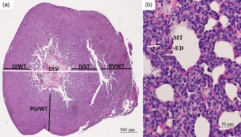
After routine deparaffinization, the sections of the lung and the ventricle were stained with hematoxylin and eosin, periodic acid-Schiff (PAS), and Masson’s trichrome, and examined by light microscopy to assess the lung and heart morphology. The heart section at 40× magnification and five randomly selected fields from each lung section at 400× magnification were captured using a digital camera and imported into a computerized image analysis system (Image-Pro Plus 6.0 for Windows, Media Cybernetics, Silver Spring, MD, USA) for morphological quantification. Right ventricular (RV) hypertrophy was assessed by measuring the ratio of the RV wall thickness (RVWT) to the left ventricular wall thickness (LVWT) and interventricular septum thickness (IVST) as follows: RV hypertrophy = RVWT/LVWT + IVST. Reference de Visser, Walther, Laghmani, Boersma, Van der Laarse and Wagenaar15 The measurement of the LV relative wall thickness (RWT) was modified from the study by Hashem et al. Reference Hashem, Kalashyan and Choy16 Calculations were performed by dividing the sum of the IVST and posterior LV wall thickness (PLVWT) by the diameter of the left ventricle (DLV). Each wall thickness was measured from the middle of the longitudinal length of its wall, and the transverse length (wall thickness) was in the range between the free edges on both sides (Fig. 1a). The medial wall thickness (MWT) of the pulmonary arteries was measured in vessels with an external diameter of 20–65 μm, and those having external diameters measured at two perpendicular planes within 33% of each other were included in the counts. In total, 20 vessels were measured in each animal. The percent medial thickness of an individual vessel was calculated using the following formula: medial thickness (MT) × 2 × 100/external diameter (Fig. 1b). Reference Masood, Yi and Lau17 The thickness of ventricular walls and interventricular septum of the heart, and the external diameter and wall thickness of the lung were measured using Image-Pro Plus (Media Cybernetics, Silver Spring, MD, USA).
Representative histological images of hematoxylin and eosin-stained heart and lung tissue sections. (a) Whole-heart cross-sectional dissection was performed to obtain transverse sections of the ventricles for morphometric analysis. (b) Photograph of lung section for morphometric analysis of medial wall thickness. RVWT: right ventricle wall thickness, LVWT: left ventricle wall thickness, IVST: interventricular septum thickness, PLVWT: posterior left ventricle wall thickness, DLV: diameter of the left ventricle, MT: medial thickness of vessel wall, ED: external diameter.

Immunohistochemistry
Immunostaining was performed on the ventricle sections using the streptavidin-peroxidase technique. After routine deparaffinization, heat-induced epitope retrieval was performed by immersing the slides in 0.01 M sodium citrate buffer (pH 6.0). For block endogenous peroxidase activity and nonspecific antibody binding, the sections were first pre-incubated for 1 h at room temperature in 0.1 M PBS containing 10% normal goat serum and 0.3% H2O2 before being incubated for 20 h at 4°C with mouse IgM monoclonal anti-human Leu-7 (HNK-1) antibody (1:80 diluted; Becton-Dickinson Immunocytometry Systems, Mountain View, CA, USA) as the primary antibody. The sections were then treated for 1 h at 37°C with biotinylated goat anti-mouse IgG (1:200, Jackson ImmunoResearch Laboratories Inc., West Grove, PA, USA). Sections were treated with reagents from an avidin-biotin complex kit (Vector Laboratories, Inc., Burlingame, CA, USA), and brown reaction products were visualized using a diaminobenzidine substrate kit (Vector Laboratories) according to the manufacturer’s recommendations. All immunostained sections were viewed and photographed using a Nikon Eclipse E600 Microscope (Nikon, Tokyo, Japan). Five randomly selected fields from each section at 400× magnification were captured using a digital camera and imported into a computerized image analysis system (Image-Pro Plus 6.0 for Windows, Media Cybernetics).
Statistical analysis
All data are presented as mean ± standard deviation with SPSS 23.0 (SPSS Inc., Chicago, IL, USA) used for statistical analyses. Differences between control and FGR groups on each postnatal day were analyzed by Student’s t-test. The differences were considered statistically significant at p < 0.05.
Results
Uteroplacental insufficiency reduced body weight in growth-restricted newborn rats
Four sham-operated dams delivered 39 control rats and four UPI-induced dams delivered 26 FGR rats. On postnatal days 0 and 7, we retrieved 12 and 8 pups from the sham-operated and UPI-induced dams, respectively. The remaining dams and pups after postnatal day 7 were not used. Compared with control rats, rats with FGR exhibited significantly lower body weights on postnatal days 0 and 7 and significantly a higher heart to body weight ratio on postnatal day 0 (Table 1). The heart weights were comparable between the control and FGR groups on postnatal days 0 and 7.
Body and heart weights and heart:body weight ratios in control and FGR rats on postnatal days 0 and 7

Values are presented as means ± SD.
* p < 0.05 vs. control group at each postnatal age.
FGR: fetal growth restriction.
Effects of uteroplacental insufficiency on ventricular histology
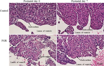
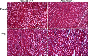
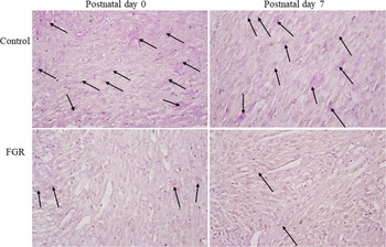
Significant hypertrophy and degeneration of ventricular cardiomyocytes were observed near the subendothelial layer in rat hearts on postnatal days 0 and 7 (Fig. 2). Cells were swollen, and organelles and nuclei were integrated to form enlarged foam cells that clumped together to form clusters within the myocardium beneath the subendothelial layer. Masson’s trichrome staining revealed that the collagen fibers in the FGR groups were deposited between cardiomyocytes (Fig. 3). However, in the control group, the results of Masson’s trichrome staining were not as clear as in the FGR group, possibly owing to an insufficient number of collagen fibers in the extracellular space. In this scenario, cardiomyocytes in the FGR group had a larger extracellular space than did those in the control group. The concentration of glycogen in cardiomyocytes was detected by PAS method. PAS-positive staining was observed in many cardiomyocytes in the ventricular myocardium of control rats as pink purple in the cytoplasm of cardiomyocytes (Fig. 4), whereas fewer PAS-positively stained cardiomyocytes were observed in the ventricular myocardium in the FGR group.
Representative ventricular sections stained with hematoxylin and eosin. Significant hypertrophy and degeneration of ventricular cardiomyocytes were observed near the subendothelial layer on postnatal days 0 and 7 (white arrow).

Representative ventricular sections stained by Masson’s trichrome. The collagen fibers were deposited between the cardiomyocytes. The FGR group exhibited more collagen deposition and larger extracellular space (black arrow) than did the control group on postnatal days 0 and 7.

Representative ventricular sections stained with periodic acid–Schiff stain. The glycogen was stained pink purple in the cytoplasm of cardiomyocytes (black arrow). The FGR group exhibited less glycogen than did the control group on postnatal days 0 and 7.

Uteroplacental insufficiency did not change ventricular morphometry in growth-restricted newborn rats
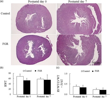
The LV RWT was assessed using the following equation: RWT = IVST + PLVWT/DLV. RV hypertrophy was assessed using the following equation: RVWT/LVWT + IVST. The indicators of LV hypertrophy (RWT) and RV hypertrophy (RVWT/LVWT + IVST) were compatible between control and FGR rats on postnatal days 0 and 7 (Fig. 5).
(a) Representative images of whole-heart transverse cross section stained by hematoxylin and eosin, (b) indicators of left ventricular hypertrophy (RWT), and (c) indicators of right ventricular hypertrophy (RVWT/LVWT + IVST) in the control and FGR rats. The RWT and RVWT/LVWT + IVST were comparable between control and FGR rats on postnatal days 0 and 7. Data are expressed as mean ± SD.

Effects of uteroplacental insufficiency on MWT
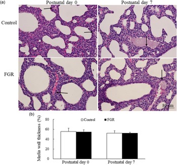
The MWT of lung vessels provides an index for evaluating pulmonary hypertension. We estimated arterial remodeling by measuring the MWT of the small pulmonary arteries (external diameter of 20–65 μm). No significant difference was observed in the percentage of pulmonary vessel lining thickening between control and FGR rats on postnatal days 0 and 7 (Fig. 6).
(a) Representative lung sections stained with hematoxylin and eosin and (b) medial wall thickness (MWT) in control and FGR rats. The MWT was comparable between control and FGR rats on postnatal days 0 and 7. Black arrow indicates the tunica media of the artery.

Uteroplacental insufficiency reduced HNK-1 expression in growth-restricted newborn rats
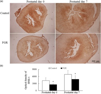
HNK-1 was immunoassayed to delineate the conducting tissues. HNK-1 immunoreactivity was detected in the subendocardium, the region between the myocardium and the endocardium, of the left and right ventricles, and more HNK-1 immunoreactivity was detected on postnatal day 7 (Fig. 7a). Less HNK-1 immunoreactivity was detected in the FGR group than in the control group on postnatal days 0 and 7. FGR rats had significantly lower optical density of HNK-1 compared with control rats on postnatal days 0 and 7 (Fig. 7b).
(a) Representative photomicrographs and (b) optical density of HNK-1. Positive immunoreactivity is indicated by brown staining. HNK-1 immunoreactivity was detected in the subendocardium, and higher HNK-1 immunoreactivity was detected on postnatal day 7. FGR rats displayed significantly lower optical density of HNK-1 compared with control rats on postnatal days 0 and 7. Data are expressed as mean ± SD. *p < 0.01 vs. control group on each postnatal day.

Discussion
Our in vivo model demonstrated that UPI through bilateral uterine ligation reduced body weight and altered ventricular histology, as evidenced by hypertrophy and degeneration of cardiomyocytes, increased intercellular spaces between the cardiomyocytes and collagen deposition, and decreased glycogen deposition and HNK-1 expression on postnatal days 0 and 7. These findings indicate that UPI during pregnancy induced FGR and ventricle histological alteration and did not induce pulmonary hypertension in the immediate neonatal period. These results suggest that neonates with FGR may have inadequate myocardial reserves at birth, which may influence subsequent cardiovascular compromise in future life. Reference Cohen, Wong, Horne and Yiallourou4–Reference Crispi, Rodríguez-López and Bernardino6
Takahashi et al. reported that four extremely-low-birthweight infants with severe FGR died 5–10 days after birth owing to heart failure, and autopsy findings revealed hypoplasia of myocardial fibers and decreased glycogen levels. Reference Takahashi, Nishida, Arai and Kaneda18 The researchers suggested that the hearts of these infants failed to adapt to postnatal hemodynamic changes because of inadequate myocardial function and inadequate glycogen reserves. Our results that FGR rats exhibited reduced glycogen in ventricles at birth support these clinical findings and suggest that infants who are small for gestational age need to be closely monitored for hemodynamic status. Tsirka et al. used a similar animal model of FGR in Sprague–Dawley rats and found no significant difference in the glycogen content between the hearts of control and FGR rats. Reference Tsirka, Gruetzmacher, Kelley, Ritov, Devaskar and Lane19 This discrepancy may be attributable to the fact that they performed bilateral uterine ligation at a gestational age of 19 days and measured glycogen content on postnatal days 21 and 120.
The effects of FGR on the incidence of pulmonary hypertension differ in human and animal studies. Reference Check, Gotteiner and Liu8–Reference Fu, Lv, Zhong, He, Liu and Du12 In the current study, we evaluated pulmonary hypertension by measuring LV and RV hypertrophy and MWT in the lungs and found that rats with FGR did not exhibit increased pulmonary hypertension. These results suggest that pulmonary hypertension was mostly due to postnatal events that influence cardiovascular adaptation during the transitional period in growth-restricted infants. Reference Abbas, Shah and Hanif20
Term infants who are small for gestational age have reduced heart rate variability and heart rate reflex responses at 1 month of age compared with term infants who are an appropriate size for gestational age. Reference Galland, Taylor, Bolton and Sayers21 FGR has been reported to be associated with prolonged time intervals between early ventricular inflow and atrial contraction. Reference Kurihara, Tachibana and Yokoi22 HNK-1 is an established marker of the cardiac conduction system development and is useful for the analysis of abnormal conduction system. Reference Kise, Nakagawa and Okamoto23 Therefore, we immunoassayed HNK-1 to evaluate the effect of FGR on cardiac conduction in rats. We found that FGR rats exhibited significantly less HNK-1 expression compared with control rats on postnatal days 0 and 7. These findings might explain the reduced cardiac reflexes in growth-restricted infants.
Our study has several limitations. First, we did not measure hemodynamic parameters such as RV systolic pressure, cardiac index, total pulmonary resistance index, and systolic arterial pressure; however, a related study has revealed that hemodynamic parameters are comparable to histologic morphometry. Reference Hessel, Steendijk, den Adel, Schutte and van der Laarse24 Second, we used the expression of these proteins in whole tissue sections for immunohistochemistry studies. Many studies have confirmed the correlation between immunohistochemistry and the tissue concentration of the antigen. Reference Matos, Trufelli, de Matos and da Silva Pinhal25 Third, we did not determine the sex of the offspring though the sex effects of FGR on cardiac development during immediate postnatal period were unknown. In maternal undernutrition, male rat offspring exhibited larger heart weight/body weight ratio on postnatal day 21 and male fetal baboon showed left ventricular fibrosis inversely correlated with birth weight. Reference Rodríguez-Rodríguez, López de Pablo and García-Prieto26,Reference Muralimanoharan, Li and Nakayasu27 Further studies are needed to determine the sex-specific differences in cardiac development of growth-restricted offspring. Despite these limitations, the results of the present study provide valuable data that demonstrate the mechanisms and potential harmful effects of UPI in growth-restricted neonates.
In conclusion, this study demonstrated that UPI, through bilateral uterine ligation, induced FGR and led to changes in ventricular histology but did not induce pulmonary hypertension in the immediate neonatal period. These results might partly explain the underlying mechanism of reduced cardiac function in infants with FGR and subsequent cardiovascular compromise in future life. Further studies are required to evaluate hemodynamic changes in these growth-restricted neonates.
Acknowledgments
None.
Financial support
This study was supported by a grant from Taipei Medical University Hospital (111TMUH-MOST-15), Taipei, Taiwan.
Conflicts of interest
None.
Ethical standards
The authors assert that all procedures contributing to this work comply with the ethical standards of the Association for Assessment and Accreditation of Laboratory Animal Care and have been approved by the Institutional Animal Care and Use Committee of Taipei Medical University.