Introduction
Cancer can metastasise to the cervical lymph nodes, and this usually indicates a poor prognosis. For primary head and neck cancers, the presence of cervical lymph node metastasis reduces the overall survival rate by 50 per cent when compared with the prognosis of cancer patients without metastatic nodes.Reference Johnson1 For cancers primarily located below the clavicle, metastatic cervical lymph nodes usually suggest incurable disease.Reference Tran, Grigsby, Dehdashti, Herzog and Siegel2, Reference Di Paolo, Tulusan, Breuel, Wilczek-Engelmann, Adam and Reitzenstein3 Therefore, detection of these nodes before treatment or during post-treatment follow up is very important in every cancer patient.
In recent years, 18F-fluorodeoxyglucose positron emission tomography (18F-FDG PET), a functional imaging modality, has been widely used for initial evaluation or post-treatment monitoring in many cancers. Imaging is based on identifying increased glycolytic activity in cancer cells.Reference Ng, Ko, Toh and Chen4–Reference Ng, Yen, Liao, Chang, Chan and Ko818F-Fluorodeoxyglucose positron emission tomography can detect small cervical lymph node metastases before they are palpable, because PET scanning can identify malignant lesions as small as 5 mm in diameter.Reference Ng, Yen, Liao, Chang, Chan and Ko8 For primary head and neck cancers, 18F-FDG PET has been shown to be useful in detection of cervical metastases, with a sensitivity ranging from 67 to 96 per cent and a specificity ranging from 82 to 100 per cent.Reference Ng, Ko, Toh and Chen4, Reference Ng, Yen, Liao, Chang, Chan and Ko8 However, false positive results are not uncommon, because some benign lymphadenopathies also have a high rate of glucose metabolism, such as granulomatous inflammation.Reference Ng, Joseph, Chan, Ko, Wang and Liao9–Reference Ho11 Therefore, pathological examination is still necessary if feasible, in order to determine the nature of these suspicious lymph nodes before treatment.Reference Cerfolio, Bryant, Ohja, Bartolucci and Eloubeidi12, Reference Eloubeidi, Cerfolio, Chen, Desmond, Syed and Ojha13
The role of ultrasonography in evaluation of cervical lymph nodes, using grey scale and power Doppler features, has been well established.Reference Ng, Ko, Toh and Chen4 Furthermore, ultrasound-guided fine needle aspiration allows cytological examination of suspicious nodes as small as 2 mm in diameter, with a reported sensitivity of 90 per cent and specificity of 100 per cent.Reference Ng, Ko, Toh and Chen4, Reference van den Brekel, Castelijns, Stel, Luth, Valk and van der Waal14 Therefore, ultrasound examination with ultrasound-guided fine needle aspiration may be a minimally invasive procedure which can enable cytological examination of lymph nodes with increased 18F-FDG uptake on PET scanning.
In this paper, we report our experience of using ultrasound-guided fine needle aspiration for examination of clinically impalpable cervical lymph nodes with positive 18F-FDG PET scanning in 10 cancer patients.
Patients and methods
Subjects
Between July 2004 and June 2006, 10 cancer patients with suspicious cervical lymph nodes revealed by 18F-fluorodeoxyglucose positron emission tomography (18F-FDG PET) scanning (performed to establish disease status) underwent ultrasound examination. Ultrasound-guided fine needle aspiration was performed for the suspicious cervical lymph nodes, irrespective of the standardised uptake value on the initial, official interpretation of the PET and computed tomography (CT) images. Clinical data for these patients were also obtained from the hospital records and retrospectively reviewed, in order to analyse demographic characteristics, results of imaging and cytological examinations, treatment plan and clinical outcomes.
Whole-body 18F-fluorodeoxyglucose positron emission tomography examination
18F-Fluorodeoxyglucose positron emission tomography examination was performed for each patient using an Advance PET scanner (General Electric Medical Systems, Milwaukee, WI). The examination protocol was as previously described.Reference Yen, Hong, Tzen, Pan and Chen15 The PET image, the noncontrast CT image and the fused PET–CT image were reviewed retrospectively by a nuclear medicine physician (RFY) and a radiologist (JKH). Both physicians were unaware of the results of the other imaging examination as well as of the final pathological results.
Ultrasonography and ultrasound-guided fine needle aspiration
The equipment and the examination setting of the ultrasound were as previously described.Reference Wu, Lee, Huang, Ko, Sheen and Hsieh16 Focussing depth was set between 1 and 3 cm, where the target lymph nodes were usually present. The lymph nodes revealed by the PET–CT scan were located under ultrasonography and evaluated for size, shape (long to short axis ratio), internal content, echogenicity, margin and intranodal vascular pattern, using the power Doppler mode. Fine needle aspiration was performed using a 22-gauge needle and 20-ml syringe under ultrasound guidance, which was used to confirm the placement of the needle into the suspicious lymph node. Each suspicious lymph node was aspirated once. Specimens were smeared on glass slides, air-dried and stained using the Papanicolaou method. Cytological diagnosis of the aspirated lesion was classified into four categories: (1) benign or reactive, (2) positive for malignancy, (3) atypical or suspicious for malignancy, or (4) nondiagnostic. The ultrasound features for malignant lymphadenopathy were documented in all nodes before cytological evaluation.
Results
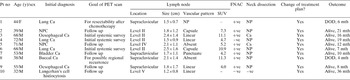
The clinical data for the 10 patients are shown in Table I. Patients comprised nine men and one woman, and their ages ranged from 32 to 72 years. Two patients (cases two and five (Figure 1)) had nasopharyngeal carcinoma, which was treated with concurrent chemoradiotherapy, achieving complete remission for two and four years before the PET scans were performed. Both these patients underwent PET scanning for monitoring of their disease status. Three patients with lung cancer underwent PET scanning to evaluate the resectability of disease after completion of induction chemotherapy (case one), or to evaluate the disease status (regarding distant metastasis) at the time of diagnosis (cases four and six (Figure 2)). Two patients with oesophageal cancer underwent PET scanning to evaluate the initial stage (case three) or to follow up the disease two years after concurrent chemoradiotherapy (case nine). One patient (case eight) with buccal cancer had undergone radical surgery plus concurrent chemoradiation five years previously; he underwent PET scanning for possible regional recurrence, due to new onset, prolonged neck pain which was not clearly diagnosed by physical examination or magnetic resonance imaging. One patient (case seven) had had bladder cancer diagnosed and treated four years previously. The final patient (case 10) had had Langerhan's histiocytosis treated one year previously, and underwent PET scanning for follow up of disease status. None of these patients had a clinically palpable neck mass.
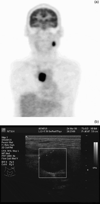
Case five, with nasopharyngeal carcinoma treated four years previously. (a) 18F-Fluorodeoxyglucose positron emission tomography scan, computed tomography assisted, revealed a ‘hotspot’ (standardised uptake value = 5.2) at level IV of the right neck near the clavicle. (b) Ultrasound examination showed an oval lymph node with slightly heterogeneous content (2.1 × 1.1 cm), just posterior to the internal jugular vein at level IV of the right neck.

Case six, with lung cancer. (a) 18F-Fluorodeoxyglucose positron emission tomography scanning revealed a ‘hotspot’ (standardised uptake value = 10.9) at level II of the left neck. (b) Ultrasound examination showed a round lymph node with capsule-type vascular pattern at level II of the left neck.

Clinical data for the 10 patients

Pt no = patient number; y = years; PET = 18F-fluorodeoxyglucose positron emission tomography; SUV = standardised uptake value; FNAC = fine needle aspiration cytology; F = female; M = male; Ca = cancer; NP = not performed; +ve = postitive for malignancy; –ve = negative for malignancy; mth = months; DOD = died of disease; NPC = nasopharyngeal carcinoma
The impalpable lymph nodes with increased 18F-fluorodeoxyglucose (18F-FDG) uptake on PET scanning were located in the supraclavicular region in four patients, the level II region in four patients, the level IV region in one patient and the accessory chain in one patient. All the nodes had a standardised uptake value of more than 4.0, except for two which were without available data. On ultrasonography, all of these suspicious lymph nodes were identified and each had at least one of the ultrasonographic features of malignancy, such as round shape (short to long axis > 0.5), present in nine nodes (Figures 1 and 2), and absent linear hilar structure or abnormal vascular pattern (capsule, punctuate or mixed types), present in five (Figure 2).
Fine needle aspiration under ultrasound guidance was successfully performed for all nodes, in order to acquire adequate specimens for evaluation. Each procedure was completed within 20 minutes. Cytological examination was positive for malignant cells in eight lymph nodes, confirmed pathologically in two (cases three and five). These eight patients received either salvage surgery with post-operative concurrent chemoradiotherapy or salvage chemotherapy. One patient (case eight) with buccal cancer died of disseminated metastasis four months later. One patient with lung cancer (case one) had originally planned to receive radical surgery after induction chemotherapy. However, the operation was halted and salvage chemotherapy was administrated because fine needle aspiration of the supraclavicular lymph nodes yielded malignant cells. Three months later, brain metastasis was discovered, and the patient died of disease six months later. Aside from these two patients, the other six patients with positive fine needle aspiration results were alive at the time of writing. The two patients with a negative fine needle aspiration result, confirmed pathologically in one patient, were alive and well, 19 and 36 months after examination, at the time of writing.
Discussion
18F-Fluorodeoxyglucose positron emission tomography (18F-FDG PET) has been widely used in the evaluation of disease status in cancer patients. Because it can scan the whole body in the one session and can identify malignant lesions as small as 5 mm in diameter, more and more clinically impalpable metastatic lymph nodes of the neck are now being identified in cancer patients.Reference Tran, Grigsby, Dehdashti, Herzog and Siegel2, Reference Ng, Ko, Toh and Chen4 Despite the high specificity of 18F-FDG PET for identifying malignant lesions, pathological verification of a ‘hotspot’ is still necessary in order to avoid patients receiving incorrect treatment because of false positive results, which are not uncommon (e.g. cases of granulomatous inflammation in Asia).Reference Ng, Joseph, Chan, Ko, Wang and Liao9–Reference Ho11 Fine needle aspiration examination and open biopsy are the conventional methods of determining the nature of these lymph nodes. Fine needle aspiration is a simple, minimally invasive, out-patient procedure with hardly any complications, which is capable of cytologically verifying suspicious lesions and is highly accurate in previously diagnosed cancer patients.Reference Kramer, van Putten, Post, van Dullemen, Bongaerts and Pruim17 In a cancer patient, the more invasive technique of open biopsy is usually unnecessary if fine needle aspiration cytology proves the diagnosis. However, if there is no imaging guidance, it is difficult for these two procedures to obtain tissue, when suspicious lymph nodes are not palpable on physical examination.
Because cervical lymph nodes are not deep in the neck space, ultrasound examinations of the neck can use higher frequency techniques with higher resolution, and can clearly identify cervical lymph nodes as small as 2 mm.Reference Wu, Lee, Huang, Ko, Sheen and Hsieh16, Reference Kramer, van Putten, Post, van Dullemen, Bongaerts and Pruim17 Ultrasonography itself can distinguish between benign and malignant lymphadenopathies, using a combination of grey scale and power Doppler techniques (despite some overlapping features).Reference Ng, Ko, Toh and Chen4, Reference Wu, Lee, Huang, Ko, Sheen and Hsieh16 Fine needle aspiration under ultrasound guidance is an easy, non-invasive and safe procedure which can be performed immediately after identifying the lymph node, and has a high yield rate.Reference Ng, Ko, Toh and Chen4 Therefore, ultrasonography may be an ideal diagnostic modality with which to detect hotspots shown by 18F-FDG PET scanning, and to enable cytological diagnosis via ultrasound-guided fine needle aspiration. As regards lung cancer staging, several reports have shown the role of endoscopic, ultrasound-guided fine needle aspiration in confirming the nature of suspicious mediastinal lymph nodes detected by 18F-FDG PET, in reducing staging costs and in avoiding invasive surgical interventions.Reference Eloubeidi, Cerfolio, Chen, Desmond, Syed and Ojha13, Reference Kramer, van Putten, Post, van Dullemen, Bongaerts and Pruim17
In this study, despite the fact that the lymph nodes with increased 18F-FDG uptake were small and clinically impalpable, all were clearly identified and were able to be evaluated using grey scale and power Doppler ultrasound techniques. All of the metastatic lymph nodes had at least one ultrasonographic feature of malignancy; such findings can complement PET scan results (in this study, nearly half of the malignant lymph nodes (three of eight) had equivocal standardised uptake values (4.0, 4.2, 5.2)).
Unlike endoscopic ultrasound fine needle aspiration, used in other regions,Reference Eloubeidi, Cerfolio, Chen, Desmond, Syed and Ojha13, Reference Kramer, van Putten, Post, van Dullemen, Bongaerts and Pruim17 ultrasound-guided fine needle aspiration of neck masses is relatively simple and is not difficult to learn. In this series, fine needle aspiration under ultrasound guidance was successfully performed, with satisfactory quality for cytological analysis, in all lymph nodes.
• 18F-Fluorodeoxyglucose positron emission tomography (PET) can detect cervical metastases before they are palpable; however, false positive results are not uncommon
• Ultrasound-guided fine needle aspiration allows cytological examination of suspicious nodes as small as 2 mm in diameter, with a reported sensitivity of 90 per cent and specificity of 100 per cent
• Ultrasound-guided fine needle aspiration is a safe, accurate, minimally invasive procedure which can enable tissue diagnosis of suspicious cervical lymph nodes detected by PET scanning
All patients with positive cytological results had a change of treatment plan as a result. Two patients received further selective neck dissection, including the suspicious lymph nodes, as a part of salvage treatment (pathological examinations of the resected nodes also revealed metastatic cancer). One patient with lung cancer avoided unnecessary radical surgery. Salvage chemotherapy was administrated in six patients and post-operative concurrent chemoradiotherapy in two patients. Of the patients with positive cytological results for malignancy, only two died of disease; the other six were alive at the time of writing. The two patients with negative cyotological results were kept under close observation, and were alive and well without the need for further treatment at the time of writing.
Conclusion
Ultrasound-guided fine needle aspiration is a simple, safe, accurate, minimally invasive procedure which can enable tissue diagnosis for suspicious cervical lymph nodes detected by PET scanning. Appropriate treatment can be instituted as a result of the information obtained from such cytological results, in addition to that from PET scanning.