Introduction
Acoustic neuromas are benign tumours with a non-linear and unpredictable growth pattern. A focused effort by the otolaryngology community to offer diagnostic imaging to patients with asymmetrical hearing loss, along with a rise in the diagnosis of incidental tumours as a result of increasing magnetic resonance imaging (MRI) utilisation, has led to an increase in the prevalence of small or asymptomatic acoustic neuroma.Reference Hoa, Drazin, Hanna, Schwartz and Lekovic1
It has been estimated that less than 1 per cent of acoustic neuromas exhibit sufficient growth to become clinically active,Reference Rosenberg2 and as many as two-thirds of acoustic neuromas do not grow,Reference Nikolopoulos, Fortnum, O'Donoghue and Baguley3 though much uncertainty still exists on the growth of these tumours. As half of patients with acoustic neuroma maintain their hearing over the first five years,Reference Hoa, Drazin, Hanna, Schwartz and Lekovic1 a reasonable initial treatment plan for patients with these small or asymptomatic tumours is monitoring with serial imaging, based on patient health, age and preference. Lifelong monitoring is necessary, as growth after long-term quiescence has been reported.Reference Rosenberg2
The ‘gold standard’ for both diagnosis and monitoring is the gadolinium-enhanced T1-weighted MRI sequence. In 2006, Marckmann et al. were the first to suspect the relationship of gadodiamide, a common gadolinium-containing contrast agent, to nephrogenic systemic fibrosis.Reference Marckmann, Skov, Rossen, Dupont, Damholt and Heaf4 Nephrogenic systemic fibrosis is an incurable disease affecting patients with renal failure; it causes skin induration, disability and increased mortality.Reference Marckmann, Skov, Rossen, Dupont, Damholt and Heaf4 Though nephrogenic systemic fibrosis has become less common with preventative measures and new gadolinium formulations, it is still a serious risk in patients with severe chronic renal disease or acute renal failure. In 2015, Gathings et al. reported two cases of patients with post-gadolinium skin changes histopathologically consistent with those seen in nephrogenic systemic fibrosis but without the systemic criteria for nephrogenic systemic fibrosis, one of whom did not have renal disease.Reference Gathings, Reddy, Santa Cruz and Brodell5 They concluded that sclerotic bodies, which are pathognomonic of gadolinium exposure, can occur in the absence of renal disease. This highlights the still-evolving risks of gadolinium, even in patients without renal failure, and should prompt clinicians to re-evaluate the marginal benefits gained from using contrast in patients monitored with serial MRI. Beyond health risks, the use of gadolinium carries increased patient and hospital costs, further weighting the balance in favour of non-contrast imaging when appropriate.
Although non-contrast, high-resolution T2-weighted sequences can enable accurate evaluation of cerebellopontine angle and internal auditory canal pathology,Reference Fortnum, O'Neill, Taylor, Lenthall, Nikolopoulos and Lightfoot6, Reference Annesley-Williams, Laitt, Jenkins, Ramsden and Gillespie7 the near-perfect sensitivity of post-contrast T1-weighted images has cemented its role within routine diagnostic protocols for acoustic neuromas. However, with an increasing proportion of acoustic neuroma patients electing for conservative management with serial observation, the value of contrast-enhanced imaging in follow-up MRI is worth investigating. The equivalence of high-resolution T2-weighted MRI scans to post-contrast T1-weighted images in monitoring the growth of previously diagnosed acoustic neuroma has not been adequately studied. This study aimed to compare the measurements of acoustic neuroma on serial T2-weighted images and post-contrast T1-weighted images, and consider a monitoring protocol based on non-contrast imaging.
Materials and methods
After approval by the institutional review board, a retrospective review was performed of all adult patients with a diagnosis of acoustic neuroma who underwent at least three high-resolution MRI scans of the internal auditory canal with and without contrast during the time period from 1 January 2008 to 11 October 2016.
At our institution, the MRI scans of the internal auditory canal are obtained with a 1.5 T Siemens Magnetom® Espree MRI machine. The T2-weighted images are based on a three-dimensional, T2-weighted, turbo-spin echo sequence, with a ‘Restore’ pulse. The post-contrast images are based on a T1-weighted turbo-spin echo sequence with fat suppression. Magnetic resonance imaging scans from other institutions were included if they involved similar post-contrast T1- and T2-weighted sequences with thin cuts through the internal auditory canal.
Exclusion criteria included cases of bilateral acoustic neuromas, other intracranial tumours and diagnosis of neurofibromatosis type 2. For patients who underwent surgical treatment during the period reviewed, post-operative images were excluded.
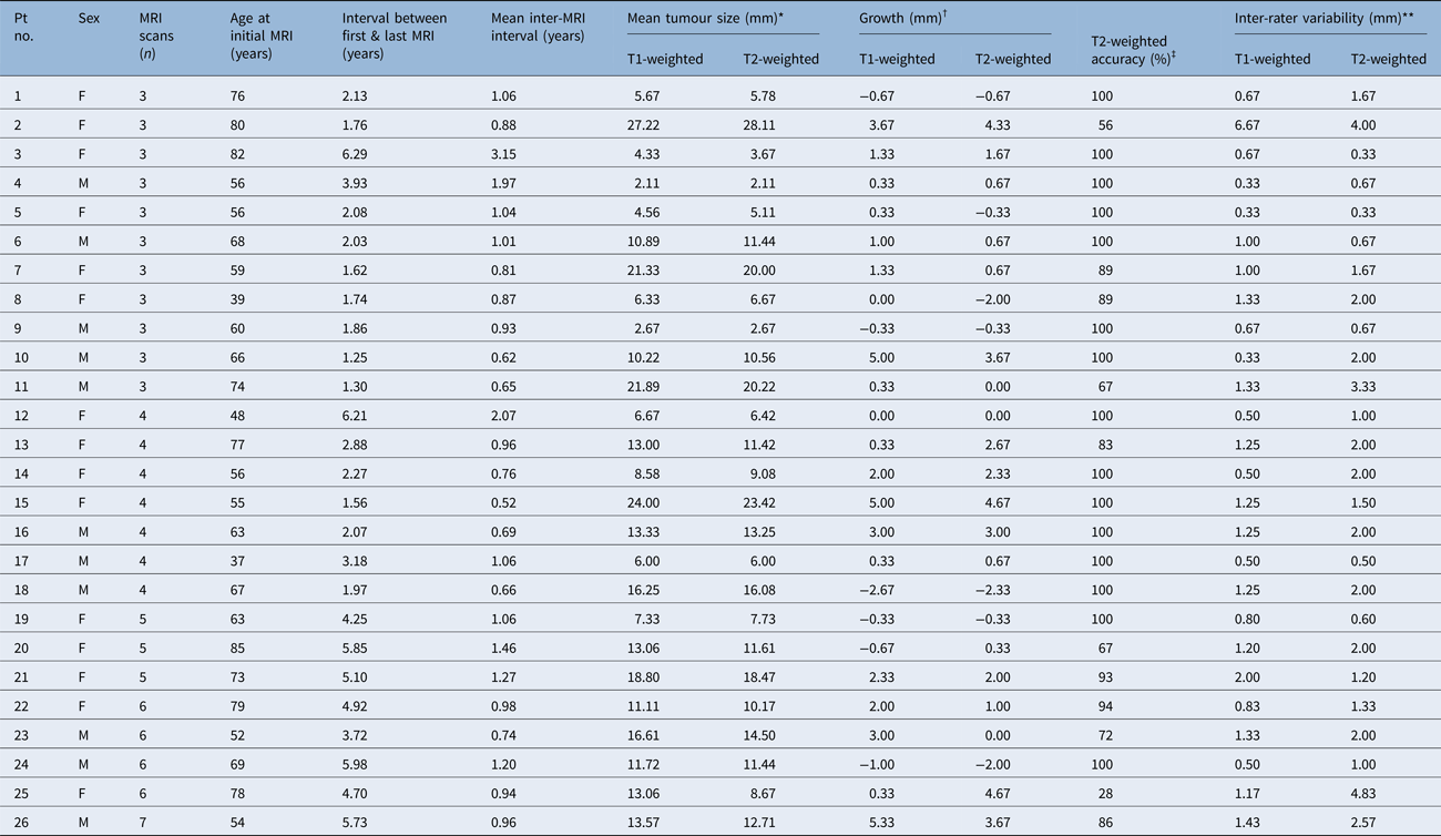
Ninety-five patients with the diagnosis of acoustic neuroma were identified in our database. After exclusion of ineligible patients and those with less than 3 eligible MRI scans, 26 patients were included. The age and sex of the patients can be found in Table 1.
Patients’ data

*Mean tumour size is defined as the mean of the maximal diameter measured in the axial and coronal planes by all three radiologists. †Growth is defined as the difference between the mean tumour measurements of the first and last magnetic resonance imaging (MRI) scans. ‡Accuracy is defined as the percentage of measurements on T2-weighted images within or equal to 2 mm of the measurement on T1-weighted images. **Inter-rater variability is defined as the mean difference between the smallest and largest measurement for each patient's T1- and T2-weighted MRI scans. Pt no. = patient number; F = female; M = male
For each patient's eligible MRI scans, axial and coronal post-contrast T1-weighted images and high-resolution non-contrast T2-weighted images were randomly and blindly reviewed separately by three radiologists. Maximal diameters of the tumours in the axial and coronal planes, including both the intracanalicular and extrameatal components, were independently measured by each radiologist and recorded. This method has been validated in previous studies, and found sufficient to follow tumour size and growth, and to allow for tumour irregularities.Reference Rosenberg2, Reference Fiirgaard, Pedersen and Lundorf8
Measurements were rounded to the nearest millimetre, and a difference of greater than 2 mm was selected as the threshold for both significant change between consecutive images and significant inter- and intra-observer variation. A 2 mm increase in the greatest diameter of an acoustic neuroma has been considered by many authors to be a marker for true tumour growth.Reference Smouha, Yoo, Mohr and Davis9–Reference Walsh, Bath, Bance, Keller, Tator and Rutka13
Results
A total of 107 MRI scans were separately reviewed by three radiologists. The patients had 3–7 MRI scans each, with a mean of 4.1 MRI scans each. Patient age at the first MRI scan ranged from 37 to 85 years. During the period reviewed, the interval between each patient's first and last MRI scans ranged from one year and three months to six years and four months, and the mean interval between MRI scans ranged from six months to three years and two months.
Tumour size, defined as the mean of the maximal measurements of the tumour diameter in the axial and coronal plane by all three radiologists, on all T1-weighted post-contrast MRI scans, ranged from 2.11 mm to 27.22 mm, and averaged 12.21 mm in our patient population.
The change in size measured on T1-weighted post-contrast MRI scans during the period reviewed ranged from 5.33 mm of growth to 2.67 mm of regression. The average tumour grew by 1.21 mm. Nine of 26 patients had 2 mm of growth or greater, nine patients had less than 2 mm of growth, and eight patients had no growth or a decrease in size.
Measurements by the radiologists for T2-weighted MRI scans differed from measurements for T1-weighted MRI by an average of 1.27 mm, or 10.4 per cent of the total size. Accuracy of tumour measurements on T2-weighted MRI, defined as a difference of less than or equal to 2 mm from the measurement on the corresponding T1-weighted image, was achieved on 88 per cent of the radiologists’ measurements.
Assessments of growth were also highly accurate on T2-weighted MRI. Of the 17 patients who exhibited less than 2 mm of growth on T1-weighted MRI, only 2 were identified as having greater than 2 mm of growth on T2-weighted MRI – a specificity of 88.2 per cent. For those patients with greater than 2 mm of growth on T1-weighted MRI, seven of nine (sensitivity of 77.8 per cent) accurately met this threshold on T2-weighted measurements.
No consistent relationship was found between T1- and T2-weighted measurements. T2-weighted measurements were smaller than their corresponding T1-weighted measurements in 47 per cent of evaluations, while the reverse was true 22.4 per cent of the time, and the measurements were equal in 30.5 per cent of cases.
The three radiologists exhibited strong inter-rater reliability. Head-to-head comparisons between the three radiologists showed a strong correlation in both T1-weighted measurements (r = 0.98, p < 0.001 for all head-to-head comparisons) and T2-weighted measurements (r = 0.97 or greater for all comparisons, p < 0.001). The average variability between measurements was 1.14 mm on T1-weighted images and 1.76 mm on T2-weighted images.
The patients’ data are summarised in Table 1.
Discussion
Our results show that measurements made with T2-weighted images are highly accurate; tumour size measurements were within 2 mm of post-contrast T1-weighted image sizes in 88 per cent of instances, and were within approximately 10 per cent of the T1-weighted sizes on average. Growth can be accurately monitored with non-contrast imaging: T2-weighted images, as compared to T1-weighted images, had a specificity of 88.2 per cent and a sensitivity of 77.8 per cent in judging growth of greater than 2 mm. These measurements were highly consistent between reviewers, as the variability in measurements on T2-weighted images differed by only 0.62 mm from the variability of measurements on T1-weighted images. These findings support the notion that non-contrast T2-weighted MRI scans can be used by radiologists to accurately monitor acoustic neuroma growth.
The prevalence of acoustic neuromas has increased dramatically in recent years, a change directly attributable to the increased availability and use of MRI,Reference Hoa, Drazin, Hanna, Schwartz and Lekovic1 along with a sharpened focus by the otolaryngology community in screening for this disease in patients presenting with asymmetric hearing loss. Demographics of this patient population have accordingly shifted following the inclusion of a greater proportion of patients with small, asymptomatic tumours. These tumours can alternate between periods of inactivity and aggressive growth, and in some cases exhibit no growth or even regress in size.
• Increased magnetic resonance imaging (MRI) utilisation has led to a rise in prevalence of small or asymptomatic acoustic neuromas
• Acoustic neuroma growth is variable
• The tumours are increasingly monitored with serial MRI, but contrast-enhanced T1-weighted imaging involves gadolinium exposure
• Acoustic neuromas can be effectively observed with serial non-contrast MRI using T2-weighted sequences
• T2-weighted sequences are highly accurate in measuring tumour size and identifying growth
• Accuracy may be further enhanced if the initial post-contrast MRI is available for comparison and guidance
Given the variable growth pattern and the rising proportion of incidental and asymptomatic tumours diagnosed, an increasing number of patients and their providers are electing to monitor their tumours with serial MRI. This conservative treatment strategy is chosen in cases where a paucity of symptoms, poor patient health or personal choice weighs against surgery or radiation. Follow-up MRI is typically performed initially at 6-month intervals and eventually distanced to 12–24-month intervals.
The use of a paramagnetic contrast material provides a heightened definition of tumour margins, and allows the radiologist to differentiate acoustic neuroma from meningiomas and other cerebellopontine angle tumours with near-perfect accuracy.Reference Lalwani and Jackler14 However, it carries significant additional financial costs over non-contrast MRI and requires approximately twice the time to perform. It also puts the patient at risk of medical complications such as nephrogenic systemic fibrosis.
Many studies in the past 25 years have investigated the feasibility of using non-contrast T2-weighted images to accurately diagnose acoustic neuromas.Reference Fortnum, O'Neill, Taylor, Lenthall, Nikolopoulos and Lightfoot6, Reference Annesley-Williams, Laitt, Jenkins, Ramsden and Gillespie7, Reference Allen, Harnsberger, Shelton, King, Bell and Miller15–Reference Zealley, Cooper, Clifford, Campbell, Potterton and Zammit-Maempel24 A review of the literature concluded that non-contrast MRI was highly sensitive and cost-effective in the diagnosis of acoustic neuroma, finding high-resolution T2-weighted sequences to be of sufficient quality to ‘permit exclusion of acoustic neuroma of any size with sufficiently high diagnostic confidence to abandon routine [contrast-enhanced T1-weighted] imaging’.Reference Fortnum, O'Neill, Taylor, Lenthall, Nikolopoulos and Lightfoot6 Clinical studies vary in regard to the risks of using non-contrast MRI as the primary screening test,Reference Curtin25, Reference Jackler26 most notably of missing small intracanalicular tumours that do not significantly alter the appearance of the nerves. Today, contrast-enhanced MRI remains part of the diagnostic investigation at most centres.
Despite a reticence to adopt a contrast-free diagnostic algorithm, an interest persists in the neurotology and neuroradiology communities for a long-term surveillance protocol for acoustic neuromas using non-contrast MRI. Patients monitored for years with serial imaging have a perpetual risk associated with using contrast agents, and thus can gain the most from avoiding it. The benefits of using T2-weighted sequences for monitoring acoustic neuroma growth, beyond decreasing risks to patient health, include reduced demands on patient time and hospital resources.
Although the use of T2-weighted MRI scans in the diagnosis of acoustic neuroma has been well studied, its efficacy in monitoring acoustic neuroma growth has not been thoroughly evaluated. In 2009, a retrospective review by Ozgen et al. compared the efficacy of high-resolution T2-weighted MRI sequences with standard contrast-enhanced T1-weighted sequences for monitoring acoustic neuroma growth over time.Reference Ozgen, Oguz and Dolgun27 Their results demonstrated good intra-rater correlation between post-contrast T1- and T2-weighted images, but revealed poor inter-rater correlation, which the authors attributed to a difference in observer experience with head and neck imaging. The generalisability of their results is limited, however, because of the undefined and qualitative assessment of growth, the limited number of follow-up studies (average of 1.8 scans per patient over a 23-month follow-up period), and the inclusion of two patients with post-surgical images.
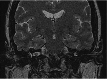
In the course of data collection for this study, the radiologists noted certain tumour characteristics that facilitated or hindered its measurement. Hyperintense cerebrospinal fluid (CSF) circumscribing the tumour provides a clear definition of tumour borders on T2-weighted images. Figure 1 shows a T2-weighted image of acoustic neuroma in patient number 12. The tumour margins are well defined by the surrounding CSF, which likely contributed to the high accuracy of the T2-weighted images and the low inter-rater variability. In the cystic degeneration of tumours, however, the hyperintense fluid within the tumour could blur with surrounding CSF to confound the tumour margins. Similarly, adjacent isointense blood vessels such as the anterior inferior cerebellar artery or veins can lead to overestimation of tumour size if erroneously included in the tumour measurement.
Surrounding cerebrospinal fluid helps define the tumour borders on this coronal T2-weighted magnetic resonance imaging scan (patient number 12).

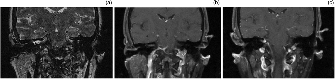
In addition, large extrameatal components of the tumour can lack sharply defined margins adjacent to a compressed cerebellum or brainstem, making it difficult to delineate tumour from brain parenchyma on T2-weighted images. Figure 2 shows T1- and T2-weighted images of patient 25. The intracanalicular portion of the tumour has a cystic component that appears bright on T2-weighted images and distracts from the cisternal component which blends with the brainstem. On post-contrast T1-weighted images, the cisternal component is bright and well-defined. These factors likely contributed to the poor accuracy and high inter-rater variability of the T2-weighted images.
Tumour (in patient number 25) with intracanalicular cystic characteristics that appear bright on the coronal T2-weighted image (a) and less distinct on the coronal T1-weighted post-contrast image (b). The cisternal component of the tumour is poorly defined on T2-weighted magnetic resonance imaging (a), leading to confounding margins. In comparison, the cisternal component is bright on the coronal T1-weighted post-contrast image (c), providing easily measurable margins.

Many of these factors can be predicted in the initial post-contrast T1-weighted images. These initial post-contrast images can be used for comparison when evaluating subsequent non-contrast follow-up studies, and prevent gross errors in measurement. We were unable to test this theory, as our radiologists were blinded to the T1-weighted measurements when evaluating T2-weighted images. We believe the accuracy of measurements on T2-weighted images would be improved, and the inter-rater and intra-rater variability reduced, if post-contrast images had been available for comparison. The decreased inter-rater variability found on post-contrast T1-weighted measurements reflects the higher precision of this modality. We believe that T2-weighted non-contrast images can be more precise when the radiologist is assisted by the initial T1-weighted post-contrast images for comparison, which can remove roadblocks to accurate measurements; additional studies may be able to quantify this benefit. Furthermore, future studies should explore whether or not radiologists can prospectively identify tumour characteristics favourable to monitoring with serial T2-weighted images.
Our findings thus bolster the argument for obtaining an initial diagnostic post-contrast MRI, which is a key part of our proposed protocol for long-term serial monitoring with non-contrast MRI. We suggest that the initial diagnostic MRI should continue to be contrast-enhanced, because of the unparalleled sensitivity of the post-contrast T1-weighted image. In addition to improved tumour detection and delineation, contrast-enhanced T1-weighted sequences can also detect cochlear infiltration and other pathologies, such as a facial nerve schwannoma. Subsequent imaging can be performed with high-resolution T2-weighted MRI without paramagnetic contrast material, unless the radiologist identifies tumour characteristics that interfere with such a strategy.
Even with the radiologist blinded to the findings of the T1-weighted contrast-enhanced MRI, this study's results indicate that T2-weighted imaging without paramagnetic contrast during follow-up imaging offers accurate growth measurements. However, it should be noted that non-contrast MRI examinations may have drawbacks in cases of cystic tumours, particularly as they bulge in the cerebellopontine angle and abut the brainstem. In addition, T2-weighted imaging alone might not be optimal in detecting post-treatment response and changes. Future studies can investigate the feasibility of using non-contrast MRI in the serial imaging of post-surgical patients, neurofibromatosis type 2 patients and other acoustic neuroma subgroups deviating from our study population.
Limitations of this study include a relatively small mean tumour size in our patient population. By excluding patients who underwent prompt surgical management after diagnosis, we selected a population unlikely to have larger tumours. Additionally, this study was limited by the possible heterogeneity of the MRI machines and sequences used, as it included MRI scans within our database that were downloaded from other hospitals and imaging centres. Though the sequences were comparable, the inability to specify the magnet strength or exact sequence used limits the replicability of our study. It is possible the inter-observer consistency would improve if a consistent sequence were used. Finally, it is our institutional protocol to measure the total size of the tumour, including both intracanalicular and cisternal components. However, it is worth noting that the cisternal component is both more relevant to clinical decisions and more difficult to measure on T2-weighted images, as described earlier.
Conclusion
In response to growing concerns over the medical risks and cost of paramagnetic contrast materials, T2-weighted sequences provide the opportunity to effectively observe acoustic neuromas with serial non-contrast MRI. This study illustrates that even when the radiologist is blinded to the results of an initial T1-weighted enhanced MRI, T2-weighted sequences are highly accurate in measuring tumour size and identifying growth. Such accuracy may be further enhanced if the initial post-contrast MRI scans are available for comparison and guidance.
Competing interests
None declared