Introduction
During the past decade, a number of studies have demonstrated the importance of innate immune recognition of microbial agents. Innate immunity specifically recognises invading pathogens with the help of germ coded receptors called pattern recognition receptors (PRRs). Toll-like receptors are a type of PRR and have been shown to have significant homology in the cytoplasmic domain to the IL-1 receptor type-1 (Yamamoto et al., Reference YAMAMOTO, SATO, MORI, HOSHINO, TAKEUCHI, TAKEDA and AKIRA2002). TLRs detect conserved molecules called ‘pathogen associated molecular patterns’ (PAMPs) and through a cascade of reactions, activate the immune system (Janeway and Medzhitov, Reference JANEWAY and MEDZHITOV2002). Because of such properties TLRs are being investigated as potential candidates for the development of an effective adjuvant system. TLR recognition of pathogens alerts the immune system to mount an immediate response to limit the spread of infection (Akira et al., Reference AKIRA, TAKEDA and KAISHO2001). Further, to combat infectious diseases both initial inflammatory and antigen specific immune responses are required. In this regard, TLRs have been shown to play an important role not only in the proinflammatory response but also in the antigen specific immune response (Akira et al., Reference AKIRA, UEMATSU and TAKEUCHI2006; Beutler et al., Reference BEUTLER, JIANG, GEORGEL, CROZAT, CROKER, RUTSCHMANN, DU and HOEBE2006).
To date, 13 TLRs have been identified in mammals (Shi et al., Reference SHI, CAI, SANCHEZ, ZHANG, WEN, WANG, YANG, FU and ZHANG2011). These TLRs recognise an array of microbial components; TLR4 recognises lipopolysaccharide (LPS) (Chow et al., Reference CHOW, YOUNG, GOLENBOCK, CHRIST and GUSOVSUREDSKY1999), TLRs 1, 2, 5, and 6 detect flagellar proteins and peptidoglycans (Schwandner et al., Reference SCHWANDNER, DZIARSKI, WESCHE, ROTHE and KIRSCHNING1999), endosomal TLR3 detects dsRNA (Alexopoulou et al., Reference ALEXOPOULOU, HOLT, MEDZHITOV and FLAVELL2001), TLR9 identifies un-methylated cytosine-guanosine containing oligonucleotides (CpG)-DNA (Takeshita et al., Reference TAKESHITA, LEIFER, GURSEL, ISHII, TAKESHITA and GURSEL2001) and the ligand for TLR7 and TLR8 has been reported to detect ssRNA (Heil et al., Reference HEIL, AHMAD-NEJAD, HEMMI, HOCHREIN, AMPENBERGER, GELLERT, DIETRICH, LIPFORD, TAKEDA, AKIRA, WAGNER and BAUER2003; Diebold et al., Reference DIEBOLD, KAISHO, HEMMI, AKIR and REIS E SOUSA2004). TLRs that detect extracellular microbial components are expressed on the cell surface (TLR1, 2, 4, 5 and 6), whereas TLRs for the detection of viral or bacterial nucleic acids are located intracellularly on endocytotic vesicles or organelles (TLR3, 7, 8 and 9) (Pathak et al., Reference PATHAK, BASU, BASU, BANERJEE, PATHAK, BHATTACHARYYA, KAISHO, KUNDU and BASU2007).
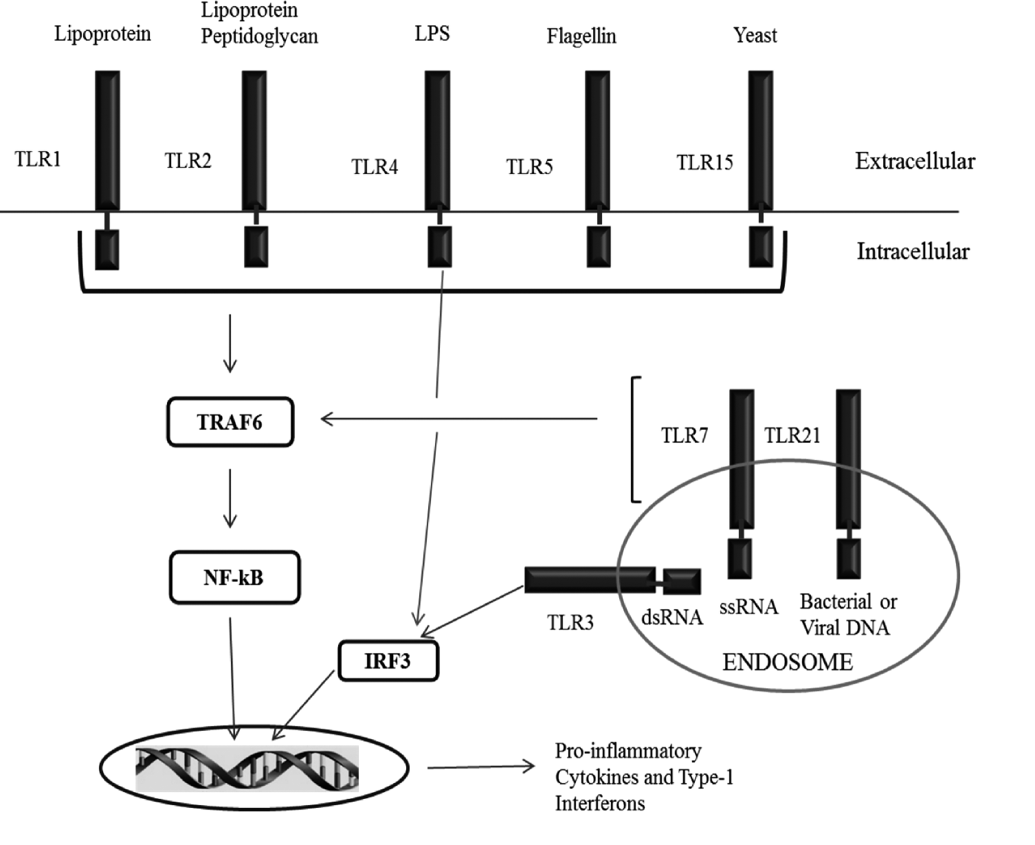
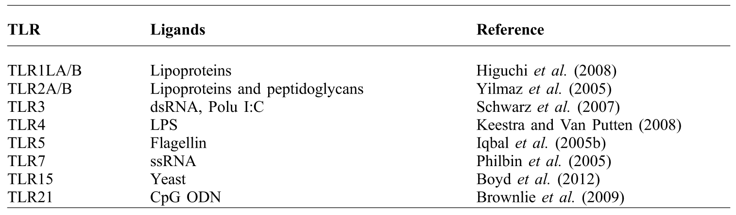
In the chicken (Figure 1), 10 TLRs have been reported (Iqbal et al., Reference IQBAL, PHILBIN and SMITH2005a; Higgs et al., Reference HIGGS, CORMICAN, CAHALANE, ALLAN, LLOYD, MEADE, JAMES, LYNN, BABIUK and O'FARRELLY2006; Ruan and Zheng, Reference RUAN and ZHENG2011) and include TLRs 3, 4, 5, 7, 15 and 21 and two isoforms of each TLR1 and TLR2. Moreover, TLR9 has not been detected in chicken, instead, in chicken, its functional homologue has been designated as TLR21 and its ligand has been reported to be CpG oligodeoxynucleotides (Brownlie et al., Reference BROWNLIE, ZHU, ALLAN, MUTWIRI, BABIUK, POTTER and GRIEBEL2009; Keestra et al., Reference KEESTRA, DE ZOETE, BOUWMAN and VAN PUTTEN2010).
An overview of chicken TLRs. TLR1 and TLR2 have two isoforms each.

Furthermore, TLR4 and TLR5 recognize lipopolysaccharide (LPS) and flagellin, respectively (Smith et al., Reference SMITH, ANDERSEN-NISSEN, HAYASHI, STROBE, BERGMAN, BARRETT, COOKSON and ADEREM2003; Keestra and Van Putten, Reference KEESTRA and VAN PUTTEN2008) (Table 1).
Chicken TLRs and their specific ligands.
Recent advancement in the knowledge of innate immune functioning has revealed that when two or more TLRs are stimulated concomitantly, as in natural microbial infection, they interact and this interaction may result in either synergistic or antagonistic regulation of their responses (Toshchakov et al., Reference TOSHCHAKOV, JONES, PERERA, THOMAS and CODY2002; Lotz et al., Reference LOTZ, EBERT, ESSELMANN, ILIEV, PRINZ and WIAZEWICZ2005; Gupta et al., Reference GUPTA, DEB, GAIKWAD, SARAVANAN, MOHAN and DEY2013). This review focuses on interactions between TLRs and their use as adjuvant in combination in chickens.
TLR signalling: molecular basis of enhanced immune responses
Ligand binding to its specific TLR, initiates the cascade of reactions leading to the activation of transcription factors and expression of genes related to cytokines, chemokines and type-1 interferon production. Two distinct pathways are reported in TLR signalling: a MyD88-dependent pathway that leads to the activation of NF-kB and MAP kinases and subsequent production of inflammatory cytokines and chemokines, and a MyD88-independent pathway or TRIF dependent pathway (TIR domain containing adaptor inducing interferon-β), associated with the stimulation of interferon inducible genes and production of type-1 interferons (IFN) like IFN-α and IFN-β (Takeda and Akira, Reference TAKEDA and AKIRA2004; Kawai and Akira, Reference KAWAI and AKIRA2006).
All TLRs, except TLR3, induce nuclear transcription factor NF-kB dependent expression of proinflammatory genes via a common signalling adaptor molecule - the myeloid differentiation protein 88 (MyD88) (Adachi et al., Reference ADACHI, KAWAI, TAKEDA, MATSUMOTO, TSUTSUI, SAKAGAMI and AKIRA1998; Yamamoto et al., Reference YAMAMOTO, SATO, MORI, HOSHINO, TAKEUCHI, TAKEDA and AKIRA2002). TLR3 signals through MyD88-independent pathway or TRIF dependent pathway leading to the activation of interferon regulatory factor-3 and 7 (IRF) and consequent type-1 interferon production (Yamamoto et al., Reference YAMAMOTO, SATO, HEMMI, HOSHINO, KAISHO, SANJO, TAKEUCHI, SUGIYAMA, OKABE, TAKEDA and AKIRA2003; Kawai and Akira, Reference KAWAI and AKIRA2006). TLR4 is unique in its action as it acts via both the pathways and induces both proinflammatory cytokines as well as interferons. Moreover, production of cytokines, chemokines, and type-1 interferons through signalling, alter many immunological processes to enhance the immune response. These events include maturation of antigen-presenting cells (APCs), significant up-regulation of expression co-stimulatory molecules (CD 80 and CD 86) as well as major histocompatibility complex (MHC) molecules (Mogensen, Reference MOGENSEN2009), increased phagocytic efficiency and increased survival of T-cells and, hence, an augmented memory response of immune cells. Furthermore, TLR ligands may also cause certain cytokines to influence the differentiation of T-helper (Th) cells towards Th1 or Th2 subset or may even induce a mixed type of reaction (both Th1 and Th2) (Mogensen, Reference MOGENSEN2009; St. Paul et al., Reference ST. PAUL, PAOLUCCI and SHARIF2012a). Taken together, these observations explain the potential role of TLRs in modulating both innate and adaptive immunity and this intrinsic property of TLRs can be exploited for their use as adjuvants in vaccines to enhance host immunity to infectious agents.
TLR agonists as adjuvants in chicken
Several studies have been conducted using TLR agonists as adjuvant in chicken in-vitro as well as in-vivo, with promising results. Assessment of the nature of the TLR-mediated responses is necessary for the rational design of prophylactic treatments and vaccine adjuvant systems. Some important studies with individual TLR agonists are described below.
TLR2
Chicken TLR2 is mainly expressed in the spleen, caecal tonsils, bursa, liver, heterophils, B cells and CD8+ cells (Iqbal et al., Reference IQBAL, PHILBIN and SMITH2005a; Kogut et al., Reference KOGUT, IQBAL, HE, PHILBIN, KAISER and SMITH2005). TLR2 has been shown to bind to a variety of ligands due to the heterodimerisation of TLR2 isoforms with the isoforms of TLR1 and different combinations of isoforms of these two TLRs mediate the response to certain ligands. In this context, the response to a synthetic triacylated lipopeptide (Pam3CSK4) in chickens is thought to be mediated by multiple combinations of TLR2/TLR1 heterodimers except for TLR2-1/TLR1-1 while the response to peptidoglycans are mediated only by the TLR2-1/TLR1-2 heterodimer (Keestra et al., Reference KEESTRA, DE ZOETE, VAN AUBEL and VAN PUTTEN2007; Higuchi et al., Reference HIGUCHI, MATSUO, SHINGAI, SHIDA, ISHII, FUNAMI, SUZUKI, OSHIUMI, MATSUMOTO and SEYA2008).
Other such TLR2 ligands include PGN (peptidoglycans) and LTA (lipoteichoic acid). These ligands (PGN and LTA), including Pam3CSK4, have been shown to stimulate chicken splenocytes and induce the production of various cytokines including IFN-γ, IL-12, IL-4 and IL-13. Cytokine production patterns suggest a mixed T-helper (Th) 1 and 2-like response by these agonists (St. Paul et al., Reference ST. PAUL, PAOLUCCI and SHARIF2012a). Nitric oxide (NO) production and oxidative burst are fee radical dependent cytotoxic mechanisms used by the immune cells to degrade the internalised pathogens. In this regard, LTA induced significant NO production in chicken monocytes when stimulated with a higher concentration (He et al., Reference HE, GENOVESE, NISBET and KOGUT2006) and an oxidative burst in chicken heterophils (Farnell et al., Reference FARNELL, CRIPPEN, HE, SWAGGERTY and KOGUT2003). Furthermore, a study where chickens were pre-treated with LTA showed higher antibody titres to KLH (Keyhole limpet haemocyanin) (Parmentier et al., Reference PARMENTIER, VAN DEN KIEBOOM, NIEUWLAND, REILINGH, HANGALAPURA, SAVELKOUL and LAMMERS2004).
TLR3
TLR3 has been shown to be expressed in various tissues including the duodenum, jejunum, ileum, caecum, colon, thymus and immune cells like heterophils, B and T cells (Iqbal et al., Reference IQBAL, PHILBIN and SMITH2005a). It recognises polyinosinic-polycytidylic acid (poly I:C) (Philbin et al., Reference PHILBIN, IQBAL, BOYD, GOODCHILD, BEAL, BUMSTEAD, YOUNG and SMITH2005; He et al., Reference HE, GENOVESE, NISBET and KOGUT2007; Schwarz et al., Reference SCHWARZ, OHNEMUS, LAVRIC, KOTHLOW, BAUER, KASPERS and STAEHELI2007; He et al., Reference HE, MACKINNON, GENOVESE and KOGUT2011). Poly(I:C), a dsRNA analogue, rapidly induced expression of INF-γ, IFN-α, IFN-β, and IL-10, but not IL-12p40, similar to that seen in mammals, but it down regulated expression of proinflammatory cytokines and produced very little NO in chicken monocytes (He et al., 2007; Karpala et al., 2008; He et al., 2012). A similar pattern was observed in chicken RBCs where poly I:C up-regulated IL-8 expression but, on the contrary, it enhanced the production of nitrite (St. Paul et al., 2013). Furthermore, double stranded RNA containing poly-UG motifs, stimulated chicken splenocytes and up-regulated expression of IFN-α, IFN-β as well as pro-inflammatory cytokines like IL-6 and IL-1β (Villanueva et al., Reference VILLANUEVA, KULKARNI and SHARIF2011). Poly I:C has successfully been used as an intranasal adjuvant for influenza virus vaccine (HA vaccine). When HA vaccine was administered with poly I:C, it up-regulated the expression of type-1 IFNs as well as Th1 and Th2 cytokines (Ichinohe et al., Reference ICHINOHE, WATANABE, ITO, FUJII, MORIYAMA, TAMURA, TAKAHASHI, SAWA, CHIBA, KURATA, SATA and HASEGAWA2005).
TLR4
TLR4 has been shown to recognise the bacterial cell wall component Lipopolysaccharides (LPS) and operates through both MyD88 dependent as well as MyD88 independent pathways (Yamamoto et al., Reference YAMAMOTO, SATO, HEMMI, HOSHINO, KAISHO, SANJO, TAKEUCHI, SUGIYAMA, OKABE, TAKEDA and AKIRA2003). Detection of LPS by the TLR4, results in a signalling cascade leading to the activation of transcription factor NF-kB and production of proinflammatory cytokines and chemokines. In the chicken, TLR4 is present on various immune cells including heterophils, macrophages, B cells and T cells as well as in tissues like the bursa and spleen (Iqbal et al., Reference IQBAL, PHILBIN and SMITH2005a). Allelic variation in TLR4 has been associated with susceptibility to systemic infection with Salmonella enterica serovar typhimurium in young chickens (Leveque et al., Reference LEVEQUE, FORGETTA, MORROLL, SMITH, BUMSTEAD, BARROW, LOREDO-OSTI, MORGAN and MALO2003). Further, the susceptibility of chickens to systemic salmonellosis has been attributed to either the lack of recognition of LPS or to the lack of binding of LPS to the LPS receptor complex resulting in a weak signal for the activation of heterophils (Kogut et al., Reference KOGUT, IQBAL, HE, PHILBIN, KAISER and SMITH2005). LPS can induce an array of cytokines and chemokines such as IL-1β, IL-6, and IL-18 and CXC, CXCLi2 and CXCLi1, CXCR1 and CCLi4 gene expression in chicken heterophils (Kogut et al., Reference KOGUT, IQBAL, HE, PHILBIN, KAISER and SMITH2005). LPS stimulated a significant increase in oxidative burst and enhanced NO production and phagocytic capabilities of chicken immune cells (Farnell et al., Reference FARNELL, CRIPPEN, HE, SWAGGERTY and KOGUT2003; He et al., Reference HE, GENOVESE, NISBET and KOGUT2006; St. Paul et al., 2012). LPS has additionally been shown to increase the expression of IFN-γ, IL-8, IL-1β, IL-10, iNOS, and MHC- II in the chicken spleen (Sijben et al., Reference SIJBEN, KLASING, SCHRAMA, PARMENTIER, VAN DER POEL, SAVELKOUL and KAISER2003; St. Paul et al., Reference ST. PAUL, MALLICK, HAQ, OROUJI, ABDUL-CAREEM and SHARIF2011). IFN-γ and IL-10 are Th1 and Th2 cytokines respectively, IL-1β is a proinflammatory cytokine and increased levels of iNOS shows enhanced macrophage function. Production of these cytokines as well as up-regulation of co-stimulatory molecules activates the immune cells of the body to fight against the invading pathogens by mounting a strong immune response.
TLR5
TLR5 detects bacterial flagellin, which may be found on both Gram-positive and Gram-negative bacteria. Flagellin has also been shown to bind to a cytoplasmic receptor Neuronal apoptosis inhibitory protein 5 (NAIP5), a cytosolic pattern-recognition receptor in the family of nucleotide-binding oligomerisation domain-leucine-rich repeat proteins (NOD-LRR), and to play an important role in defence against Legionella infections. Through cytoplasmic receptors (NAIP5), it has been shown to produce inflammatory cytokines as well as to activate caspase -1via Ipaf when cells fail to restrain the infection (Hawn et al., Reference HAWN, VERBON, LETTINGA, ZHAO, LI, LAWS, SKERRETT, BEUTLER, SCHROEDER, NACHMAN, OZINSKY, SMITH and ADEREM2003; Franchi et al., Reference FRANCHI, AMER, BODY-MALAPEL, KANNEGANTI, ÖZÖREN, JAGIRDAR, INOHARA, VANDENABEELE, BERTIN, COYLE, GRANT and NUNEZ2006; Molofsky et al., Reference MOLOFSKY, BYRNE, WHITFIELD, MADIGAN, FUSE, TATEDA and and SWANSON2006). Nevertheless, most of the adjuvant action of flagellin is TLR5 dependent and, in this context has been studied extensively. It has been shown to have potent immunostimulatory properties (Gupta et al., Reference GUPTA, BAJWA, DEB, MOHAN and DEY2014a). In this regard, flagellin stimulates heterophils, enhances oxidative burst and causes significant up-regulation of proinflammatory cytokines like IL-1β, IL-6 and IL-8 and chemokines CXCLi2 (Kogut et al., Reference KOGUT, IQBAL, HE, PHILBIN, KAISER and SMITH2005; Reference KOGUT, SWAGGERTY, HE, PEVZNER and KAISER2006). Nitric oxide production was up-regulated when chicken monocytes were stimulated with flagellin which suggests an enhanced macrophage function (He et al., Reference HE, GENOVESE, NISBET and KOGUT2006). Chicken cells like HD11, CKC (chicken kidney cells) and chicken embryo fibroblasts (CEF) have been shown to up-regulate the production of proinflammatory cytokine IL-1β when exposed to flagellin (Iqbal et al., Reference IQBAL, PHILBIN, WITHANAGE, WIGLEY, BEAL, GOODCHILD, BARROW, MCCONNELL, MASKELL, YOUNG, BUMSTEAD, BOYD and SMITH2005b). An increased proinflammatory response was observed in HeLa cells expressing chicken TLR5 due to enhanced production of NF-kB (Keestra et al., Reference KEESTRA, DE ZOETE, VAN AUBEL and VAN PUTTEN2008). Recent studies have reported a mixed induction of Th1 and Th2 immune responses in chicken immune cells (St. Paul et al., Reference ST. PAUL, PAOLUCCI and SHARIF2012a; Gupta et al., Reference GUPTA, DEB, GAIKWAD, SARAVANAN, MOHAN and DEY2013). We have observed that when chicken peripheral blood mononuclear cells (PBMCs) were stimulated with recombinant flagellin, both Th1 and Th2 cytokines (IL-12 and IL-4) as well as proinflammatory cytokine IL-6 were induced (Gupta et al., Reference GUPTA, DEB, GAIKWAD, SARAVANAN, MOHAN and DEY2013). Similar mixed types of immune responses have also been observed in chicken splenocytes (St. Paul et al., Reference ST. PAUL, PAOLUCCI and SHARIF2012a). Additionally, flagellin, being protein in nature, can effectively be used as a recombinant fusion protein with the antigen of interest or in a DNA vaccine encoding flagellin and an antigen (Gupta et al., Reference GUPTA, BAJWA, DEB, MOHAN and DEY2014a).
TLR7
TLR7 is reported to be present in the duodenum, caecum, colon and caecal tonsils as well as on immune cells including heterophils, macrophages, B and T cells (Iqbal et al., Reference IQBAL, PHILBIN and SMITH2005a). TLR7 is localised in the endosome where it may detect viral infection with single stranded RNA genomes. Ligands for TLR7 are single stranded RNA molecules and synthetic compounds like loxoribine (LOX) and R848. Stimulation of splenocytes with LOX results in IFN and IL-6 production (Jenkins et al., Reference JENKINS, LOWENTHAL, KIMPTON and BEAN2009). In heterophils, stimulation with LOX results in a significant induction of both an oxidative burst and a degranulation response but at the same time, it causes down regulation of IL-1β, IL-6, and IL-8 production (Kogut et al., Reference KOGUT, IQBAL, HE, PHILBIN, KAISER and SMITH2005). This contrasting characteristic may be due the fact that the level of TLR7 expression in heterophils is low and so is hypo-responsive to its ligand (Kogut et al., Reference KOGUT, IQBAL, HE, PHILBIN, KAISER and SMITH2005). Furthermore, LOX did not induce NO production in chicken monocytes (He et al., Reference HE, GENOVESE, NISBET and KOGUT2006). Another TLR7 ligand R848 was found to induce IL-1β, IL-6 and IL-8 expression in chicken splenocytes (Phibin et al., 2005). LOX has been shown to protect chicks from Salmonella spp. infection. In this regard, pre-treatment of chicks with LOX resulted in a significant reduction in the invasion by Salmonella enteritidis in the liver and spleen of birds (Swaggerty et al., Reference SWAGGERTY, HE, GENOVESE, DUKE and KOGUT2012).
TLR15
A new TLR has been reported in chickens which has not yet been detected in mammals and recent studies have characterised the new TLR and designated it as TLR15 (Boyd et al., Reference BOYD, PEROVAL, HAMMOND, PRICKETT, YOUNG and SMITH2012). It has been found to be significantly up-regulated in the caecum following Salmonella enterica serovar typhimurium infection (Nerren et al., Reference NERREN, HE, GENOVESE and KOGUT2010). Ligands for all TLRs were tried however none of them were found to stimulate TLR15, suggesting a new novel ligand for it. Later, it was reported that TLR15 recognises a unique and heat-stable component of bacteria capable of causing disease (Nerren et al., Reference NERREN, HE, GENOVESE and KOGUT2010). Boyd et al. (Reference BOYD, PEROVAL, HAMMOND, PRICKETT, YOUNG and SMITH2012) used yeast lysate and found it to be able to activate TLR15 dependent NF-kB and the production of pro-inflammatory cytokines like IL-1β. Furthermore, this signalling was abolished when lysate was exposed to heat prior to stimulation, suggesting the compound is heat labile (Boyd et al., Reference BOYD, PEROVAL, HAMMOND, PRICKETT, YOUNG and SMITH2012). Subsequent studies have reported the heat labile component to be proteases in yeast lysate (De Zoete et al., Reference DE ZOETE, BOUWMAN, KEESTRA and VAN PUTTEN2011). The parasite E. tenella sporozoites have also been reported to up-regulate the expression of TLR15 in heterophils and monocytes (Zhou et al., Reference ZHOU, WANG, CAO, HU, ZHANG, QIN, GUO and NIE2013). The presence of TLR15 in chickens may contribute towards species specific defence mechanisms.
TLR21
A mammalian counterpart of TLR9 has been reported to be missing in the chicken. Instead, a functional homologue of TLR9 is present, designated TLR21 which recognises the TLR9 ligand CpG-ODN (Brownlie et al., Reference BROWNLIE, ZHU, ALLAN, MUTWIRI, BABIUK, POTTER and GRIEBEL2009). It is one of the most extensively studied TLRs in the chicken and expression of this TLR has been found in several cells including macrophages and B cells (Brownlie et al., Reference BROWNLIE, ZHU, ALLAN, MUTWIRI, BABIUK, POTTER and GRIEBEL2009). CpG-ODN possesses immunostimulatory properties and has been shown to stimulate chicken monocytes to up-regulate the expression of INF-γ, IL-10, and IL-12p40, NO but not IFN-α and IFN-β (He et al., Reference HE, GENOVESE, NISBET and KOGUT2007; Reference HE, GENOVESE, SWAGGERTY, MACKINNON and KOGUT2012). Chicken splenocytes when stimulated with CpG-ODN induced proinflammatory cytokine production and increased cell proliferation (Jenkins et al., Reference JENKINS, LOWENTHAL, KIMPTON and BEAN2009). CpG-ODN also induced pro-inflammatory responses in thrombocytes and up-regulated IL-1β, IL-6 and IL-8 expression (St. Paul et al., Reference PAUL, PAOLUCCI, BARJESTEH, WOOD, SCHAT and SHARIF2012b). CpG-ODN seems to promote the Th1 immune response. Patel et al. (Reference PATEL, GOMIS, DAR, WILLSON, BABIUK, POTTER, MUTWIRI and TIKOO2008) demonstrated that administration of CpG-ODN induced the expression of IL-1β, IL-6, IL-8, IL-10, IL-18 and IFN-γ in the spleen and IL-10 and IFN-α in the Bursa of Fabricious. Up-regulation of IL-18 and IFN-γ but not IL-4 expression indicate that CpG-ODN induces a Th1 type immune response (Patel et al., Reference PATEL, GOMIS, DAR, WILLSON, BABIUK, POTTER, MUTWIRI and TIKOO2008). Furthermore, CpG-ODN administration has been shown to protect chicks from infectious bronchitis virus (IBV) by up-regulating IFN-γ, interleukin IL-1β, IL-6, IL-8 and oligoadenyl synthetase A expressions (Dar et al., Reference DAR, POTTER, TIKOO, GERDTS, LAI, BABIUK and MUTWIRI2009).
summary of TLR action
Taken together, these studies indicate that most TLRs, when stimulated, lead to the production of inflammatory cytokines and can direct the immune response towards either Th1 or Th2 types through the production of specific cytokines. However, a mixed type of immune response (both Th1 and Th2) has been observed (St. Paul et al., Reference ST. PAUL, PAOLUCCI and SHARIF2012a; Gupta et al., Reference GUPTA, DEB, GAIKWAD, SARAVANAN, MOHAN and DEY2013). Clearance of deadly infectious agents not only requires humoral immunity but also a strong cell mediated immunity (Khalifeh et al., Reference KHALIFEH, AMAWI, ABU-BASHA and YONIS2009; Gurjar et al., Reference GURJAR, GULLEY and VAN GINKEL2013). New generation vaccines, including recombinant DNA based vaccines, which contain an antigenic protein as an immunogen, are weakly immunogenic compared to attenuated or killed vaccines and often fail to elicit a strong cell mediated immunity and, hence, provide incomplete protection against pathogens (Gupta et al., 2014b). Therefore, the inclusion of certain combinations of TLR agonists can provide the selective (either Th1 or Th2) as well as a strong immune response due to a synergistic action of the ligands. Furthermore, the antagonistic effects of using a combination of TLR ligands have been seen and this observation may be exploited where there is a need to avoid over immuno-stimulation (Marshall et al., Reference MARSHALL, HEEKE, GESNER, LIVINGSTON and VAN NEST2007; Booth et al., Reference BOOTH, BUZA, POTTER, BABIUK and MUTWIRI2010). However, apart from the TLR mediated actions of ligands, some ligands are also detected by the cytoplasmic receptors, for example flagellin by NAIP5, dsRNA by RIG-1 (Retinoic acid Inducible Gene 1), and MDP (muramyldipeptide) and DAP (diaminopimelic acid) by NOD receptors. The action mediated through these receptors must be taken into account when designing these agonists for their use in vaccines as adjuvants (Yoneyama et al., Reference YONEYAMA, KIKUCHI, NATSUKAWA, SHINOBU, IMAIZUMI, TAIRA, AKIRA and FUJITA2004; Reference YONEYAMA, KIKUCHI, MATSUMOTO, IMAIZUMI, MIYAGISHI, TAIRA, FOY, LOO, GALE, AKIRA, YONEHARA, KATO and FUJITA2005; Fritz et al., Reference FRITZ, FERRERO, PHILPOTT and GIRARDIN2006; Shaw et al., Reference SHAW, REIMER, KIM and NUNEZ2008).
Interaction between TLRs
During the past decade, our knowledge about innate immune functioning has increased tremendously. TLRs have emerged as one of the most important innate immune receptors linking innate with adaptive immunities. Pathogens have many components that can activate many TLRs simultaneously to produce final effector immune responses which are a characteristic of that specific pathogen. A number of studies have demonstrated that when two or more TLRs are stimulated simultaneously they interact and produce either a synergistic or an antagonistic response (He et al., Reference HE, GENOVESE, NISBET and KOGUT2007; Reference HE, MACKINNON, GENOVESE and KOGUT2011; St. Paul et al., Reference ST. PAUL, PAOLUCCI, BARJESTEH, WOOD and SHARIF2013). To design an effective adjuvant system for use in vaccines, we need to elucidate the pattern of cytokines produced as a result of interaction between TLRs (Th1/Th2 or mixed type).
adjuvant effect of combinations of TLR ligands in humans and mice
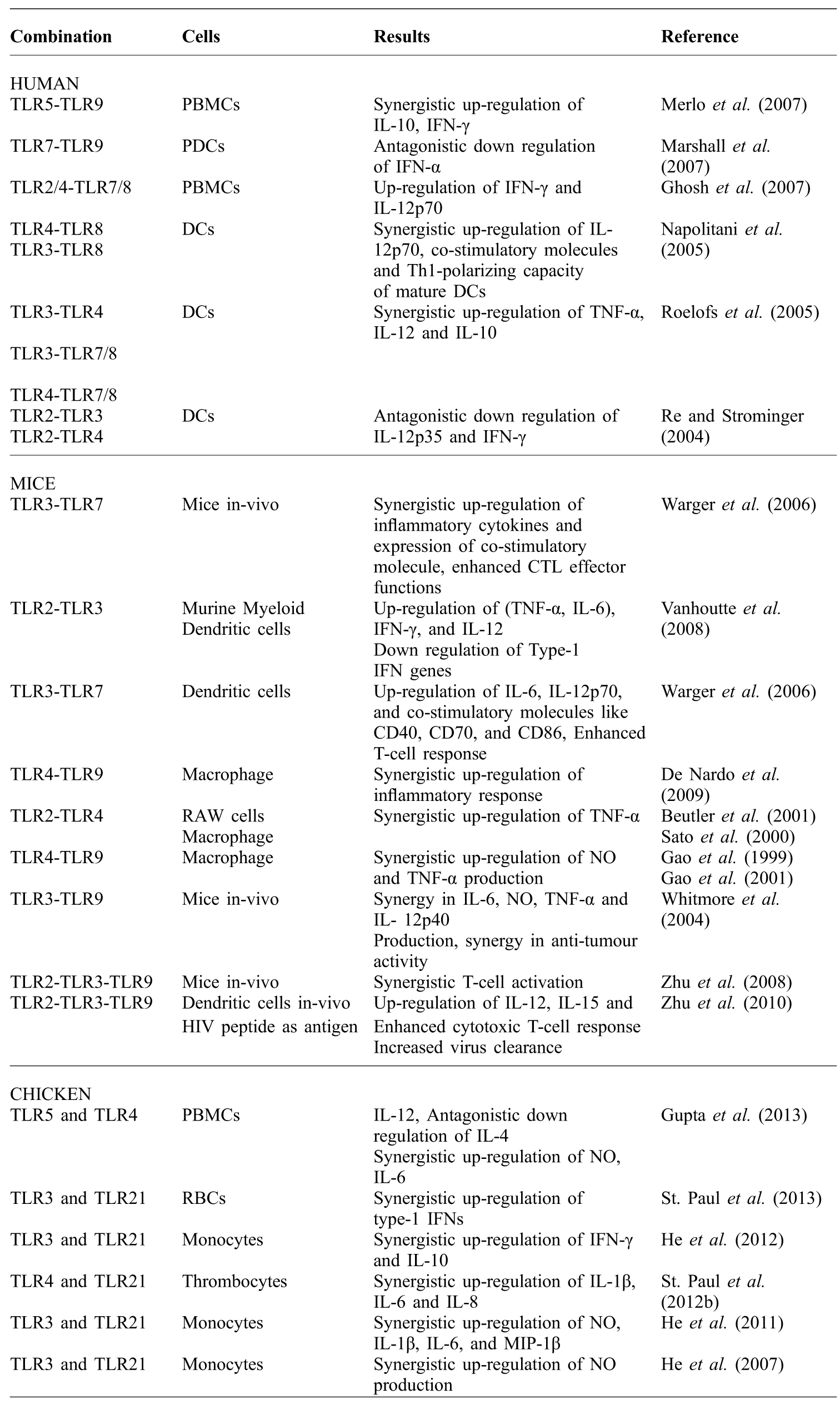
TLR combinations can produce a stronger and selective immune response and a number of studies have been undertaken in mice as well as in human immune cells (Table 2). Interaction between TLR5 and TLR9 has been shown to up-regulate the production of IL-10 and IFN-γ in human PBMCs (Merlo et al., Reference MERLO, CALCATERRA, MÈNARD and BALSARI2007). Another study with the human PBMCs reported the production of IFN-γ and IL-12p70 when stimulated with a combination of TLR2 or TLR4 with either TLR7 or TLR8, suggesting these combinations can promote Th1 type of immune response (Ghosh et al., Reference GHOSH, MICKELSON, SOLBERG, LIPSON, INGLEFIELD and ALKAN2007). Many other investigations with combinations like TLR3 or TLR4 with either TLR7 or TLR8 have also reported up-regulation of expression of co-stimulatory molecules, TNF-α and IL-12 in human dendritic cells (DCs) (Napolitani et al., Reference NAPOLITANI, RINALDI, BERTONI, SALLUSTO and LANZAVECCHIA2005; Roelofs et al., Reference ROELOFS, ABDOLLAHI-ROODSAZ, VAN LIESHOUT, SPRONG, VAN DEN HOOGEN, VAN DEN BERG and RADSTAKE2005). Roelofs et al. (Reference ROELOFS, ABDOLLAHI-ROODSAZ, VAN LIESHOUT, SPRONG, VAN DEN HOOGEN, VAN DEN BERG and RADSTAKE2005) tried a combination of TLR3 and TLR4 and found up-regulation of IL-10, TNF-α and IL-12 in human DCs. TLR7 and TLR9, which detects RNA and CpG-ODN, seem to antagonise the production of IFN-α when used simultaneously which can be seen as a way to avoid the overstimulation of immune system (Marshall et al., Reference MARSHALL, HEEKE, GESNER, LIVINGSTON and VAN NEST2007).
Combinations of TLR ligands: summary of applications.
*RAW cells: Mouse leukaemic monocyte macrophage cell line, PDCs: Plasmacytoid Dendritic Cells
The effects of various combinations of TLRs have been investigated both in-vitro as well as in-vivo in mice. Combinations of TLR2 -TLR4 and TLR4-TLR9, synergistically up-regulated the production of TNF-α and NO in mouse macrophage and RAW cells (Mouse leukemic monocyte macrophage cell line) (Sato et al., Reference SATO, NOMURA, KAWAI, TAKEUCHI, MÜHLRADT, TAKEDA and AKIRA2000; Beutler et al., Reference BEUTLER, GELBART and WEST2001; Gao et al., Reference GAO, XUE, PAPASIAN and MORRISON2001; De Nardo et al., Reference DE NARDO, DE NARDO, NGUYEN, HAMILTON and SCHOLZ2009). Interaction between TLR2-TLR3 and TLR3-TLR7 has been shown to significantly up-regulate the production of proinflammatory cytokines, IL-12 and T-effector cells response, but down regulated type-1 IFN genes with TLR2-TLR3 combination in mice dendritic cells (Warger et al., Reference WARGER, OSTERLOH, RECHTSTEINER, FASSBENDER, HEIB, SCHMID, SCHMITT, SCHILD and RADSAK2006; Vanhoutte et al., Reference VANHOUTTE, PAGET, BREUILH, FONTAINE, VENDEVILLE, GORIELY, RYFFEL and FAVEEUW2008). Promising results encouraged the researchers to take on in-vivo investigations and in this regard, combinations like TLR3-TLR7 and TLR3-TLR9 showed enhanced production of pro-inflammatory cytokines, IL-12, up-regulation of expression of co-stimulatory molecules, enhanced CTL effector functions and a synergy in anti-tumour activity (Whitmore et al., Reference WHITMORE, DEVEER, EDLING, OATES, SIMONS, LINDNER and WILLIAMS2004; Warger et al., Reference WARGER, OSTERLOH, RECHTSTEINER, FASSBENDER, HEIB, SCHMID, SCHMITT, SCHILD and RADSAK2006). Subsequent studies even tried triplet combinations of TLRs and reported encouraging results. A triplet combination of TLR2-TLR3-TLR9 has been shown to synergise the activation of T- cells, enhanced cytotoxic response and up-regulate IL-12 and IL-15 (Zhu et al., Reference ZHU, EGELSTON, VIVEKANANDHAN, UEMATSU, AKIRA, KLINMAN, BELYAKOV and BERZOFSKY2008; Reference ZHU, EGELSTON, GAGNON, SUI, BELYAKOV, KLINMAN and BERZOFSKY2010). When this combination was used along with HIV peptide antigen, an increased virus clearance was seen indicating the potential of combination against infectious viral diseases (Zhu et al., Reference ZHU, EGELSTON, GAGNON, SUI, BELYAKOV, KLINMAN and BERZOFSKY2010).
Overall, these studies in humans and mice suggest a pattern of immune response when combinations of TLRs are used. In humans the above combinations showed their potential in inducing an inflammatory response as well as a selective Th1 type immune response as seen in PBMCs and DCs (Napolitani et al., Reference NAPOLITANI, RINALDI, BERTONI, SALLUSTO and LANZAVECCHIA2005; Ghosh et al., Reference GHOSH, MICKELSON, SOLBERG, LIPSON, INGLEFIELD and ALKAN2007; Merlo et al., Reference MERLO, CALCATERRA, MÈNARD and BALSARI2007). In mice, in-vivo studies suggest these combinations induce the production of inflammatory cytokines, up-regulation of co-stimulatory molecules and the effector T-cell functions and an increased production of IL-12 indicating a Th1 type of immune response (Whitmore et al., Reference WHITMORE, DEVEER, EDLING, OATES, SIMONS, LINDNER and WILLIAMS2004; Zhu et al., Reference ZHU, EGELSTON, GAGNON, SUI, BELYAKOV, KLINMAN and BERZOFSKY2010).
adjuvant effect of combination of TLR ligands on chicken cells
In an effort to prevent the huge losses in the poultry industry associated with infectious diseases a new generation of vaccines has been developed; however, these vaccines require adjuvant to enhance the immune response. TLR based adjuvants have emerged as one of the important candidates and various studies have reported that combinations of TLRs can promote a stronger and selective immune response in chickens (Table 2). In this regard, a combination of TLR3 and TLR21 has been shown to up-regulate the expression of pro-inflammatory cytokines including IL-6, IL-8 and IL-1β, type-1 IFNs, IFN-γ, Th1 cytokines like IL-12 and production of NO in chicken monocytes and erythrocytes (He et al., Reference HE, GENOVESE, NISBET and KOGUT2007; Reference HE, MACKINNON, GENOVESE and KOGUT2011, St. Paul et al., Reference ST. PAUL, PAOLUCCI, BARJESTEH, WOOD and SHARIF2013). Flagellin, a TLR5 ligand, in combination with TLR4 ligand LPS synergistically up-regulated production of NO, pro-inflammatory cytokine IL-6 and Th1 cytokine IL-12 while at the same time this combination antagonistically down regulated Th2 cytokine IL-4 expression in chicken PBMCs (Gupta et al., Reference GUPTA, DEB, GAIKWAD, SARAVANAN, MOHAN and DEY2013). Similarly, TLR21 ligand, CpG-ODN synergistically up regulated the production of inflammatory cytokines (IL-6, IL-8 and IL-1β) in chicken thrombocytes when combined with LPS, a TLR4 ligand (St. Paul et al., Reference PAUL, PAOLUCCI, BARJESTEH, WOOD, SCHAT and SHARIF2012b). Taken together, these studies indicate a definite pattern of cytokine production in chicken cells when stimulated with a combination of TLR agonists. The combination of TLR4 and TLR5 promotes inflammatory responses as well as Th1 type immunity (Gupta et al., Reference GUPTA, DEB, GAIKWAD, SARAVANAN, MOHAN and DEY2013) while the combination of TLR3 and TLR21 activates chicken immune cells for the synergistically upregulated production of proinflammatory cytokines, type-1 IFN and IL-12 (He et al., Reference HE, GENOVESE, NISBET and KOGUT2007; Reference HE, MACKINNON, GENOVESE and KOGUT2011). Increased NO production indicates enhanced macrophage functions and hence greater killing capacity against pathogens.
advantages of using combinations of TLR ligands as adjuvant
A number of studies have explored the potential of TLR based adjuvants and these have shown very encouraging results. TLR agonists enhance the immune response to the antigen when given together through the production of cytokines and chemokines which in turn activate and shape the adaptive immune response. However, clearance of deadly infectious diseases requires a strong and direct immune response (Th1 or Th2). In this regard, combinations of TLR ligands have shown great potential. Certain combinations of TLRs can selectively promote cell mediated immunity over humoral. For example, it has been shown that the TLR5 ligand flagellin stimulates chicken PBMCs to produce a mixed type of immune response (both th1 and Th2) (Gupta et al., Reference GUPTA, DEB, GAIKWAD, SARAVANAN, MOHAN and DEY2013). A similar pattern has been observed in chicken splenocytes (St. Paul et al., Reference ST. PAUL, PAOLUCCI and SHARIF2012a). However, when flagellin is combined with LPS, it synergistically up-regulated the production of IL-12 and antagonistically down regulated IL-4, indicating an interaction between the two TLR pathways (Gupta et al., Reference GUPTA, DEB, GAIKWAD, SARAVANAN, MOHAN and DEY2013). Such Th1 inducing combinations can be used when selective cell mediated immunity is needed. However, flagellin, LPS and other TLR ligands impose a risk of toxicity when used in high doses. In this context, TLR combinations can help decrease the dose of an agonist and, hence, may alleviate toxicity related concerns associated with the usage of higher dosage (Gupta et al., Reference GUPTA, DEB, GAIKWAD, SARAVANAN, MOHAN and DEY2013). TLRs are also known to have antagonistic actions and TLR9 has been shown to interact with TLR7 and antagonise the immune activation in human PDCs as well as in sheep PBMCs (Marshall et al., Reference MARSHALL, HEEKE, GESNER, LIVINGSTON and VAN NEST2007; Booth et al., Reference BOOTH, BUZA, POTTER, BABIUK and MUTWIRI2010). These interactions in nature exist to avoid overstimulation of the immune system and induction of autoimmunity (Booth et al., Reference BOOTH, BUZA, POTTER, BABIUK and MUTWIRI2010; Nickerson et al., Reference NICKERSON, CHRISTENSEN, SHUPE, KASHGARIAN, KIM, ELKON and SHLOMCHIK2010; Santiago-Raber et al., Reference SANTIAGO-RABER, DUNAND-SAUTHIER, WU, LI, UEMATSU, AKIRA, REITH, MOHAN, KOTZIN and IZUI2010). This interaction can also be used to achieve a desired level of protection against a particular pathogen in cases where a TLR ligand is known to over stimulate the immune system.
Conclusions
TLRs exhibit a range of immunological effects on immune cells. Activation of TLRs results in the production of various cytokines, chemokines and interferons which in turn direct the immune response towards either Th1 or Th2 or a mixed immune response (St. Paul et al., Reference ST. PAUL, PAOLUCCI and SHARIF2012a; Gupta et al., Reference GUPTA, DEB, GAIKWAD, SARAVANAN, MOHAN and DEY2013). It has been shown that TLR5 ligand flagellin induced a mixed type of immune response in chicken PMBCs. However, when flagellin is used in combination with LPS it down regulated IL-4 and induced a strong Th1 response (Gupta et al., Reference GUPTA, DEB, GAIKWAD, SARAVANAN, MOHAN and DEY2013). These properties make TLR5 ligand an attractive option for the induction of both humoral as well as cell mediated immunity. When an elevated antibody-mediated immune response is desired, ligand alone might be used and in cases where cell mediated immunity is required a combination with LPS might be tried (Gupta et al., Reference GUPTA, DEB, GAIKWAD, SARAVANAN, MOHAN and DEY2013). However, the particular type of response may further be influenced by several factors including the type of combination, dose of TLR ligands or antigens, cell subtypes and route of immunisation (Gupta et al., Reference GUPTA, DEB, GAIKWAD, SARAVANAN, MOHAN and DEY2013). An effective strategy to avoid toxicity related concerns associated with the use of certain ligands may be the use of a combination of TLR ligands, such combinations will help in reducing the quantity of the agonists used. Although many of the studies undertaken suggest TLR combinations can be a potent adjuvant system in chicken vaccines (He et al., Reference HE, GENOVESE, NISBET and KOGUT2007; Reference HE, MACKINNON, GENOVESE and KOGUT2011; St. Paul et al., Reference PAUL, PAOLUCCI, BARJESTEH, WOOD, SCHAT and SHARIF2012b, Gupta et al., Reference GUPTA, DEB, GAIKWAD, SARAVANAN, MOHAN and DEY2013; St. Paul et al., Reference ST. PAUL, PAOLUCCI, BARJESTEH, WOOD and SHARIF2013), further detailed in-vivo investigations of the use of combinations of TLR ligands in chickens are warranted to measure their long term safety and efficacy, as well as for the determination of factors such as the route of administration and dose. The results of such studies will provide a foundation for the possible use of TLR combinations as adjuvant in future chicken vaccines.