Introduction
In patients with cyanotic CHD, chronic cyanosis can lead to the development of aortopulmonary collateral arteries. Reference Triedman, Bridges, Mayer and Lock1,Reference Salim, Case, Sade, Watson, Alpert and DiSessa2 This process represents a compensatory physiological response aimed at maintaining pulmonary blood flow and alleviating cyanosis. However, the presence of aortopulmonary collateral arteries contributes to increased pulmonary arterial pressure and pulmonary vascular resistance and also augments preload to the systemic ventricle. These effects can impede the establishment of Fontan circulation in patients with functionally single ventricles who are candidates for the Fontan procedure. Moreover, the volume of blood flow through aortopulmonary collateral arteries has been reported to correlate with adverse outcomes after Fontan procedure, such as prolonged pleural effusion and extended hospitalisation. Reference Grosse-Wortmann, Drolet and Dragulescu3–Reference Odenwald, Quail and Giardini6 Therefore, in patients with a functionally single ventricle, coil embolisation of aortopulmonary collateral arteries is often performed prior to Fontan procedure. Reference Parakash, Satiroglu and Porras7–Reference Dori, Glatz and Hanna9
Various types of coils are used for embolisation of aortopulmonary collateral arteries, but based on their embolisation mechanisms, they can be broadly classified into platinum coils and hydrogel coils. Reference Ding, Dai, Lewis, Cloft and Kallmes10 Platinum coils occlude the target vessel through both the mechanical obstruction of the coil itself and thrombus formation within the vessel. In contrast, hydrogel coils achieve embolisation through the expansion of the hydrogel material. Hydrogel coils consist of a radio-opaque platinum-based framework coated with bioactive polymers that expand upon contact with blood. Reference Killer, Arthur, Barr, Richling and Cruise11,Reference Nambiar, Bozlar, Angle, Jensen and Hagspiel12 The advantages of hydrogel coils include increased volumetric filling of the target vessel and a reduced number of devices required compared with platinum coils. Reference Maleux, Deroose and Fieuws13
A common issue observed after coil embolisation of aortopulmonary collateral arteries is the occurrence of post-procedural fever. Post-embolisation fever may prompt us to reconsider the treatment strategy, including the use of antibiotics and further investigation of potential causes. In particular, when coil embolisation is performed prior to Fontan procedure, the presence of fever and inflammation may necessitate postponement of the operation itself. However, to date, there have been no reports investigating the factors associated with post-embolisation fever following coil embolisation of aortopulmonary collateral arteries in patients with CHD. Therefore, in this study, we retrospectively examined the factors contributing to the development of fever after coil embolisation of aortopulmonary collateral arteries.
Methods
This was a single-centre, retrospective study approved by the Institutional Review Board of Saitama Medical University (No. 2025-074). A total of 180 patients who underwent coil embolisation for CHD at the Department of Pediatric Cardiology, Saitama Medical University International Medical Center, between 2007 and 2023 were retrospectively reviewed.
To ensure clear comparison of results, cases of veno-venous shunt (n = 20), pulmonary arteriovenous fistula (n = 8), and coronary artery fistula (n = 2) were excluded. Thus, 150 patients who underwent coil embolisation for aortopulmonary collateral arteries were initially included. Of these, 53 patients were excluded due to incomplete records regarding the type and number of coils used, leaving 97 patients for final analysis. These 97 patients were divided into three groups according to the type of coil used: P group (n = 52): embolisation performed using platinum coils only, P–H group (n = 29): embolisation performed using both platinum and hydrogel coils, and H group (n = 16): embolisation performed using hydrogel coils only.
For each group, we examined patient characteristics (age, gender, and underlying diagnosis), procedural variables (contrast volume and procedure time), coil characteristics (type, number, diameter, and total length), and the presence or absence of post-embolisation fever (defined as ≥38.0°C). In cases with fever, inflammatory markers from blood tests at the time of fever – specifically white blood cell count, C-reactive protein and D-dimer levels – were also evaluated. In addition, we assessed the histopathological findings after coil embolisation using platinum and hydrogel coils, evaluating the presence and degree of inflammatory cell infiltration.
All continuous data are expressed as the mean± standard deviation. Categorical data were examined with a Pearson’s chi-square test or Fisher’s exact test. Comparisons between the 2 groups were performed using a Mann–Whitney’s U test. Comparisons within groups were made using an analysis of variance. A p < 0.05 was considered statistically significant. All statistical analyses were performed using JMP version 11.2.0 (SAS, Cary, NC) and GraphPad PRISM version 5.01 (GraphPad Software, Inc., La Jolla, CA) software.
Results
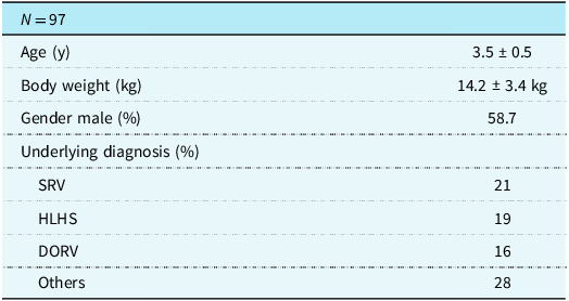
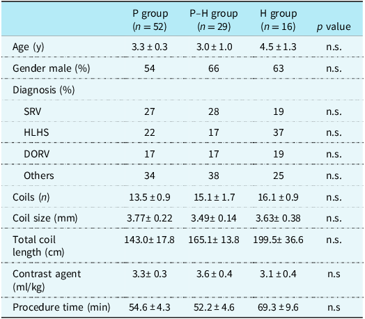
The characteristics of the 97 patients (a total of 1,412 coils) included in the study are shown in Table 1. The mean age was 3.5 ± 0.5 years, and the mean body weight was 14.2 ± 3.4 kg; 58.7% of the patients were male. The underlying diagnoses include single right ventricle (21%), hypoplastic left heart syndrome (19%), and double outlet right ventricle (16%; subpulmonic VSD 7%, subaortic VSD type 4%, doubly committed VSD 4%, and remote VSD 1%). Of the 97 patients, 88 underwent coil embolisation immediately before the Fontan procedure, while 9 underwent embolisation as treatment for excessive pulmonary blood flow. Table 2 summarises the patient characteristics by group. No significant differences were observed among the three groups in age, sex, number of coils used, coil diameter, total coil length, contrast volume, or procedure time.
Patient characteristics

SRV = single right ventricle; HLHS = hypoplastic left heart syndrome; DORV = double outlet right ventricle.
Patient characteristics by group

SRV = single right ventricle; HLHS = hypoplastic left heart syndrome; DORV= double outlet right ventricle.
Procedural success, defined as complete angiographic occlusion of the target vessel, did not differ significantly among groups (P group: 31/52; P–H group: 15/29; H group: 10/16; p = 0.64). Similarly, no significant difference was observed in re-intervention rates (P group: 6/52; P–H group: 2/29; H group: 3/16; p = 0.71).
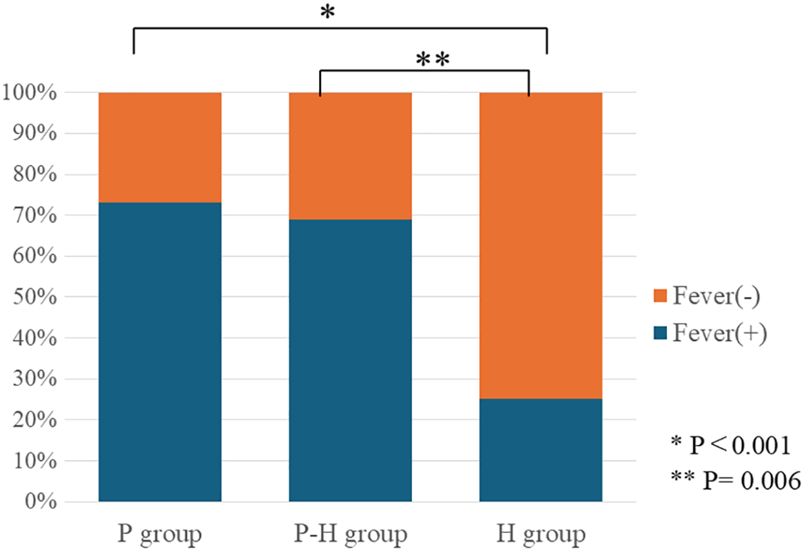
Post-embolisation fever occurred in 64% of all patients. The incidence of fever by group was 73.0% (38/52) in the P group, 68.9% (20/29) in the P–H group, and 25.0% (4/16) in the H group. The incidence of fever was significantly lower in the H group compared with the other two groups (H group versus P group: p < 0.001; H group versus P–H group: p = 0.006; Figure 1). In the P–H group, the overall proportion of platinum coils was 57.1 ± 3.5%. When this group was further divided into patients who developed fever after coil embolisation and those who did not, the proportion of platinum coils was 62.1 ± 2.7% in the febrile group and 45.9 ± 8.9% in the afebrile group (p = 0.031). Most cases of fever developed within 24 hours after coil embolisation (P group: 92%, P–H group: 85%, and H group: 100%). The duration of fever was 1.9 ± 0.2 days in the P group, 2.1 ± 0.3 days in the P–H group, and 1.0 day in the H group. Although the difference was not statistically significant, the H group tended to have a shorter duration of fever. Regarding the relationship between the number of coils used and the presence of fever, patients with fever received 14.9 ± 0.7 coils, while those without fever received 13.8 ± 1.1 coils, showing no significant association between the number of coils used and the occurrence of fever (p = 0.378).
Comparison of the incidence of fever among each group. The H group showed a significantly lower incidence of fever compared with the other two groups.

Regarding the haematologic laboratory findings, the white blood cell count and C-reactive protein levels were compared before and after the onset of fever. The white blood cell count showed no significant change following fever onset (8,999 ± 456/µL versus 10,321 ± 604/µL, p = 0.084). In contrast, C-reactive protein levels increased markedly after fever onset, demonstrating a statistically significant elevation (0.53 ± 0.17 mg/dL versus 4.21 ± 0.49 mg/dL, p < 0.0001). Also, D-dimer levels were measured in 15 of the 62 patients who developed fever. Among the patients in whom D-dimer levels were measured, all but one showed elevated values (0.89–15.59μg/mL). The mean time interval from coil embolisation to blood sampling was 1.5 ± 0.6 days. Among the three groups, there was no statistical significance of mean time interval (P group; 1.5 ± 0.3 days, P–H group; 1.8 ± 0.5 days, H group; 1.3 ± 1.5days, p = 0.18). Among the 62 patients who developed fever, blood cultures were obtained in 11 cases, and all results were negative. Antibiotics were administered in 56 of the 62 patients. The overall duration of antibiotic therapy was 4.1 ± 0.3 days. In comparisons among the three groups, the duration was 3.9 ± 0.4 days in P group, 4.3 ± 0.4 days in P–H group, and 2.5 ± 0.5 days in H group (p = 0.09). Although there was a tendency towards a longer duration in P–H group, the difference did not reach statistical significance.
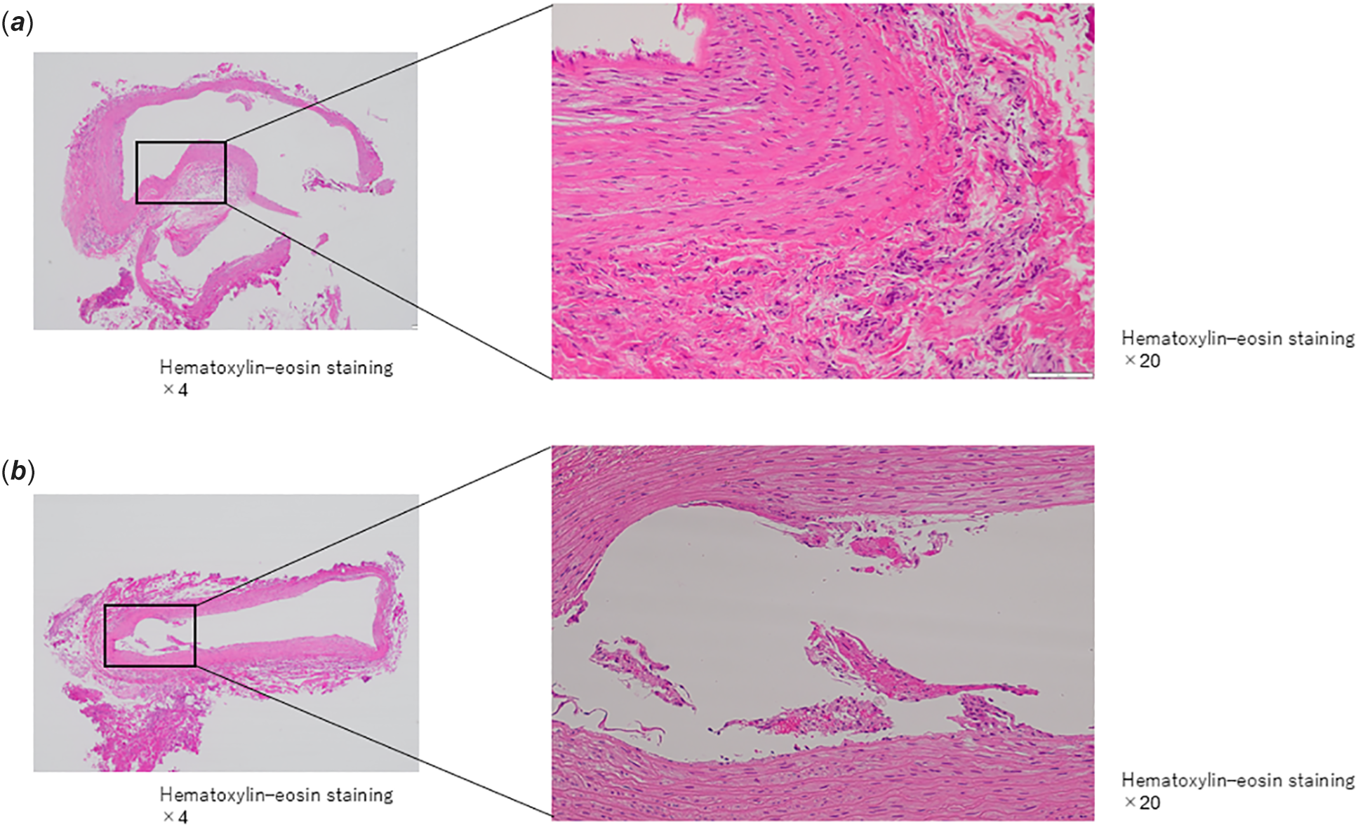
In the pathological examination of the internal thoracic artery obtained during the Fontan procedure in patients from the P–H group, fibrin and inflammatory cells were observed adhering to the vascular lumen at the site of platinum coil embolisation. Inflammatory cell infiltration, including neutrophils, was mainly seen around the vessel (Figure 2 a ). In contrast, in vessels embolised with hydrogel coils, attachment of fibrin and inflammatory cells to the vascular lumen was not evident, and only mild infiltration of inflammatory cells, including neutrophils, was observed (Figure 2 b ). This patient underwent coil embolisation using a total of 17 coils (platinum coil proportion: 58.8%). The patient developed a fever on the day following the procedure, which resolved two days later. The white blood cell count showed no significant change before and after the procedure (6220/µL to 5750/µL), whereas C-reactive protein increased from 0.024 mg/dL to 3.18 mg/dL. Blood cultures were negative; however, antibiotics were administered for five days. After discussion with the cardiac surgery team, the Fontan procedure was postponed by one week.
( a ) Histopathological findings of the aortopulmonary collateral vessel after platinum coil. Fibrin and inflammatory cells are observed adhering to the vascular lumen. Also, Infiltration of inflammatory cells, including neutrophils, is seen mainly around the vessel. ( b ) Histopathological findings of the aortopulmonary collateral vessel after hydrogel coil. Attachment of fibrin and inflammatory cells to the vascular lumen is not evident. On the other hand, Mild infiltration of inflammatory cells, including neutrophils, is observed in the vessel.

Discussion
In this study, we demonstrated that hydrogel coils significantly suppressed post-procedural fever and inflammation compared with platinum coils following coil embolisation of aortopulmonary collateral arteries. As there were no significant differences among the three groups in patient characteristics, coil characteristics other than coil type (number of coils, coil diameter, and total coil length), contrast volume used, or procedural time, coil type appears to be the strongly associated factor determining the occurrence of post-procedural fever. In coil embolisation, it is important to consider factors such as the embolic efficacy of the coil, its operability, and the economic aspects of medical care. In the present analysis, no significant difference in the vascular occlusion rate was observed among the three groups. In addition, no significant difference in the post-treatment reintervention rate was observed among the three groups. Regarding recanalisation after vascular embolisation, platinum coils have been reported to be associated with a lower recanalisation rate compared with fibered coils. Reference Fohlen, Namur, Ghegediban, Laurent, Wassef and Pelage14,Reference Cortese, Ghozy and Zarrintan15 On the other hand, particularly in paediatric patients, the presence or absence of post-embolisation fever may also become an important factor when considering subsequent treatment strategies. As for the cause of fever, bacterial infection appears unlikely, given that blood cultures were negative in all cases, defervescence occurred within a few days, and no leukocytosis was observed on laboratory testing. Furthermore, the elevation of D-dimer levels in all but one patient supports the thrombus-forming embolisation mechanism, particularly associated with fibered coils. This thrombotic process may have triggered an inflammatory response, thereby resulting in fever. To the best of our knowledge, this is the first report to discuss post-embolisation fever and inflammatory response following coil embolisation in paediatric patients with CHD.
Regarding inflammation after coil embolisation, numerous studies—particularly in the field of neuro-endovascular therapy and in animal models—have reported similar findings. Fohlen et al. investigated the histopathological changes in renal arteries embolised with fibered coils and hydrogel coils using an animal model. Reference Fohlen, Namur, Ghegediban, Laurent, Wassef and Pelage16 They showed that the percentage of surface area occupied by thrombus was significantly lower with hydrogel coils than with fibered coils. The authors suggested that hydrogel coils prevent thrombus formation by inhibiting fibrin network formation through the properties of the hydrogel material. Furthermore, Killer et al. Reference Killer, Arthur, Barr, Richling and Cruise11 reported that the foreign-body response to platinum coils consisted predominantly of macrophages and giant cells, whereas in the case of hydrogel coils, mononuclear macrophages were aligned along the hydrogel surface. They suggested that this difference in the inflammatory reaction may be attributed to the hydrophilic nature of the hydrogel. In our study as well, histopathological findings showed that, compared with platinum coils, hydrogel coils exhibited only mild infiltration of inflammatory cells around the embolised vessels. In the case of hydrogel coils, the activation of inflammatory cells is considered to be mild, which may account for the difference in the frequency of fever compared with platinum coils. Platinum coils exert their embolic effect through mechanical occlusion by the coil itself and through thrombus formation. Therefore, the induction of an inflammatory response associated with thrombosis is unavoidable. In contrast, hydrogel coils achieve embolisation through the hydrophilic hydrogel material itself, and because this mechanism does not require the process of thrombus formation, it is considered less likely to induce an inflammatory reaction.
This study has several limitations. First, the number of patients included was small, and the study was a single-centre retrospective analysis. Therefore, larger prospective studies are warranted. In addition, since the target vessels in this study were limited to aortopulmonary collateral arteries, it remains unclear whether similar findings can be observed in other types of target vessels.
In conclusion, this study is the first to investigate post-embolisation fever following aortopulmonary collateral artery coil embolisation in children with CHD. Compared with platinum coils, hydrogel coils are less likely to induce an inflammatory response, resulting in a lower incidence of fever. In the future, when performing coil embolisation, it is recommended that coil selection be made with consideration of the potential for post-procedural fever.
Acknowledgements
The authors used ChatGPT (OpenAI) to improve the readability and linguistic quality of the manuscript. No AI tools were involved in data analysis, interpretation, or drawing scientific conclusions. The authors are fully responsible for the final content.