Introduction
Trichinellosis is a parasitic infection caused by Trichinella spiralis (T. spiralis), which is a serious parasitic zoonosis and a globally endemic disease (Pavel et al. Reference Pavel, Ursoniu, Lupu and Olariu2023). Trichinella larvae are frequently found in raw or undercooked pork meat, which can infect humans (Kim et al. Reference Kim, Choi, Kim, Kang, Jeon, Jung, Lee and Oh2011). Human trichinellosis outbreaks occur frequently throughout the world, which emphasises the necessity of creating a potent vaccine (Tang et al. Reference Tang, Li, Li, Xie, Guan, Zhao, Yang, Liu and Xu2022). Various vaccines, including whole worm extract, excretory-secretory antigen, recombinant DNA vaccine, and protease vaccines, have been developed to prevent T. spiralis (Zhang et al. Reference Zhang, Li and Fu2018).
Secretory cathepsin B protease is a major papain-like cysteine protease which plays key roles in parasite survival, host invasion, and host immune response. It has been documented that immunising mice infected with T. spiralis with a recombinant T. spiralis cathepsin B-like protein (TsCBP) causes a T-helper2 (Th2) response, so Trichinella cathepsin B proteins appear to be promising targets for vaccine development (Yang et al. Reference Yang, Li, Yang, Pan, Liao and Zhou2018). Because of their distinct physiochemical characteristics, nanovaccines offer an advantageous tool for vaccine delivery. They also have an efficient loading capacity, excellent biocompatibility, and a suitable size for antigen-based immunotherapy nanoparticle-based systems (Jeevanandam et al. Reference Jeevanandam, Barhoum, Chan, Dufresne and Danquah2018).
IgG, IgM, and IgA antibodies play a vital role in preventing Trichinella infections by ensnaring and quickly expelling infectious first-stage rhabditiform larvae (L1 larvae), decreasing adult worm fecundity, and aiding in the death of newborn larvae (Zhai et al. Reference Zhai, Sun, Liu, Liu, Bai, Wang, Wu and Chen2019).
Adjuvants have been used extensively in various vaccines because of their capacity to modify cellular and humoral immune responses without causing local or widespread reactions. To assess the protective immunity against T. spiralis infection, different antigens were emulsified using aluminium hydroxide in a murine model of trichinellosis. It acts through activating numerous receptors and ligands in the host, and it secretes Th2-type cytokines that promote mast cell activation and hyperplasia, both of which are essential for intestinal epithelial cell permeability and worm expulsion. In addition, Th2-type cytokines like interleukin-4 (IL-4) and IL-13 can activate macrophages in a different way, which mediates host defence against the parasite infection (Zhang et al. Reference Zhang, Li and Fu2018).
Despite the promise of nanotechnology in vaccine development, nano-based vaccines also face significant challenges that must be acknowledged. Nanovaccine platforms can suffer from low antigen loading efficiency, difficulties in long-term storage, high production costs, and complexities in large-scale manufacturing, which may limit their practical application in resource-limited settings (Cheng et al. Reference Cheng, Chai, Rong, Xin, Gu, Zhou and Hong2025).
Additionally, concerns about biocompatibility and long-term safety of nanoparticle carriers remain, as their physicochemical properties may influence toxicity and immune interactions that are not yet fully understood (engineered nanovaccines challenges). These limitations highlight the need for continued research into optimising nanovaccine design and evaluating alternative delivery strategies to ensure both efficacy and safety (Arabiyat Reference Arabiyat2025).
The purpose of this study was to investigate the protective effects of cathepsin B protein and cathepsin B protein transported on nanoliposomes with or without aluminium hydroxide as an adjuvant in murine trichinellosis.
Materials and methods
The experimental study started in March 2021 and was completed in July 2022. This study was carried out in the Medical Parasitology Department, the Central Laboratory and Pathology Laboratories of Menoufia University’s Faculty of Medicine, as well as in the Medical Parasitology Laboratories of the Theodore Bilharz Research Institute in Giza, Egypt. All applicable institutional guidelines for the care and use of animals were followed with IRB NO 12/2020PARA35.
Study design: Mice were divided into two main groups: group I, (the control group) (20 mice) which then divided into control negative Ia (10 mice and control positive Ib (10 mice), and group II, (vaccinated group) (40 mice) which divided into 4 subgroups (IIa, IIb, IIc, and IId) according to the given vaccine. Every subgroup was split into two equal subgroups based on scarification. On 8th day post infection (p.i.) (subgroups: Ia1, Ib1, IIa1, IIb1, IIc1, and IId1) and on 35th day p.i. (subgroups: Ia2, Ib2, IIa2, IIb2, IIc2, and IId2) to assess the protective effect of TsCBP in intestinal and muscular phases, respectively, as shown in Table 2.
Experimental animals: In this study, 60 Swiss male albino mice weighing an average of 25 ± 0.2 grams were kept in a controlled environment with a temperature of 25°C and a humidity of 70%. The mice were kept in accordance with the National Guidelines for Experimental Animal Welfare and were acquired from the Theodore Bilharz Research Institute (TBRI) in Giza, Egypt.
Drugs and chemicals: As in Table 1.
Drugs and chemicals used in the study

Study groups: Table 2 showed the characteristics and time of sacrification of each subgroup
Characteristics of the study groups and subgroups

Mice infection: The strain of T. spiralis was kindly provided by Tanta University’s Medical Parasitology Department in Egypt. For an oral infection, 300 T. spiralis larvae were administered to each mouse.
Preparation of TsCBPA: To obtain the crude antigen, adult T. spiralis were placed in 1 millilitre of phosphate-buffered saline (PBS), homogenised for 10 minutes at 4°C at 1500 rpm, and then diluted with 3 millilitres of PBS. To isolate TsCB, the crude antigen was purified by two methods: ion exchange chromatography method using diethyl amino-ethyl-cellulose (DEAE) (sephadex chromatography A-50), and gel filtration chromatography method using Sephadex-G-200 HR column (Kueakhai et al. Reference Kueakhai, Changklungmoa, Cheukamud, Osotprasit, Chantree, Preyavichyapugdee, Sobhon and Meemon2021). Protein content was adjusted (5 μg/ml) in PBS. Part of the pure TsCB was conjugated later with nanoliposomes (Tang et al. Reference Tang, Li, Li, Xie, Guan, Zhao, Yang, Liu and Xu2022).
Preparation of nano-TsCBPA: Liposome nanoparticles were created using standard wet chemical methods, which involved incubating 150 ml of liposome nanoparticles with 600 μg of CB. After that, the conjugates underwent a one-hour ultra-centrifugation at 2500 rpm and 10°C (Liu et al. Reference Liu, Wen, Zhan, Li, Shen, Yang, Zhou and Liu2015).
Drug regimens
TsCBPA and TsCBPA-nanoliposome: At zero, seven, and fourteen days, mice received a subcutaneous injection of 40 μg of cathepsin B in PBS. On the 21st day, they were given an oral infection with 300 T. spiralis muscle larvae (Swilam et al. Reference Swilam, Nasreldin, Abd-Elrahman and Nasr2022).
Aluminium hydroxide: It was kindly obtained from TBRI. It was used as adjuvant at 1.2 mg/dose in subgroups IIc (TsCBPA) and IId (nanoliposomes-TsCBPA) as shown in Table 2. Its optimal adjuvanticity was obtained at a concentration of 2% (Lindblad, Reference Lindblad2004).
Tissue specimens’ collection: Mice of all groups were euthanised by cervical dislocation on the 8th dpi, and at the end of the experiment (35th dpi) (Swilam et al. Reference Swilam, Nasreldin, Abd-Elrahman and Nasr2022). Small intestine and muscle specimens were collected on the 8th and 35th dpi, respectively and were divided into two parts. One part was fixed in formalin 10% for further H&E staining and histopathological examination. The 2nd part was frozen at −80°C for the detection of T. spiralis larval DNA by real-time PCR.
Blood samples collection: The serum was separated by centrifuging 2 ml of blood that had been extracted from the submandibular vein in plain tubes and allowed to clot. The serum was stored at −20°C for subsequent immunological measurement of IgM, IgG1 and estimation of serum T. spiralis circulating larval antigen using ELISA.
Isolation and counting of T. spiralis adults: According to Shi et al., adult worms were isolated and counted from the small intestine of all infected and vaccinated subgroups (Ib1, IIa1, IIb1, IIc1, and IId1) on the 8th dpi (Shi et al. Reference Shi, Sang, Beom, Ki, Mo, Joo, Cho, Lee, Nakayama and Sekine2008).
The reduction rate was calculated = [(average number in the positive control group − the average number in the treatment group)/average number in the control group] × 100% (Saad et al. Reference Saad, Ashour, Abou Rayia and Bedeer2016).
Isolation and counting of T. spiralis larvae: On the 35th day after vaccination, larvae from abdominal muscles of the vaccinated and infected subgroups (Ib2, IIa2, IIb2, IIc2, and IId2) were isolated by artificially digesting muscle tissue. In warm tap water, 1% concentrated HCL (volume/volume) and 1% pepsin (weight/volume) were added to create artificial gastric juice. At 37°C, the mixture was constantly stirred for two hours using an electric stirrer. A 50-mesh/inch copper sieve was used to filter out the coarse particles from the digested product. To gather excysted larvae, the filtered fluid ran through a 200 mesh/inch sieve. Under a microscope, both live (coiled and motile) and dead (coma-shaped and non-motile) larvae were counted (Wang et al. Reference Wang, Cui, Hu, Liu and Wang2014). The reduction rate was calculated as discussed before.
Estimation of serum IgM and IgG1 levels by ELISA: T. spiralis crude antigen at 125 ng/ml concentration was coated on Maxsorp 96-well microtitre plates overnight at 4°C in carbonate-bicarbonate buffer with a pH of 9.4. Plates were blocked for two hours at room temperature with 250 μl/well of 2% bovine serum albumin diluted in PBS, pH 7.2, and 0.05% Tween 20 (PBS/BSA/T-20). Sera were diluted 1:100 in PBS/T20, added to plates (100 μl/well), and left to incubate for one hour at room temperature. After washing, 100 μl/well of diluted anti-mouse IgM and IgG1 (1 mg/ml each; Southern Biotech, USA) in PBS/T20 (1:5000 for IgG1) were added for 90 min. Reaction developed by adding 100 μl/well of P-nitrophenyl phosphate substrate and incubation until appearance of yellow color. Using an ELISA reader (Bio-Rad, UK), the absorbance was read at 405 nm (El-Aswad et al. Reference El-Aswad BE, Harba, Moharm and Mahmoud2019).
Estimation of serum T. spiralis antigen level by indirect ELISA: Polyclonal antibodies were produced by immunisation of a rabbit with crude Trichinella antigen. Sandwich ELISA was performed by subsequent addition of the coated unlabeled antibodies (primary), tested mice serum, and enzyme-labeled polyclonal antibodies specific to the antigen. Addition of a suitable substrate resulted in hydrolysis, the degree of which was proportional to the amount of attached enzyme. Absorbance of Trichinella antigen was measured at 492 nm using an ELISA reader (Bio-Rad microplate reader, Richmond, CA) (Liu et al. Reference Liu, Jing, Cui, Fu and Wang2013).
Detection of T. spiralis larva DNA in intestinal and muscular tissue by real-time PCR (Cuttell et al. Reference Cuttell, Corley, Gray, Vanderlinde, Jackson and Traub2012): Genomic DNA was extracted from frozen tissues (intestinal and muscular) using JET™ Genomic DNA Purification Mini Kit (Thermo Scientific, EU/Lithuania). The study utilised a forward primer (‘5-CATGGTTAGGTGAGATATTG-CCTGC-3’) and reverse primer (‘5-GGTCCTCC-TTCCAGAAGATCTACTTTG-3’). Fluorescence was measured in the green channel and data was collected at the extension step. Cycle threshold (Ct) values were individually calculated by internal software using a manual threshold setting of 0.2, and ‘dynamic tube’ and ‘ignore first 10 cycles’ functions were activated. Each run was followed by a melt-curve analysis to verify specificity and match amplicons with positive control melt curve peaks. The amount of larval DNA identified by real-time PCR was inversely correlated with the cycle threshold in muscle tissue.
Histopathological study: At Menoufia University’s Faculty of Medicine, the Pathology Department, specimens from the small intestine and muscles had been fixed and processed. After being cleaned and dehydrated, the specimens were cleared in xylene and then embedded in paraffin blocks. H&E was used to stain paraffin sections (5 μm thick), which were subsequently inspected under a microscope (Bancroft and Gamble Reference Bancroft and Gamble2008).
Statistical analysis: Using SPSS (20), IBM Corp. released in 2011 (IBM SPSS Statistics for Windows, Version 20.0, Armonk, New York), data were coded, tabulated, and analysed. Quantitative type data were expressed as mean and standard deviation (SD). The comparison of quantitative variables between two groups was determined using the Mann-Whitney test. The difference between the two means of the two distinct groups was evaluated using Post Hoc Value. A statistically significant difference was considered when P = 0.05 or lower.
Ethical consideration: The current study used experimental animals in accordance with international guidelines. The study was approved by the Ethical Committee of the Faculty of Medicine, Menoufia University, Egypt (IRB;12/2020PARA35).
Results
Parasitological results
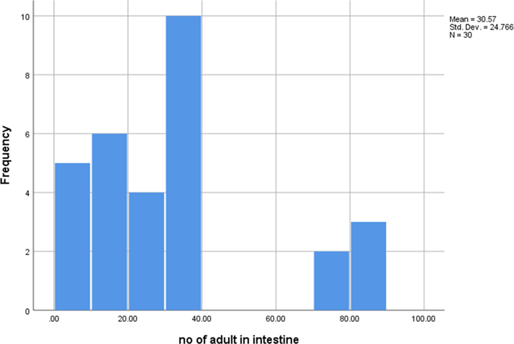
Intestinal adult count: According to Table 3, graphs 1 and 2, GIId1 (vaccinated with liposome-TsCBPA with aluminium hydroxide) had the highest reduction rate (77.2%), while GIIa1 (vaccinated with TsCBPA) had the lowest reduction rate (56%) with a high statistically significant difference between the two vaccinated groups (P7 <0.001**).
Comparison of T. spiralis adult count among vaccinated mice groups (intestinal phase), sacrificed on the 8th dpi

GIa1: Control negative; GIb1: Control positive; GIIa1: Vaccinated by TsCBPA; GIIb1: Vaccinated by nanoliposome-TsCBPA; GIIc1: Vaccinated by TsCBPA with aluminium hydroxide; GIId1: Vaccinated by nanoliposome-TsCBPA with aluminium hydroxide; F: One-way ANOVA test; *: Significant (P <0.05); **: Significant (P <0.001).
The number of T. spiralis adult in intestinal phase.

The number of T. spiralis adult in intestinal phase (subgroups).

In addition, the reduction rate was 59.6% in GIIc1 (vaccinated with TsCBPA with aluminium hydroxide) with a highly statistically significant difference compared to GIId1 (vaccinated with nanoliposome-TsCBPA with aluminium hydroxide) (P10 <0.001**) (Table 3).
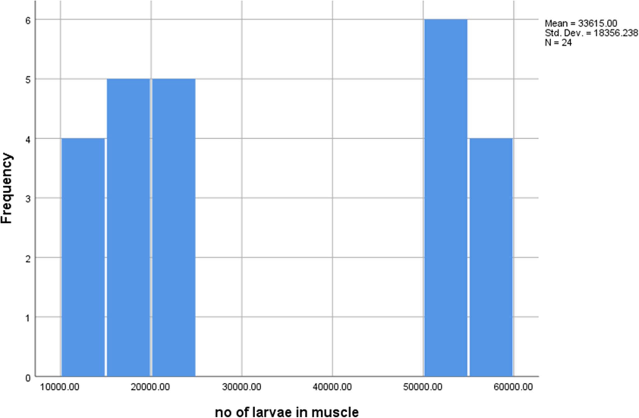
Muscle larval count: Mice sacrificed on the 35th day post-immunisation (dpi) had the highest larval reduction rate (73.8%) in GIId2 (vaccinated with nanoliposome-TsCBPA with aluminium hydroxide) and the lowest larval reduction rate (34.9%) in GIIa2 (vaccinated with TsCBPA). The difference between the two vaccinated groups was statistically significant (P7 <0.001**). In addition, there was a high statistically significance difference (P10 <0.001**) between GIIc2 (vaccinated with TsCBPA with aluminium hydroxide) with reduction 56.8% and GIId2 (vaccinated with nanoliposome-TsCBPA with aluminium hydroxide) with reduction 73.8% (P10 <0.001**) (Table 4) (graphs 3 and 4).
Comparison of larval count among vaccinated mice groups during the muscular phase, sacrificed on the 35th dpi

GIa2: Control negative; GIb2: Control positive; GIIa2: Vaccinated by TsCBPA; GIIb2: Vaccinated by nanoliposome-TsCBPA; GIIc2: Vaccinated by TsCBPA with aluminium hydroxide; GIId2: Vaccinated by nanoliposome-TsCBPA with aluminium hydroxide; F: One-way ANOVA test; *: Significant (P <0.05); **: Significant (P <0.001).
The number of T. spiralis larvae in muscular phase.

The number of T. spiralis larvae in muscular phase (subgroups).

Immunological results
IgM level: The intestinal phase showed a highly statistically significant difference between the two vaccinated subgroups (GIIa1 and GIId1) (P7 <0.001**), with the highest IgM level in GIId1 (vaccinated with liposome-TsCBPA with aluminium hydroxide) being 0.85 ± 0.04 and the lowest IgM level in GIIa1 (vaccinated with TsCBPA) being 0.49 ± 0.04. Also, there was a highly significant difference between both groups vaccinated with liposome-TsCBPA alone or with adjuvant (GIIb1 and GIId1) (P9 <0.001**) (Table 5).
Comparison of serum IgM level among vaccinated groups (intestinal)

F = One-way ANOVA test.
* = statistically significant.
** = statistically highly significant.
P1: between group GIb1 and GIIa1.
P2: between group GIb1 and GIIb1.
P3: between group GIb1 and GIIc1.
P4: between group GIb1 and GIId1.
P5: between group GIIa1 and GIIb1.
P6: between group GIIa1 and GIIc1.
P7: between group GIIa1 and GIId1.
P8: between group GIIb1 and GIIc1.
P9: between group GIIb1 and GIId1.
P10: between group GIIc1 and GIId1.
In the muscular phase, the highest IgM value was 0.48 ± 0.06 in GIId2 (vaccinated with liposome-TsCBPA with aluminium hydroxide), while the lowest IgM level was 0.29 ± 0.04 in GIIa2 (vaccinated with TsCBPA), with a highly statistically significance difference between both vaccinated groups (P7 <0.001**). Also, there was a significant difference between both groups vaccinated with liposome-TsCBPA alone or with adjuvant (GIIb2 and GIId2) (P9 = 0.003*) (Table 6).
Comparison of serum IgM level among vaccinated groups (muscular)

F = One-way ANOVA test.
* = statistically significant.
** = statistically highly significant.
P1: between group GIb2 and GIIa2.
P2: between group GIb2 and GIIb2.
P3: between group GIb2 and GIIc2.
P4: between group GIb2 and GIId2.
P5: between group GIIa2 and GIIb2.
P6: between group GIIa2 and GIIc2.
P7: between group GIIa2 and GIId2.
P8: between group GIIb2 and GIIc2.
P9: between group GIIb2 and GIId2.
P10: between group GIIc2 and GIId2.
IgG1 level: In the intestinal phase, the highest level of IgG1 among vaccinated groups was 0.3 ± 0.03 in GIId1 (vaccinated with liposome-TsCBPA), while the lowest level was 0.21 ± 0.08 in GIIa1 (vaccinated with TsCBPA) with statistically significance difference between both subgroups (P7 <0.001**). There was no significant difference (P9 = 0.802) between both groups vaccinated with nanocathepsin alone or with adjuvant (GIIb1 and GIId1) (Table 7).
Comparison of serum IgG1 level among vaccinated groups (intestinal)

F = One-way ANOVA test.
* = statistically significant.
** = statistically highly significant.
P1: between group GIb1 and GIIa1.
P2: between group GIb1 and GIIb1.
P3: between group GIb1 and GIIc1.
P4: between group GIb1 and GIId1.
P5: between group GIIa1 and GIIb1.
P6: between group GIIa1 and GIIc1.
P7: between group GIIa1 and GIId1.
P8: between group GIIb1 and GIIc1.
P9: between group GIIb1 and GIId1.
P10: between group GIIc1 and GIId1.
In the muscular phase, the highest IgG1 serum level was 0.85 ± 0.04 in GIId2 (vaccinated with liposome-TsCBPA with aluminium hydroxide), while the lowest level was 0.48 ± 0.04 in GIIa2 (vaccinated with TsCBPA), with highly statistically significance difference between both vaccinated groups (P7 <0.001**). Also, there was a significant difference between both groups vaccinated with liposome-TsCBPA alone or with adjuvant (GIIb2 and GIId2) (P9 = 0.006*) (Table 8).
Comparison of serum IgG1 level among vaccinated subgroups (muscular)

F = One-way ANOVA test.
* = statistically significant.
** = statistically highly significant.
P1: between group GIb2 and GIIa2.
P2: between group GIb2 and GIIb2.
P3: between group GIb2 and GIIc2.
P4: between group GIb2 and GIId2.
P5: between group GIIa2 and GIIb2.
P6: between group GIIa2 and GIIc2.
P7: between group GIIa2 and GIId2.
P8: between group GIIb2 and GIIc2.
P9: between group GIIb2 and GIId2.
P10: between group GIIc2 and GIId2.
T. spiralis circulating larval antigen level: In the intestinal phase, the lowest level was 0.25 ± 0.04 in GIId1 (vaccinated with liposome-TsCBPA with aluminium hydroxide), while the highest level was 0.54 ± 0.05 in GIIa1 (vaccinated with TsCBPA), with statistically highly significant difference (P7 <0.001**). Also, there was a highly significant difference between both groups vaccinated with liposome-TsCBPA alone or with adjuvant (GIIb1 and GIId) (P9 < 0.001**) (Table 9).
Serum level of T. spiralis circulating larval antigen in vaccinated groups (intestinal)

F = One-way ANOVA test.
** = statistically highly significant.
P1: between group GIb1 and GIIa1.
P2: between group GIb1 and GIIb1.
P3: between group GIb1 and GIIc2.
P4: between group GIb1 and GIId1.
P5: between group GIIa1 and GIIb1.
P6: between group GIIa1 and GIIc1.
P7: between group GIIa1 and GIId1.
P8: between group GIIb1 and GIIc1.
P9: between group GIIb1 and GIId1.
P10: between group GIIc1 and GIId1.
In the muscular phase, GIId2 (vaccinated with liposome-TsCBPA) had the lowest level of Trichinella antigen (0.61 ± 0.07), while GIIa2 (vaccinated with TsCBPA) had the highest level (0.86 ± 0.03) with a statistically significant difference (P7 <0.001**). Furthermore, neither the adjuvant nor the liposome-TsCBPA-vaccinated groups differed significantly (GIIb2 and GIId2) (P9 = 0.613) (Table 10).
Serum level of T. spiralis circulating larval antigen in vaccinated groups (muscular)

F = One-way ANOVA test.
* = statistically significantly.
** = statistically highly significant.
P1: between group GIb2 and GIIa2.
P2: between group GIb2 and GIIb2.
P3: between group GIb2 and GIIc2.
P4: between group GIb2 and GIId2.
P5: between group GIIa2 and GIIb2.
P6: between group GIIa2 and GIIc2.
P7: between group GIIa2 and GIId2.
P8: between group GIIb2 and GIIc2.
P9: between group GIIb2 and GIId2.
P10: between group GIIc2 and GIId2.
Real-time PCR: The intestinal phase revealed a highly statistically significant difference between the two groups (P7 <0.001**), with the lowest cycle threshold value in GIIa1 (vaccinated with TsCBPA) being 35.5 ± 0.52, and the highest value in GIId1 (vaccinated with liposome-TsCBPA with aluminium hydroxide) being 38.35 ± 0.42. Also, there was a statistically significant difference (P9 = 0.003) between both groups vaccinated with nanocathepsin alone or with adjuvant (GIIb1 and GIId1) (Table 11) (graph 5).
Comparison between the cycle threshold values of RT-PCR among vaccinated groups (intestinal)

N.B: Cycle threshold is indirectly proportional to the amount of larval DNA detected by real-time PCR
F = One-way ANOVA test.
* = statistically significant.
** = statistically highly significant.
P1: between group GIb1 and GIIa1.
P2: between group GIb1 and GIIb1.
P3: between group GIb1 and GIIc1.
P4: between group GIb1 and GIId1.
P5: between group GIIa1 and GIIb1.
P6: between group GIIa1 and GIIc1.
P7: between group GIIa1 and GIId1.
P8: between group GIIb1 and GIIc1
P9: between group GIIb1 and GIId1.
P10: between group GIIc1 and GIId1.
Shows comparison between the cycle threshold values of RT-PCR among vaccinated groups (intestinal).

In the muscular phase, the GIIa2 (vaccinated with TsCBPA) group had the lowest cycle threshold value (34.42 ± 0.52), while the GIId2 (vaccinated with liposome-TsCBPA with aluminium hydroxide) group had the highest value (37.15 ± 0.34). The difference between the two groups was highly statistically significant (P7 <0.001**). There was no statistically significant difference (P9 = 0.486) between both groups vaccinated with liposome-TsCBPA alone or with aluminium hydroxide (GIIb2 and GIId2, respectively) with cycle threshold 36.95 ± 0.59 and 37.15 ± 0.34, respectively (Table 12) (graph 6).
Comparison between the cycle threshold values of RT-PCR among vaccinated groups (muscular).

N.B: Cycle threshold is indirectly proportional to the amount of larval DNA detected by real-time PCR. **means highly significant difference.
P1: between group GIb2 and GIIa2.
P2: between group GIb2 and GIIb2.
P3: between group GIb2 and GIIc2.
P4: between group GIb2 and GIId2.
P5: between group GIIa2 and GIIb2.
P6: between group GIIa2 and GIIc2.
P7: between group GIIa2 and GIId2.
P8: between group GIIb2 and GIIc2.
P9: between group GIIb2 and GIId2.
P10: between group GIIc2 and GIId2.
shows comparison between the cycle threshold values of RT-PCR among vaccinated groups (muscular).

Histopathological results: In the intestinal phase (Table 13), GIa1 (control negative) mice displayed no inflammation and normal intestinal tissue with a preserved villous/crypt ratio (Fig. 1a). In contrast, edema, ulceration, intestinal villi destruction, and a dense inflammatory infiltrate in the lamina propria were observed in the infected untreated subgroup (GIb1) (Fig. 1b, c, and d). Group GIIa1 (vaccinated with TsCBPA) showed a moderate decrease in villous/crypt ratio (black and blue arrows), blunting of villi (short black arrows) with moderate inflammation ++ (black box), and mild edema (H&E ×40) (Fig. 1e).
Comparison of degree of inflammation among Studied groups (intestinal phase).

** means highly significant difference.
(a) Transverse section (TS) in small intestine of group GIa1 (non-infected non-treated group) showed preserved villous (continuous black arrows)/crypt (interrupted blue arrows) ratio, no inflammation, and no edema (H&E ×40 with camera zooming). (b) TS in small intestine of group GIb1 (infected non-treated) showed severe inflammation with lymphoid aggregates (black arrows) (H&E ×40). (c) TS in small intestine of group GIb1 (infected non-treated) showed severe inflammation with lymphoid aggregates (arrows) (H&E ×100). (d) TS in small intestine of group GIb1 (infected non-treated) showed marked blunting of villi (black short arrows), a decrease in villous (black arrows)/crypt (blue arrows) ratio, and moderate edema (green arrows) (H&E ×40). (e) TS in small intestine of group GIIa1 (vaccinated with TsCBPA) showed a moderate decrease in villous/crypt ratio (black and blue arrows), blunting of villi (short black arrows) with moderate inflammation ++ (black boxes), and mild edema (green arrows) (H&E ×40). (f) TS in small intestine of GIIb1 (vaccinated with liposome-TsCBPA) showed a mild decrease in villous (black arrow)/crypt (blue arrow) ratio with moderate inflammatory cells ++ (black boxes), and mild edema (green arrows) (H&E ×40). (g) TS in small intestine of GIIc1 (vaccinated with TsCBPA with aluminium hydroxide) showed a moderate ++ decrease in villous/crypt ratio (black and blue arrows), blunting of villi (short black arrows) with mild inflammation +, and mild edema (green arrows) (H&E ×40). (h) TS in small intestine of GIId1 (vaccinated with liposome-TsCBPA with aluminium hydroxide) showed a mild decrease + in villous (black arrow)/crypt (blue arrow) ratio with mild inflammatory cells + (black boxes), and mild edema (green arrows) (H&E ×40).

Additionally, subgroup GIIb1 (vaccinated with liposome-TsCBPA) displayed mild edema and a moderate reduction in the villous (black arrow)/crypt (blue arrow) ratio along with moderate inflammatory cells ++ (black box) (Fig. 1f). Subgroup GIIc1 (vaccinated with TsCBPA with aluminium hydroxide) showed a moderate decrease in villous/crypt ratio (black and blue arrows), blunting of villi (short black arrows) with mild inflammation, and mild edema (H&E ×40) (Fig. 1g).
GIId1 (vaccinated with liposome-TsCBPA with aluminium hydroxide) displayed mild edema and a mild reduction in the villous (black arrow)/crypt (blue arrow) ratio along with mild inflammatory cells + (black box) (H&E ×40) (Fig. 1h).
Regarding the muscular phase (Table 14), the infected control group (GIb2) had a dense larval deposition with thick intact capsule with an intense inflammatory infiltrate primarily composed of lymphocytes and macrophages, along with fat deposition (Fig. 2b, c, and d). In contrast, the non-infected control group (GIa2) displayed normal muscle without inflammation and the absence of larvae (Fig. 2a).
Comparison of degree of inflammation among Studied groups (muscular phase).

** means highly significant difference.
(a) TS in skeletal muscle of non-infected non-treated group (GIa2) showed normal muscle with no inflammation and no larvae (H&E ×100). (b, c) TS in skeletal muscle of the infected non-treated group (GIb2) showed encysted T. spiralis larvae (black arrows) (H&E ×100). (d) TS in skeletal muscle of the infected non-treated group (GIb2) showed a large density of encysted T. spiralis larvae (black arrows) associated with severe inflammation (blue arrow) (H&E ×400). (e) TS in skeletal muscle of the GIIa2 group (vaccinated with TsCBPA) showed a moderate (++) density of encysted T. spiralis larvae associated with moderate inflammation (++), mild fragmentation (+) of internal structures (black arrows), and moderate degeneration of surrounding muscle (white arrows) (H&E ×100). (f) TS in skeletal muscle of GIId2 group (vaccinated with liposome-TsCBPA with aluminium hydroxide) showed encysted T. spiralis larvae associated with mild inflammation (+) (black circle), and mild fragmentation of internal structures (black arrows) (H&E ×100). (g) TS in skeletal muscle of the GIIb2 group (vaccinated with liposome-TsCBPA) showed moderate eosinophilic infiltrate (++) (black circles) (H&E ×400). (h) TS in skeletal muscle of the GIIc2 group (treated with TsCBPA with aluminium hydroxide) showed encysted T. spiralis larvae associated with mild inflammation (+) (yellow arrow), mild fragmentation of internal structures (+) (black arrows), and moderate degeneration of surrounding muscle (++) (green arrow) (H&E ×100).

The TsCBPA-vaccinated GIIa2 group displayed a moderate density of encysted T. spiralis larvae along with mild internal structure fragmentation (black arrows), moderate inflammation, and moderate surrounding muscle degeneration (H&E ×100) (Fig. 2e).
Additionally, transverse section in the skeletal muscle of the TsCBPA-vaccinated GIIa2 group displayed a moderate eosinophilic infiltrate (circles) (H&E ×400) (Fig. 2g). The GIIc2 group (treated with TsCBPA with aluminium hydroxide) showed encysted T. spiralis larvae associated with mild inflammation, mild fragmentation of internal structures (black arrows), and moderate degeneration of surrounding muscle (H&E ×100) (Fig. 2h). In addition, GIId2 group (vaccinated with nanoliposome-TsCBPA with aluminium hydroxide) showed encysted T. spiralis larvae associated with mild inflammation (circle), and mild fragmentation of internal structures (black arrows) (H&E ×100) (Fig. 2f).
Discussion
Effective anti-helminthic drugs, such as mebendazole and albendazole, are limited in efficacy against encysted T. spiralis or newborn larvae, highlighting the need for vaccine development (El-Sayed et al. Reference El-Sayed, Mahmoud, El-Shenawy and Yousof2023; Saad et al. Reference Saad, Ashour, Abou Rayia and Bedeer2016).
The global focus on pandemic preparedness has emphasised the importance of vaccines that activate both humoral and cellular immunity and induce long-term immune memory.
Cathepsins, particularly secretory cathepsin B (TsCBP), are promising vaccine candidates due to their role in parasite survival, host invasion, and immune modulation, inducing Th2-biased responses and specific antibody production (Hassan et al. Reference Hassan, Zelt, Perera, Ndao and Ward2019; Yang et al. Reference Yang, Li, Yang, Pan, Liao and Zhou2018; Yuthithum et al. Reference Yuthithum, Phuphisut, Reamtong, Kosoltanapiwat, Chaimon, Kobpornchai, Thawornkuno, Malaithong, Sawatdichaikul and Adisakwattana2024).
Nanoparticle-based delivery systems, including liposomes, enhance antigen uptake by phagocytic and mucosa-associated lymphoid tissues, improving antigen presentation and eliciting targeted immune responses (Gheibi Hayat and Darroudi Reference Gheibi Hayat and Darroudi2019; Jeevanandam et al. Reference Jeevanandam, Barhoum, Chan, Dufresne and Danquah2018). TsCBP expression occurs across all T. spiralis life stages and is localised in the stichosome and cuticle (Cui et al. Reference Cui, Han, Yue, Liu, Song, Yan, Lei, Zhang, Jiang and Wang2019). Antibodies (IgG, IgM, IgA) play a critical role in controlling infection by trapping larvae, reducing worm fecundity, and mediating larval killing (Zhai et al. Reference Zhai, Sun, Liu, Liu, Bai, Wang, Wu and Chen2019). Aluminium hydroxide as an adjuvant promotes Th2-type cytokine responses, mast cell activation, and macrophage-mediated defense, enhancing vaccine efficacy (Zhang et al. Reference Zhang, Li and Fu2018).
In the present study, TsCBPA and cathepsin B protein conjugated with nanoliposomes, with or without aluminium hydroxide, showed strong protective and therapeutic effects against enteral and muscular phases of infection, consistent with previous reports of cathepsin B-induced reductions in adult worms and muscle larvae (Cui et al. Reference Cui, Han, Yue, Liu, Song, Yan, Lei, Zhang, Jiang and Wang2019; Hassan et al. Reference Hassan, El-Rahman, El-Hamed, Fattah, Harb, Mohamed and Sarhan2019, Reference Hassan, Houle, Labrie, Perera, Dozois, Ward and Ndao2023). Nanoparticles and adjuvants synergistically improved immunogenicity, antibody production, and parasite clearance (Darwish et al. Reference Darwish, Hussien, Fahmy and Bayaumy2022; Jin et al. Reference Jin, Liu, Ding, Zhang, Yang, Wang, Yang and Liu2020; Lu and Gao, Reference Lu and Gao2023).
A mixed Th1/Th2 immune response was also demonstrated by Buffoni et al. and Villa-Mancera et al. who found that vaccination of sheep with Fasciola hepatica cathepsin resulted in a significant decrease in fluke length and width, wet weights, and egg output, as well as an increase in specific IgG1 and IgG2 (Buffoni et al. Reference Buffoni, Garza-Cuartero, Pérez-Caballero, Zafra, Javier Martínez-Moreno, Molina-Hernández, Pérez, Martínez-Moreno and Mulcahy2020; Villa-Mancera et al. Reference Villa-Mancera, Olivares-Pérez, Olmedo-Juárez and Reynoso-Palomar2021).
Additionally, Darwish et al. found that when zinc nanoparticles were administered to T. spiralis mice at the intestinal and muscle larvae stages, there was a significant improvement in pathological features along with a decrease in the number of larvae and almost 100% adult worm mortality (Darwish et al. Reference Darwish, Hussien, Fahmy and Bayaumy2022).
Serine and cysteine proteases are essential for mediating larval invasion, growth, and survival in hosts as well as for rupturing the integrity of the gut epithelium. In order to prevent larval invasion and development, T. spiralis cysteine proteases may be the primary target molecules for vaccines and treatments (Song et al. Reference Song, Lu, Han, Yan, Zhang, Liu, Long, Cui and Wang2022).
The administration of recombinant T. spiralis cathepsin B protein (rTsCB) as a vaccine produces anti-rTsCB antibodies that partially impede the larval intrusion of enterocytes, providing a therapeutic effect, as a study found that rTsCB facilitated the larval invasion of intestinal epithelium (Han et al. Reference Han, Yue, Hu, Liu, Liu, He, Long, Cui and Wang2020). The current findings highlight the importance of cysteine and serine proteases as vaccine targets, given their role in larval invasion, growth, and survival (Han et al. Reference Han, Yue, Hu, Liu, Liu, He, Long, Cui and Wang2020; Song et al. Reference Song, Lu, Han, Yan, Zhang, Liu, Long, Cui and Wang2022).
Immunisation with recombinant cathepsin B proteins, particularly when delivered via nanoliposomes, elicited strong Th2 responses and increased IgG1 and IgM levels, reinforcing the intestinal stage as a key target for infection control (Cui et al. Reference Cui, Han, Yue, Liu, Song, Yan, Lei, Zhang, Jiang and Wang2019, Sun et al. Reference Sun, Lei, Ren, Zhang, Guo, Long, Liu, Jiang, Wang and Cui2019; Yang et al. Reference Yang, Li, Yang, Pan, Liao and Zhou2018; Zeng et al. Reference Zeng, Zhang, Zhang, Yan, Song, Long, Dan Liu, Wang and Cui2021).
Furthermore, Hassan et al. (Reference Hassan, Houle, Labrie, Perera, Dozois, Ward and Ndao2023) found that administration of chromosomally integrated cathepsin B to Schistosoma mansoni-infected mice reduced intestinal egg burden by 78.4%, worm burden by 80.4%, and hepatic egg counts by 75.2% (Hassan et al. Reference Hassan, Houle, Labrie, Perera, Dozois, Ward and Ndao2023).
Additionally, Buffoni et al. found that vaccinating sheep against fascioliasis with F. hepatica cathepsin L (FhCL1) resulted in a sharp and significant increase in IgG1 after vaccination (Buffoni et al. Reference Buffoni, Garza-Cuartero, Pérez-Caballero, Zafra, Javier Martínez-Moreno, Molina-Hernández, Pérez, Martínez-Moreno and Mulcahy2020).
However, Zhang et al. and Sun et al. found no statistically significant difference in the levels of anti-Trichinella spiralis IgG between all groups prior to and following treatment with albendazole and ivermectin packed with nitazoxanide on solid lipid nanoparticles (NTZ-SLNs), respectively. This could be because liposome nanoparticles work on Th2 cells, causing them to release cytokines that encourage B cells to produce antibodies more than solid lipid nanoparticles do (Sun et al. Reference Sun, Lei, Ren, Zhang, Guo, Long, Liu, Jiang, Wang and Cui2019; Zhang et al. Reference Zhang, Li and Fu2018).
Molecular evaluation using real-time PCR in the current study supported the parasitological findings and concurred with those of Cuttell et al., who measured cycle threshold using the same primers and chose to target a conserved gene region that permitted the amplification of a 195 bp Trichinella fragment (Cuttell et al. Reference Cuttell, Corley, Gray, Vanderlinde, Jackson and Traub2012). Similarly, Al-Attar et al. (Reference Al-Attar TA, El-Kersh, Sadek, Harba, Osheiba and Brakat2020) reported that vaccination with adult and larval excretory-secretory (ES) antigens significantly reduced T. spiralis DNA in muscle tissues, with the greatest reduction observed in groups treated with larval ES, confirming the efficacy of antigen-based immunisation (Al-Attar et al. Reference Al-Attar TA, El-Kersh, Sadek, Harba, Osheiba and Brakat2020).
Histopathological analyses confirmed reduced intestinal damage, larval deposition, and worm burden, aligning with prior studies using nanoparticles or recombinant antigens (Abo-Hussien et al. Reference Abo-Hussien, Soliman, Sweed, Mandour, Abokhalil and Atia2023; Atia et al. Reference Atia, Abokhalil, Sweed, Moaz and Abou Hussien2021; Darwish et al. Reference Darwish, Hussien, Fahmy and Bayaumy2022; El-Melegy et al. Reference El-Melegy, Ghoneim, El-Dien and Rizk2019; Hassan et al. Reference Hassan, El-Rahman, El-Hamed, Fattah, Harb, Mohamed and Sarhan2019; Khalifa et al. Reference Khalifa, Ramadan, Youssef, Auda, El-Bahy and Taha2023; Liu et al. Reference Liu, Wen, Zhan, Li, Shen, Yang, Zhou and Liu2015; Nassef et al. Reference Nassef, Moharm, Atia, Brakat, Abou Hussien and Shamseldeen2019; Taha et al. Reference Taha, Abdel-Radi, Youssef, Auda, El-Bahy and Ramadan2022; Youssef et al. Reference Youssef, Mohamed, Ismail, Elzorba, Galal and Elbanna2021).
This was attributed to the increased solubility, bioavailability, and efficiency of nanoparticles, leading to damage in ultrastructural morphology and decreased worm count as it was able to cause irreversible genotoxic damage (Youssef et al. Reference Youssef, Mohamed, Ismail, Elzorba, Galal and Elbanna2021).
Liposome-TsCBPA with aluminium hydroxide provided the most consistent protective effect, likely due to enhanced cellular uptake, antigen presentation, and synergistic immunomodulatory activity without host toxicity (Gogoi et al. Reference Gogoi, Mani and Bhatnagar2025; Terhune and Deth Reference Terhune and Deth2018; Vassoudevane et al. Reference Vassoudevane, Mariebernard and Rajendran2023).
Cysteine peptidases have inherent adjuvanticity, which may explain why our results differ from those of Abdel Aziz et al., who found vaccination without adjuvant to be less effective. Furthermore, using CFA to induce type 1 immunity can counteract cysteine peptidase immunogenicity (Abdel Aziz et al. Reference Abdel Aziz, Tallima, A and El Ridi2020). Nano liposome-TsCBPA combined with aluminium hydroxide achieved the best outcomes, likely due to enhanced cellular uptake, synergistic immunostimulatory effects, absence of host toxicity, and the ability of liposomes to target multiple parasite life stages (Vassoudevane et al. Reference Vassoudevane, Mariebernard and Rajendran2023). Aluminium hydroxide enhanced Th2-mediated responses, promoting mast cell activation, eosinophil recruitment, and intestinal worm expulsion (Gogoi et al. Reference Gogoi, Mani and Bhatnagar2025; Terhune and Deth Reference Terhune and Deth2018; Zhang et al. Reference Zhang, Li and Fu2018). Similar findings were reported in Schistosoma and Fasciola models using cathepsin plus adjuvants (Kueakhai et al. Reference Kueakhai, Changklungmoa, Cheukamud, Osotprasit, Chantree, Preyavichyapugdee, Sobhon and Meemon2021; Perera et al. Reference Perera, Hassan, Jia, Ricciardi, McCluskie, Weeratna and Ndao2020).
Overall, these findings support the potential of cathepsin-based nanovaccines, combined with appropriate adjuvants, as an effective strategy for controlling trichinellosis and reducing both intestinal and muscular parasite burdens, while highlighting the value of nanoparticles for targeted antigen delivery and immune modulation (Abdel Aziz et al. Reference Abdel Aziz, Tallima, A and El Ridi2020; Kueakhai et al. Reference Kueakhai, Changklungmoa, Cheukamud, Osotprasit, Chantree, Preyavichyapugdee, Sobhon and Meemon2021; Perera et al. Reference Perera, Hassan, Jia, Ricciardi, McCluskie, Weeratna and Ndao2020; Ssemaganda et al. Reference Ssemaganda, Giddam, Zaman, Skwarczynski, Toth, Stanisic and Good2019; Tallima and El Ridi Reference Tallima and El Ridi2020).
In the present study, liposome-TsCBPA with aluminium hydroxide had the best results as a vaccine agent in all groups and all assessment parameters. This was attributed to the delivery of anti-parasitic drugs using liposome vesicles as drug delivery has enhanced their efficacy as it enhances cellular uptake of drugs and due to the synergistic impacts without causing any toxicity on the host cells. In addition, the delivery of antigens as vaccine candidates using liposomes elicits a pronounced immune response in clearing the infection. Also, liposomes alone elicit a strong anti-parasitic activity with immunomodulatory potential. Additionally, the liposome possesses significant inhibitory potential on multiple life stages of the parasite cycle (Vassoudevane et al. Reference Vassoudevane, Mariebernard and Rajendran2023).
Conclusion
Vaccination with cathepsin B, particularly in nanoliposome form, provided effective protection against experimental trichinellosis by reducing adult and larval burdens, limiting intestinal and muscular pathology, increasing serum IgM and IgG1 levels, elevating muscle tissue cycle threshold values, and decreasing T. spiralis antigen levels. Aluminium hydroxide further enhanced vaccine efficacy.
A deeper understanding of T. spiralis biology is essential for optimising antigen-based vaccines. Further studies are needed to validate candidate antigens, clarify immune mechanisms, and explore alternative nanoparticles and adjuvants to improve vaccine effectiveness.