Introduction
Spermatogenesis in mammals is an unique differentiation process that requires a fine and precise control of gene expression. Male germ cell development is governed by extensive epigenetic changes that result in reprogramming of the paternal genome to function in the future embryo. If epigenetic errors occur in the germline, they may have consequences for fertility and the health of future offspring (Goldman et al., Reference Goldman, Bailey, Pfaffle, Al Masri, Christian and Fleming2009). Epigenetic reprogramming involves the incorporation of testis-specific histone variants, a high level of DNA methylation in the final stages of spermiogenesis and multiple chemical modifications of histone tails (Burlibaşa and Gavrilă, Reference Burlibaşa, Gavrilă, Niculescu and Haggarty2011).
A major event in male meiosis is the replacement of canonical histones with specific variants, some of these testis-specific. Meiotic I division (reductional division) yields secondary spermatocytes that rapidly undergo meiotic II division (equational division), generating haploid, round spermatids. During post-meiotic maturation (spermiogenesis) the nuclei of spermatids undergo a dramatic remodelling process. There is a widespread reorganization of the haploid nucleus followed by DNA compaction, global chemical modifications and gene expression is completely shut off in the spermatozoon. Many investigations have revealed the critical role of histone variants and post-translational modifications of core histones in these processes (Caron et al., Reference Caron, Govin, Rousseaux, Khochbin and Jeanteur2005; Burlibaşa and Zărnescu, Reference Burlibaşa and Zărnescu2013). The replacement of canonical histones with specialized histone variants and post-translational histone modifications, provide complex levels of epigenetic regulation to cellular processes such as DNA repair, cell differentiation, DNA recombination.
Post-translational modifications of histones are associated with different transcriptional states and represent heritable epigenetic markers for chromatin structure and function (Berger, Reference Berger2002; Burlibaşa and Suciu, Reference Burlibaşa and Suciu2015).
In recent years, a histone code hypothesis was issued. This hypothesis postulated that combinations of histone modifications could define specific signals and serve as an interface language between histones and chromatin-modifying machinery to assign particular structure and function to specific chromatin domains (Strahl and Allis, Reference Strahl and Allis2000). Each histone has several sites of potential modifications recognized by the specific cellular factors (Jenuwein and Allis, Reference Jenuwein and Allis2001).
The histone code also acts in male gametes as stage-specific histone modifications, therefore a testis-specific histone code has been postulated (Govin et al., Reference Govin, Caron, Lestrat, Rousseaux and Khochbin2004).
Many works highlighted that the dynamics of histone modifications are essential for the normal spermatogenesis and early embryogenesis. Among the modifications, acetylation of lysine residues is well characterized and known to be involved in chromatin remodelling and gene expression changes (Natsume-Kitatani et al., Reference Natsume-Kitatani, Shiga and Mamitsuka2011; Burlibaşa and Suciu, Reference Burlibaşa and Suciu2015). Spermatogenesis is a suitable model to study the roles of acetylation in cellular functions associated with cell proliferation and differentiation. Two sets of events are important in spermatogenesis: one is shutting off the gene transcription in final stages of this process and the other is the replacement of histones by sperm nuclear basic proteins (protamines). The mechanism of histone displacement is not known in detail, but there is evidence that it involves a strong link between some kind of histone modifications (especially histone H4 acetylation) and the subsequent replacement of histones by protamines or protamine-like proteins in condensing spermatids (Burlibaşa and Gavrilă, Reference Burlibaşa and Gavrilă2005; Caron et al., Reference Caron, Govin, Rousseaux, Khochbin and Jeanteur2005). The treatment of spermatogenic cell suspension with trichostatin A, a histone-deacetylase inhibitor, demonstrated the role for this kind of enzymes in the control of histone hyperacetylation and removal of histones and protamine-like/protamine replacement proteins in condensing spermatids (Fenic et al., Reference Fenic, Sonnack, Failing, Bergman and Steger2004; Burlibaşa and Gavrilă, Reference Burlibaşa and Gavrilă2005). According to the histone code hypothesis, switch of acetylation/deacetylation events would serve as a marker for the recruitment of remodelling enzymes acting on modified histones (Caron et al., Reference Caron, Govin, Rousseaux, Khochbin and Jeanteur2005; Burlibaşa and Gavrilă, Reference Burlibaşa, Gavrilă, Niculescu and Haggarty2011). Histone acetylation seems to be tightly associated with histone replacement. The mechanisms of histone displacement are not fully discovered, but there could be a link between extensive acetylation of histones in elongating spermatids and the replacement of histones by protamines or protamine-like proteins in condensing spermatids (Burlibaşa and Gavrilă, Reference Burlibaşa and Gavrilă2005, Caron et al., Reference Caron, Govin, Rousseaux, Khochbin and Jeanteur2005).
Another important epigenetic modification in male gametogenesis is represented by global DNA methylation in spermiogenesis. The crucial role of DNA methylation in male germ cells is supported by the presence of distinct methylation profiles (Shiota et al., Reference Shiota, Kogo, Ohgane, Imamura and Urano2002; Eckhardt et al., Reference Eckhardt, Lewin, Cortese, Rakyan and Attwood2006; Oakes et al., Reference Oakes, La Salle, Smiraglia, Robaire and Trasler2007a). The unique state of DNA methylation might arise in primordial germ cells (PCGs) of the developing embryo between 10.5 and 12.5 days post-coitus (Reik et al., Reference Reik, Dean and Walter2001), when the DNA methylation patterns are erased and re-established (Hajkova et al., Reference Hajkova, Erhardt, Lane, Haaf and El-Maarri2002; Maatouk et al., Reference Maatouk, Kellam, Mann, Lei and Li2006).
Epigenetic processes are essential for development and differentiation, but they can also arise in individuals either by random change or under the influence of the environment (Gavrilă et al., Reference Gavrilă, Burlibaşa, Usurelu, Radu, Magdalena, Ardelean and Carabas2008).
In the recent years, much research into the role of epigenetic modifications, especially in DNA methylation in the male germ cells, has been made. Despite this huge package of information, the interplay between different kinds of epigenetic modifications remains poorly understood. To gain insight into the mechanisms controlling histone hyperacetylation/histone replacement and DNA methylation, we investigated the in vivo effect of 5-aza-2′-deoxycytidine (5-azadC; Decitabine) on murine spermatogenesis. 5-azadC is a DNA methyltransferase (DNMT) inhibitor, used as an anticancer agent for the treatment of different types of cancer. It has the ability to demethylate tumour-suppressor genes and cause replication-dependent cytotoxicity (Gabbara and Bhagwat, Reference Gabbara and Bhagwat1995).
Materials and Methods
Animals
BALB/c mice (Mus musculus) were chosen as the mammalian model organism. Adult mice (22–25 g weight) were kept under constant environmental conditions (12:12 light:dark cycles, at 22 ± 3°C, and in 30% humidity). The animals were fed with standard granules and had access to drinking water ad libitum.
5-AzadC treatment
Here, 14 male mice were divided into two groups. 10 individuals were injected with 5azadC and four individuals were injected with phosphate-buffered saline (PBS) and used as control animals.
Treated male adults were injected intraperitoneally two times a weak for 6 weeks with 0.1 mg/kg body weight 5-azadC to target male germ cells throughout their development.
Electron microscopy
Testes from the two groups of mice were collected, fragmented and fixed in 2.5% glutaraldehyde in 0.1 M sodium cacodylate buffer and post-fixed in 1% osmium tetraoxide in the same buffer. Electron microscopy was performed as described previously (Gavrilă and Mircea, Reference Gavrilă and Mircea2001). The samples were examined using a Philips 201 transmission electron microscope (TEM).
Immunocytochemistry (ICC)
Acetylated histone H4 labelling was performed using a rabbit polyclonal antibody against a penta-acetylated peptide in histone H4 (H4hac) (ab10807, Abcam) as primary antibody and Alexa Fluor™ 488 goat anti-rabbit (ab150077, Abcam) as secondary antibody. Testes and epididymis were fragmented and washed with 0.05 M potassium citrate solution. Hypotonia was achieved using the same solution for 40 min and then the samples were centrifuged for 10 min at 1000 rpm. Pellets were fixed in 4% paraformaldehyde in PBS for 24 h at 4°C. Pellets were resuspended and dropped onto slides previously covered with poly-l-lysine. The slides were then washed with PBS for 15 min, and treated with PBS containing 0.25% Triton X-100 for 10 min at room temperature (RT) for permeabilization. Slides were rinsed three times (5 min/wash) with PBS and then incubated in 1% BSA in PBST (PBS with 0.1% Tween 20) with 0.3M glycine for 30 min to block non-specific antibody binding. Slides were incubated with primary antibody, anti-histone H4 [EPR 16606] (ab10807) at a 1:200 dilution in 1% BSA in PBS from a stock solution (0.5mg/ml), overnight at 4°C. The solution was decanted and then slides washed three times in PBS, 5 min for each wash. Cells were then incubated with the secondary antibody, Alexa Fluor 488 (ab150077), diluted 1:500 in 1% BSA in PBS and left for 1 h in the dark at RT. Cells were washed three times in PBS, counterstained and then mounted in Fluoroshield mounting medium containing 4′,6-diamidino-2-phenylindole (DAPI; ab104139). All experiments were performed using the same comparative study of a negative control (slides without primary antibody). Digital images were obtained using an Olympus BX40 fluorescence microscope and processed with QuickPhoto Micro 2.3 software.
Quantification of immunostaining and statistical analysis
Immunocytochemistry (ICC) can be used to assess changes in specific testicular populations following treatment with chemicals or hormones. It can be challenging to determine whether a cell should be marked ‘positive’ or ‘negative’ for the protein marker (H4hac). To accomplish this, the stains were quantified using a threshold tool in ImageJ software program (US National Institutes of Health, Bethesda, MD, USA) (Niedenberger et al., Reference Niedenberger, Busada and Geyer2015). All images were captured using the same microscope settings, so that the same threshold settings were used. Statistical analysis was carried out using one-way analysis of variance (one-way ANOVA). Using ImageJ software the staining intensity for each cell stage (control versus 5-azadC treatment) was computed. Histograms of the staining were plotted at a range from zero to 255, where zero represented maximum intensity and 255 value indicated no stain. Ten mean values were recorded for each cell stage and the mean staining intensities were calculated. One-way ANOVA was carried out to determine statistical significance between the mean values of the control and tested groups. Differences were considered significant at a P-value < 0.05.
Chromatin immunoprecipitation assay
Chromatin immunoprecipitation (ChIP) studies were performed using a Quick ChIP kit (Imgenex) according to an in-house adapted variant from the Quick ChIP user manual, published previously (Burlibaşa et al., Reference Burlibaşa, Zărnescu, Cucu and Gavrilă2008). Two cell fractions (spermatogonia and primary spermatocytes/elongated spermatids and spermatozoa) were obtained according to a method described by Bellvé (Reference Bellvé1993). Chromatin from cross-linked cells was sheared using 50 UI/ml micrococcal nuclease (MNase; Biomol), incubated overnight with anti-H4hac and the precipitated proteins were captured with protein A–agarose saturated with salmon sperm DNA. After elution and reversion of crosslinks, the precipitated DNA was amplified by PCR as follows: 25 cycles of 94°C/60 s, 53°C/60 s, 72°C/60 s and final elongation step for 7 min at 72°C. PCR amplification of a 424-bp region from the 28S ribosomal gene was performed using primers with sequences chosen from NCBI (accession number GU372691, version GU372691.1). Primer sequences used here are available on request. Two control samples: positive control (input) sample and negative control sample were run through each step of the ChIP procedure. PCR products were separated by electrophoresis on 1% agarose gels stained with ethidium bromide.
Results and Discussion
Increasing evidence points to an important role for DNA methylation in the process of male germ cell development.
Methylation of deoxycytosine residues in CpG dinucleotides requires the activity of DNA methyltransferases (DNMTs), namely the maintenance DNMT1 (Bestor et al., Reference Bestor, Laudano, Mattaliano and Ingram1988) or de novo DNMTs 3a and 3b (Okano et al., Reference Okano, Xie and Li1998), which catalyzes the transfer of a methyl group from S-adenosyl-methionine to the 5′ position of cytosine. Expression of different types of DNMTs is highly regulated throughout spermatogenesis (La Salle and Trasler, Reference La Salle, Trasler, De Jonge and Barratt2006). Inactivation of DNMTs results in male infertility (Kelly et al., Reference Kelly, Li and Trasler2003; Kaneda et al., Reference Kaneda, Okano, Hata, Sado, Tsujimoto, Li and Sasaki2004). In this context, the use of 5-azadC is a useful alternative to further investigate the role of DNA methylation in germ cells. 5-AzadC is a cytidine analogue that is incorporated into replicating DNA but, because of the presence of a nitrogen moiety at the fifth position of the pyrimidine ring, DNMTs remain bound as covalent adducts causing indirect genomic hypomethylation (Gabbara and Bhagwat, Reference Gabbara and Bhagwat1995).
Previous studies in rats into the effect of 5-azacytidine (Doerksen et al., Reference Doerksen, Benoit and Trasler2000) and 5-azadC in mice (Kelly et al., Reference Kelly, Li and Trasler2003) have demonstrated the disruption of spermatogenesis.
To assess the cytological consequences of 5-azadC administration in mouse testis, transmission electron microscopy (TEM) was conducted. Body weight was monitored throughout the experiment. No effect on weight or behaviour was seen during treatment and initial and final weight did not differ significantly between the treated and the control groups (Table 1).
Mean ± standard deviation (SD) of body weight after 6 weeks of treatment with 5-aza-2′-deoxycytidine

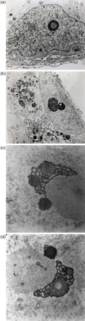
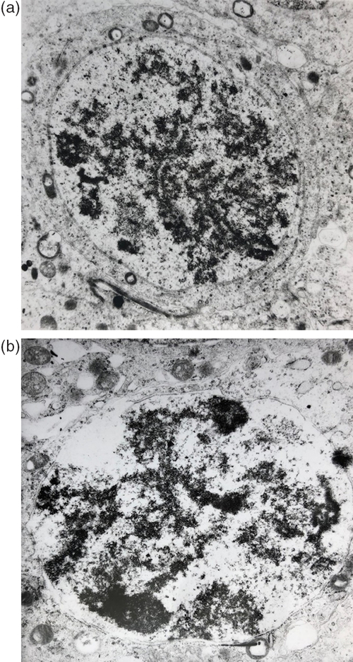
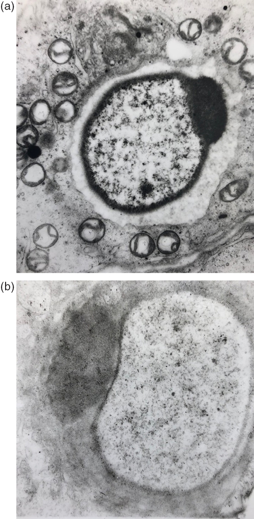
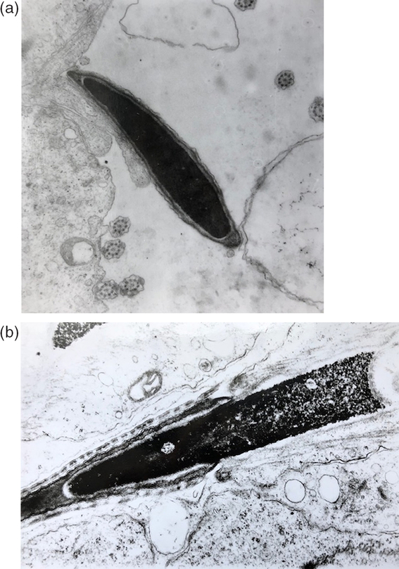
During spermatogenesis, chromatin from gonadal stem cells undergoes dynamic modifications associated with changes in its physical conformation, structure and composition. In spermatozoa, in the final differentiated level of spermatogenesis, chromatin becomes highly compacted and gene expression is completely shut off. Chromatin condensation is facilitated by the formation of DNA–sperm nuclear basic proteins complexes. Gradual replacement with transition proteins, and then with protamines, involves discrete steps evidenced by TEM (Fig. 1a–6a). In the control mice, spermatogonia had a finely granulated chromatin structure with a centrally located active nucleolus (Fig. 1a). Complete nucleolar disorganization and cytoplasmic accumulation of ribonucleoprotein were observed in the primary spermatocytes at the pachytene stages. The presence of bivalents consisting of homologous chromosomes joined by synaptonemal complexes could be evidenced in many sections through spermatocyte nuclei (Fig. 2a). Fully organized nucleoli were observed again in the nucleus of late secondary spermatocytes (Fig. 3a). In the cells formed at the end of the second division, ultrafine sections evidenced intermediate stages of chromatin condensation, preparing such meiotic products to become round (Fig. 4a) and elongated (Fig. 5a) spermatids. Spermatozoa during the maturation process (Fig. 6a) appear to have a high electron dense nucleus because of the extreme chromatin compaction.
Spermatogonia. (a) Spermatogonia nucleus from the control group (×2180 magnification). (b) Transmission electron microscopy image of spermatogonia and primary spermatocyte from the treated group (×5250 magnification). (c, d) Nucleolus details of spermatogonia from the treated group (×4800 magnification). Spermatogonia from the treated group displayed an unusual nucleolar structure with a reticulated aspect.

Primary spermatocytes. (a) Primary spermatocyte from the control group (×7990 magnification). (b) Primary spermatocyte from the treated group (×7990 magnification). The presence of bivalents consisting of homologous chromosomes joined by synaptonemal complexes could be evidenced in both groups.

Secondary spermatocytes. (a) Secondary spermatocyte from the control group (×7345 magnification). (b) Secondary spermatocyte from the treated group (×7345 magnification). Transmission electron microscopy analysis revealed the fine structure of secondary spermatocytes with fully organized nucleoli in both groups.

Round spermatids. (a) Round spermatid from the control group (×16,320 magnification). (b) Round spermatid from the treated group (×17,280 magnification). Transmission electron microscopy analysis evinced intermediate stages of chromatin condensation, preparing such meiotic products to become round spermatids.

Elongated spermatids. (a) Elongated spermatid from the control group. Discrete steps in chromatin compaction can be noticed (×17,280 magnification). (b) Elongated spermatid from the treated group (×17,280 magnification). Elongated spermatids have characteristic features of apoptosis with changes in chromatin architecture.

Spermatozoa. (a) Spermatozoon from the control group in the maturation process appear with a high electron density of nucleus because of extremely chromatin compaction (×17,760 magnification). (b) Ultrastructural image of spermatozoon shows the non-compaction of the sperm nuclei and the appearance of low electron density areas (×17,250 magnification).

Chromatin compaction and replacement of histones with sperm nuclear basic proteins (SNBPs; e.g. protamines 1 and 2 in mouse) may be gradual, involving discrete steps evidenced by TEM analysis. The precise mechanisms of histone replacement are still uncertain. A set of experiments involved epigenetic markers such as histone acetylation in chromatin composition changes during spermiogenesis (Christensen et al., Reference Christensen, Rattner and Dixon1984; Hazzouri et al., Reference Hazzouri, Rousseaux, Mongelard, Usson, Pelletier, Faure, Vourc’h and Sele2000; Fenic et al., Reference Fenic, Sonnack, Failing, Bergman and Steger2004; Burlibaşa and Gavrilă, Reference Burlibaşa and Gavrilă2005; Burlibaşa and Zărnescu, Reference Burlibaşa and Zărnescu2013). Other authors (Eden et al., Reference Eden, Hashimshony, Keshet, Cedar and Thorne1998; Nan et al., Reference Nan, Ng, Johnson, Laherty, Turner, Eisenman and Bird1998; Jones et al., Reference Jones, Veenstra, Wade, Vermaak, Kass, Landsberg, Strouboulis and Wolffe1998) proposed that DNA hypermethylation could cause a decrease in the level of histone acetylation and, in fact, could be a trigger for histone replacement.
Under these circumstances, we also proceed to study the effect of DNA hypomethylation in spermatogenesis.
Previous studies on effects of 5-azacytidine in rats (Doerksen et al., Reference Doerksen, Benoit and Trasler2000) and 5-azadC in mice (Kelly et al., Reference Kelly, Li and Trasler2003; Oakes et al., Reference Oakes, Kelly, Robaire and Trasler2007b) have demonstrated that this treatment results in spermatogenesis disruption.
Our TEM analysis confirmed the cytotoxicity of 5-azadC. Treatments were of sufficient duration to expose developing germ cells from the gonadal stem cell stage through to spermatogenesis. Hypomethylation occurred during successive rounds of DNA replication because the depleted cellular DNMTs were insufficient to maintain established methylation patterns. Electron microscopy images revealed the cytotoxicity of treatment and the subsequent apoptosis of cells. Spermatogonia displayed an unusual nucleolar structure with a reticulated aspect. Nucleolar components seem to be segregated (Fig 1b–d). Primary spermatocytes from treated testes revealed a normal ultrastructure with no evident changes in chromatin morphology (Fig. 2b), but a decreased number of cells could be noticed. The same unchanged ultrastructural features were revealed for secondary spermatocytes (Fig. 3b). Instead, the round, and especially the elongated, spermatids had characteristic features of apoptosis with changes in the chromatin architecture (Figs 4b and 5b). Ultrastructural analysis showed the non-compaction of the sperm nuclei and the appearance of low electron density areas that could be interpreted as apoptotic features because of DNA hypomethylation, resulting in chromatin relaxation, DNA fragmentation and modulation of gene expression (Fig. 6b). A low number of mature spermatozoa were observed. This finding is consistent with other experiments in which 5-azadC treatment caused a dose-dependent decrease in testis weight, reduced sperm counts, abnormalities in testicular histology and increased preimplantation loss after mating treated male mice with normal females (Oakes et al., Reference Oakes, Kelly, Robaire and Trasler2007b).
Mammalian spermatogenesis is a differentiation process governed by genetic and epigenetic factors. During spermatogenesis, each spermatogenic cell undergoes erasure and re-establishment of epigenetic marks to confer a paternal imprint. This process of epigenetic preprogramming involves DNA methylation, histone variants, histone modifications and effector enzymes that catalyze these modifications (Khalil and Wahlested, Reference Khalil and Wahlested2008). Over recent years, intense efforts have been made to understand the function of histone modifications and to identify the enzymes that are responsible for adding various modifications to histone tails.
The transition of histones to protamines is a crucial step in the maturation of spermatids and is thought to be facilitated by histone H4 hyperacetylation in mammals, similar to that found in other species (Hazzouri et al., Reference Hazzouri, Rousseaux, Mongelard, Usson, Pelletier, Faure, Vourc’h and Sele2000; Burlibaşa and Zărnescu, Reference Burlibaşa and Zărnescu2013). Our previous studies demonstrated the DNA hypomethylating effect of trichostatin A, a histone-deacetylase inhibitor during spermatogenesis (Burlibaşa and Gavrilă, Reference Burlibaşa and Gavrilă2005; Burlibaşa and Zărnescu, Reference Burlibaşa and Zărnescu2013). As published information regarding the interplay between epigenetic modifications in advances stages of spermatogenesis is unclear and poor, we considered it appropriate to study the effect of DNA demethylating agent (5-azadC) on histone H4 acetylation status during spermatogenesis. In this context, we proceeded to study the distribution of H4 hyperacetylation in control and treated spermatogenic cells by ICC. This study is the first report that attempts to correlate the distribution of highly acetylated histone H4 with testicular cell types in control and in DNA demethylating agent-treated groups.
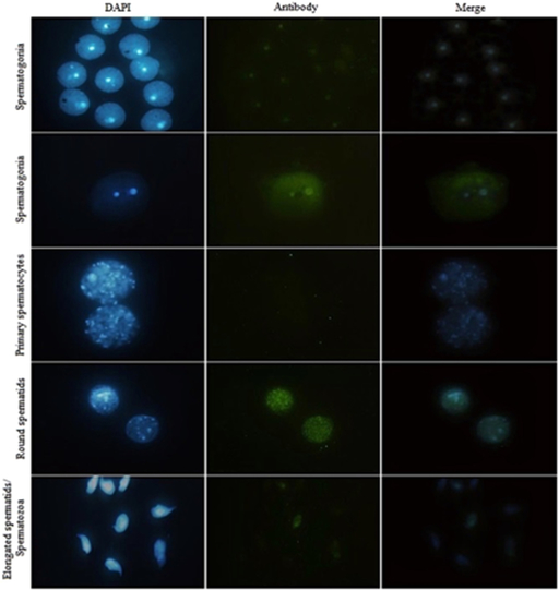
The primary antibody used in this experiment recognized hyperacetylated isoforms of histone H4. The specificity of primary antibody has been previously tested (Liu and Zhang, Reference Liu and Zhang2015). Immunostaining with the secondary antibody (Alexa Fluor 488) of the hyperacetylated H4 in mouse spermatogenic cells produced a nuclear pattern dependent on the cell type and stage of reproductive cycle. ICC analysis with anti-H4hac revealed a selective localization of this epigenetic marker. Global hyperacetylation was highlighted in spermatogonia (Fig. 7a,b) and in round spermatids (Fig. 7d). In the other differentiation stages the presence of this modification is more subtle, being located in certain regions (chromatin domains) such as the nucleolar region of the spermatogonia nuclei. In primary spermatocytes, the anti-H4hac signal is absent (fig. 7 c). Also, in mature sperm, the anti-H4hac signal is very low (Fig 7e).
Immunofluorescence detection of histone H4 (H4hac) in control mouse testes. As a negative control for immunocytochemistry, cells were reacted only with secondary antibody (without anti-H4hac) (×100 magnification).

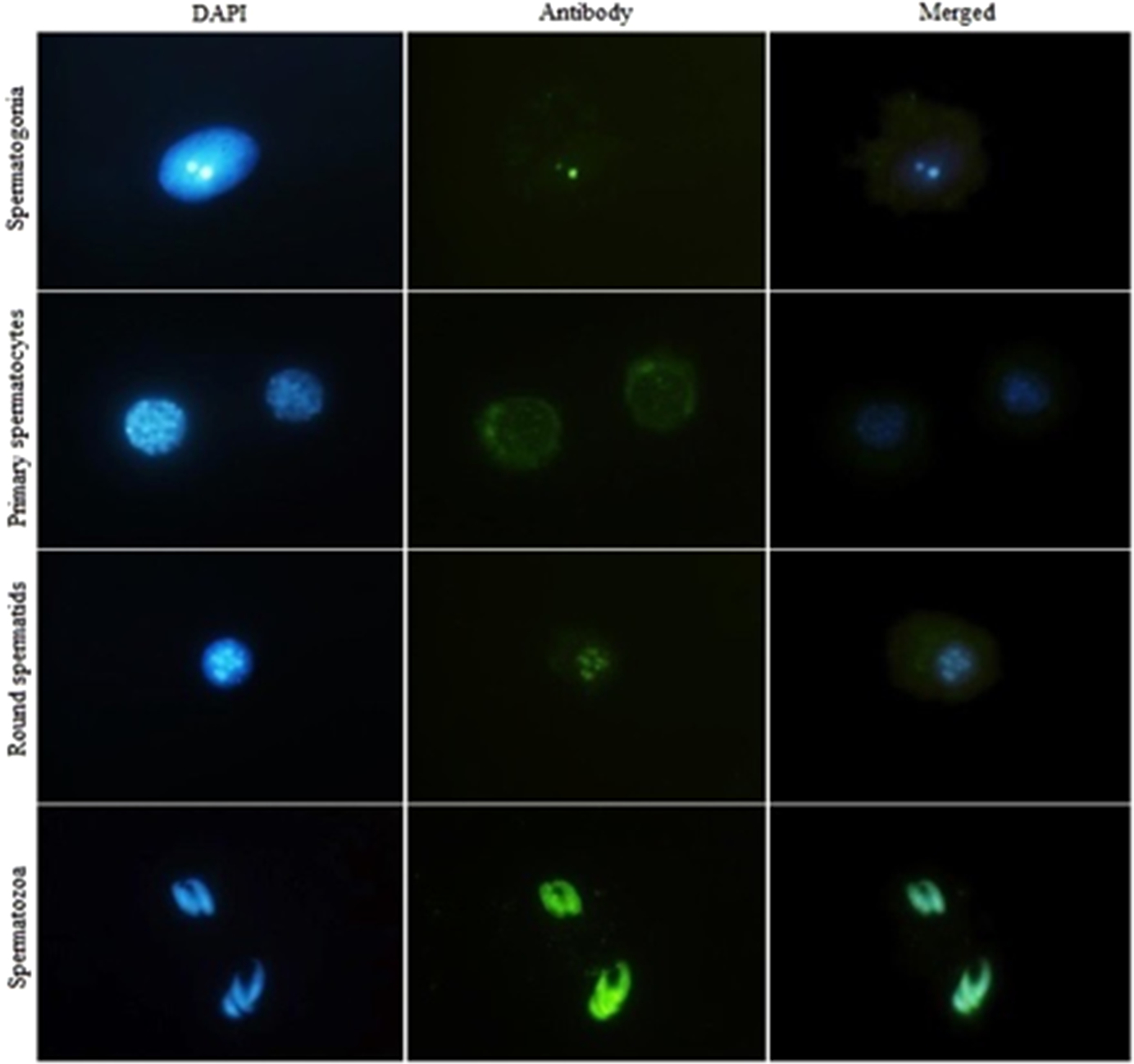
ICC analysis with anti-H4hac following 5-azadC treatments revealed the presence of the signal at several stages of male germ cells differentiation. In the spermatogonia, the H4hac signal was localized in the nucleolus region but also in the cytoplasm (Fig 8a). In primary spermatocytes, the signal had an inner surface nuclear membrane localization (Fig 8b). In round spermatids, the signal was concentrated both in certain chromatin domains and at a significant amount in the cytoplasm (Fig. 8c). Surprisingly, the signal for hyperacetylated H4 was detected in mature spermatozoa (Fig 8d).
Immunofluorescence detection of histone H4 (H4hac) in the 5-aza-2′-deoxycytidine-treated mouse group. As a negative control for immunocytochemistry, cells were reacted only with secondary antibody (without anti-H4hac) (×100 magnification).

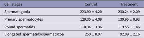
Mean pixels value was calculated from each histogram (Table 2). Results showed a significant increase in the mean reactivities of H4 hyperacetylation for primary spermatocytes and round spermatids in the control group, respectively, in spermatocytes and in elongated spermatids/spermatozoa in the treated group.
Mean pixel values for H4 hyperacetylation

P < 0.05.
Histone acetylation is an important epigenetic marker in male germ line differentiation, both in mouse and human, as well as in many other vertebrate species (Burlibaşa and Zărnescu, Reference Burlibaşa and Zărnescu2013). The H4 hyperacetylation signal has a selective localization. An explanation for this phenomenon has been suggested by Christensen and Dixon (1983) based on trout studies that showed that transcriptionally active spermatogonia and spermatocytes have almost undetectable H4 hyperacetylation levels. This situation changes dramatically during spermiogenesis, when the amount of H4 hyperacetylated increases by about 50%. In this case, researchers concluded that most of the hyperacetylated H4 could not be associated with transcriptional activity, but rather with a later event in spermiogenesis, replacing histones with protamines.
The connection between histone acetylation and gene activity has also been studied in rooster spermatogenesis. As in trout, the amount of hyperacetylated H4 could not be correlated with transcriptional activity because it was detected in both testicular active cells and in inactive elongated spermatids. In elongated spermatids, during the transition from nucleohistones to nucleoprotamines, chromatin undergoes structural changes, therefore exposing DNA binding sites. Hyperacetylation of H4 may be correlated with the specific chromatin conformation of elongating spermatids, and could be a necessary condition for binding of SNBPs to DNA (Oliva and Mazquita, Reference Oliva and Mazquita1982).
The study of functional information has emerged in the new and powerful domain of epigenetics that could explain the dynamics of gene expression observed during cell differentiation and organism development. One widely used assay to study this is chromatin immunoprecipitation (ChIP), which enables analysis of the association of regulatory molecules to specific DNA sequences and histone modifications. ChIP-derived technologies such as ChIP-seq, ChIP-on-chip, along with microarrays, represent the most important tools to study functional genomics (Banu et al., Reference Banu, Simion, Ratiu, Popescu, Romanitan, Danila, Radoi, Ecovoiu and Kusko2015).
In this context, in our next set of experiments, the study of ribosomal gene acetylation patterns would be an excellent model to better understand the dynamics of chromatin structure during gamete differentiation.
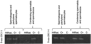
ChIP was used to evaluate the association of penta-acetylated histone H4 with specific DNA sequences. For this study, only cells from fractions 1 and 3 (spermatogonia and primary spermatocytes fraction 1; and condensing spermatids and spermatozoa) were used.
Some ChIP experiments have shown that histone H4 is hyperacetylated in the promoter regions of active genes (Zhao et al., Reference Zhao, Herrera-Diaz and Gross2005; Workman, Reference Workman2006).
Our ChIP experiments performed for the first time on chromatin from spermatogonia/spermatocytes and condensing spermatids and spermatozoa from mice revealed the dynamics of histone H4 hyperacetylation status at the level of 28S rRNA genes during spermiogenesis.
Interestingly, PCR analyses revealed a positive result for ChIP with anti-hyperacetylated H4 antibody in spermatogonia and pachytene spermatocytes in both control and 2-azadC-treated groups of mice. For the condensing spermatids and spermatozoa fraction, a change was noticed in the acetylation level following 5-azadC treatment. All experiments were performed using the same comparative study of a negative control and positive (input) sample for the hyperacetylated H4 (Fig. 9a, b).
Chromatin immunoprecipitation experiments. (a) Control group: PCR analysis of hyperacetylated histone H4 (H4hac) immunoprecipitates from spermatogonia/pachytene spermatocytes and from condensing spermatids/spermatozoa. (C+) Input sample; (C−) negative control (without antibodies). (b) 5-Aza-2′-deoxycytidine treatment group: PCR analysis of H4hac immunoprecipitates from spermatogonia/pachytene spermatocytes and from condensing spermatids/spermatozoa. (C+) Input sample; (C−) negative control (without antibodies).

ChIP experiments revealed that histone H4 at the level of target sequence was presented in a highly acetylated state in spermatogonia/pachytene spermatocytes before and after treatment. Hyperacetylation of H4 in the ribosomal genes from spermatogonia may be required to keep chromatin in a potentially relaxed or accessible state in which transcription can be induced as required. Inhibition of DNA–MTase did not affect the level of acetylation in this type of sperm cells. Our intriguing result, which indicated a hyperacetylated status in condensing spermatids and spermatozoa after 5azadC treatment, may be explained by the fact that chromatin from this cell type remains in a higher acetylation status during spermiogenesis due to DNA–MTase inhibition. In chromatin from normally condensing spermatids and spermatozoa, ChIP revealed a negative signal for hyperacetylated H4.
Hyperacetylation of H4 could be involved in two major events that affect male germ line: (i) chromatin assembly in premeiotic cells/transcription in spermatogonia; and (ii) replacement of histones with transition proteins and subsequently with protamines in round spermatids. Our results are consistent with published literature, suggesting an important role for H4 hyperacetylation in the replacement of histones in murine spermiogenesis.
The pattern of hyperacetylated H4 distribution after 5-azadC correlated with information obtained from ultrastructural evidence that the regulation of DNA–methyltransferase activity plays a major role in controlling histone hyperacetylation and probably histone replacement during spermiogenesis. There is an obvious cross-talk between acetylation/deacetylation of histones and methylation/demethylation of DNA. Cytoplasmic detection of the anti-H4hac signal in treated cells can be explained by the initiation of apoptosis in these cells. A particularly surprising result is that 5-aza2DC treatment led to the expansion of H4 hyperacetylation in mature sperm. This result demonstrated the link between DNA–methyltransferase and histone deacetylases (HDAC). Most probably, these two enzymes are part of the same chromatin remodelling complex. Inhibition of DNA–MTase activity by 5-azadC treatment resulted in inhibition of HDAC activity.
Concomitant with hyperacetylation extension, there is extensive apoptosis of different cell types, which leads to infertility. Regarding this hypothesis, a study conducted by Zhu et al. (Reference Zhu, Lakshmanan, Beal and Otterson2001) demonstrated that apoptosis induced by histone-deacetylase inhibitors can be accelerated by inhibiting DNA–methyltransferase.
Conclusions
Germinal cells show a particular architecture of genetic material with functional implications. The major events in nuclear modification during spermatogenesis are chromatin condensation, and its morphological and composition changes.
Taken together, the results of this study allowed us to propose a critical role for chromatin-modifying enzymes in the control of chromatin architecture and composition during spermatogenesis. Our studies evinced an interplay between epigenetic markers to achieve a higher order structure in mature sperm, with implication not only for its organization and function but especially for zygotic chromatin function.
The sperm chromatin state retains important attributes that facilitate proper progression of spermatogenesis, as well as being a potential contributor to early embryonic developmental processes. A highly dynamic and continuous epigenetic remodelling state could make sperm cells susceptible to environmental factors, causing some degree of infertility; altered epigenetic markers could be inherited and further result in phenotypic defects in the offspring. For this reason, the study of specific details of the dynamics of chromatin structure and composition during spermatogenesis is essential for the development of new methods of diagnosis and treatment, especially in the context of assisted reproductive technologies. Over the last decade, male infertility issues have been given special attention, including studies focusing on how extrinsic factors influence epigenetic processes.
An important challenge for the next years will be to understand how various epigenetic modifications interact to achieve a sperm-specific epigenetic code with consequences for fertility and health of future offspring.
Ethical standards
To collect biological samples, animals were sacrificed in compliance with the European Council (86/609/CEE/24.11.2004) recommendations regarding the protection of animals used for experimental and scientific purposes.
Financial support
This work was supported by an IDEI-UB-08 grant.
Conflict of interest
None of the authors of this paper had any financial relationship with any other person or organization that might inappropriately influence or bias the content of the paper.