Introduction
Dementia with Lewy bodies (DLB) is the second most common type of neurodegenerative dementia in late life after Alzheimer's disease (AD) and accounts for 10.9%–22.0% of all causes of dementia (Stevens et al., Reference Stevens, Livingston, Kitchen, Manela, Walker and Katona2002; Rahkonen et al., Reference Rahkonen, Eloniemi-Sulkava, Rissanen, Vatanen, Viramo and Sulkava2003). DLB has a more malignant course in terms of the rate of cognitive decline (Williams et al., Reference Williams, Xiong, Morris and Galvin2006), mortality (Williams et al., Reference Williams, Xiong, Morris and Galvin2006), quality of life (Boström et al., Reference Boström, Jönsson, Minthon and Londos2007a), and resource utilization compared to AD (Boström et al., Reference Boström, Jönsson, Minthon and Londos2007b). The accurate antemortem diagnosis of DLB is particularly important because of the development of interventions and specific pharmacologic treatments and outcome evaluations.
The clinical diagnostic criteria for DLB were first published in 1996 (McKeith et al., Reference McKeith, Galasko and Kosaka1996), and were modified in 2005 (McKeith et al., Reference McKeith, Dickson and Lowe2005). The central or core symptoms in DLB are progressive cognitive decline, marked fluctuations in cognition, recurrent visual hallucinations, and spontaneous features of Parkinsonism (McKeith et al., Reference McKeith, Galasko and Kosaka1996). However, a recent study exploring the early symptoms of DLB reported that memory impairment was the most common presenting symptom (57%) in DLB, followed by visual hallucinations (44%) (Auning et al., Reference Auning, Rongve, Fladby, Booij, Hortobágyi, Siepel, Ballard and Aarsland2011). Therefore, when the initial presentation of DLB is impaired cognition, it is difficult to differentiate DLB from AD during the early course of the illness. Although Single Photon Emission Computed Tomography (SPECT) and Iodine-123 Metaiodobenzylguanidine (123I-MIBG) myocardial scintigraphy are useful in the differential diagnosis of DLB (Lobotesis et al., Reference Lobotesis, Fenwick, Phipps, Ryman, Swann, Ballard, McKeith and O'Brien2001; Yoshita et al., Reference Yoshita, Taki and Yamada2001), these examinations are too expensive to be utilized generally.
Rapid eye movement (REM) sleep behavior disorder (RBD) is a parasomnia characterized by a loss of normal skeletal muscle atonia during REM sleep with prominent motor activity accompanying dreaming (American Academy of Sleep Medicine, 2005). This condition is considered to be frequently associated with an underlying synucleiopathy such as DLB, Parkinson's disease, or multiple system atrophy and only rarely with other neurodegenerative disorders (Boeve et al., Reference Boeve, Silber and Parisi2003a). Revised criteria for the clinical diagnosis of DLB have included RBD in suggestive features (McKeith et al., Reference McKeith, Dickson and Lowe2005). Furthermore, Ferman et al. (Reference Ferman, Boeve, Smith and Lin2011) reported that the inclusion of RBD as a core feature improved the diagnostic accuracy of autopsy-confirmed DLB. Polysomnography (PSG) is necessary to confirm the diagnosis of RBD. However, it is impractical to perform PSG routinely on patients suspected of having DLB.
Sleep talking is a major symptom of RBD. A questionnaire concerning sleep talking could easily be asked from all caregivers of patients with dementia in daily medical practice. However, there have been few studies of sleep talking in patients with DLB and other types of dementia. We hypothesized that patients with DLB would exhibit a higher frequency of sleep talking compared with other demented patients, including AD, and examined the usefulness of the questionnaire for the differential diagnosis of DLB.
Methods
Subjects
The whole procedure followed the 2010 Clinical Study Guidelines of the Ethics Committee of Kumamoto University Hospital and was approved by the Internal Review Board. After a complete description of all procedures of the present study, written informed consent was obtained from the patients or their caregivers.
This study was a prospective dementia referral center-based cohort study. A total of 317 patients with dementia were selected from a consecutive series of 573 patients who underwent a medical examination at the Dementia Clinic of the Department of Neuropsychiatry, Kumamoto University Hospital between January 2010 and December 2011. All patients were examined comprehensively by senior neuropsychiatrists with sufficient experience in examining patients with dementia, and all patients underwent routine laboratory tests, standard neuropsychological examinations, including the Mini-Mental State Examination (MMSE). Brain magnetic resonance imaging (MRI) or computed tomography (CT) and SPECT were also performed. The following patients were excluded from the current study: (1) those with developmental abnormalities, serious psychiatric diseases, such as schizophrenia or major depression, or substance abuse before the onset of dementia; (2) those living alone or in a nursing home; (3) those whose caregivers had hearing loss; and (4) those without a reliable informant.
Dementia was diagnosed according to the Diagnostic and Statistical Manual of Mental Disorders, 3rd edition-revised (DSM-III-R). Probable DLB was diagnosed on the basis of the international working group criteria (McKeith et al., Reference McKeith, Galasko and Kosaka1996). To avoid circularity, we did not use the revised criteria in 2005 (McKeith et al., Reference McKeith, Dickson and Lowe2005), in which RBD was included as a suggestive feature. Brain SPECT was also used to support the clinical diagnosis of DLB. In this study, patients whose dementia developed 12 months or later after the onset of Parkinson's disease (Parkinson's disease with dementia) were classified into the DLB group because they usually have underlying Lewy body pathology and their number was too small to analyze separately (n = 3). Patients were diagnosed as having AD if they met the criteria of the National Institute of Neurological Disorders and Stroke–Alzheimer's Disease and Related Disorders Association (NINCDS–ADRDA) guidelines (McKhann et al., Reference McKhann, Drachman, Folstein, Katzman, Price and Stadlan1984). Patients who fulfilled both probable AD and possible DLB were classified into the AD group (n = 6). Diagnoses of frontotemporal lobar degeneration (FTLD) were based on the consensus clinical diagnostic criteria in an international workshop on FTLD (Neary et al., Reference Neary, Snowden and Gustafson1998) and brain SPECT was used to support the clinical diagnosis of FTLD as described earlier (Pickut et al., Reference Pickut, Saerens and Mariën1997). Probable vascular dementia (VaD) was diagnosed on the basis of the Criteria for the Diagnosis of Ischemic Vascular Dementia proposed by the State of California Alzheimer's Disease Diagnostic and Treatment Centers (ADDTC) (Chui et al., Reference Chui, Victoroff, Margolin, Jagust, Shankle and Katzman1992). Patients who fulfilled the criteria of probable AD and, in addition, displayed cerebrovascular disease (CVD) on brain MRI or CT that did not meet the criteria of VaD were classified in the AD group. Diagnostic categories consisted of probable DLB (n = 55), probable AD (n = 191), FTLD (n = 16), VaD (n = 18), and other/unspecified dementias (O/U dementias) (n = 37). The O/U dementia group consisted of patients with probable progressive supranuclear palsy (PSP) (n = 5; Litvan et al., Reference Litvan, Mangone, McKee, Verny, Parsa, Jellinger, D'Olhaberriague, Chaudhuri and Pearce1996), probable corticobasal degeneration (CBD) (n = 2; Boeve et al., Reference Boeve, Lang and Litvan2003b), and unspecified etiology (n = 30).
Normal elderly subjects
Thirty-two normal elderly subjects (NE subjects) were recruited from the community (14 males and 18 females). They showed normal cognitive functions (25 or above on the MMSE), normal findings in the physical and neurologic examinations, no history of psychiatric disorders, and no risk factors for CVD (hypertension, heart disease, and diabetes mellitus). All NE subjects had their family members in the same household.
Evaluation of sleep talking
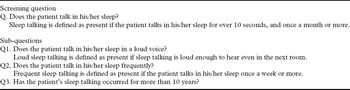
We evaluated sleep talking in dementia patients and NE subjects by using a questionnaire that had originally been designed to assess sleep talking easily (Table 1). The questionnaire asks a screening question to confirm whether the subject has any sleep talking or not. If the screening question is answered in affirmative, three sub-questions about the feature of sleep talking (volume, frequency, and duration of sleep talking) are asked. The questionnaire is based on the International Classification of Sleep Disorders, diagnostic criteria for RBD (American Academy of Sleep Medicine, 2005), and some questionnaires that adequately screen for RBD (Boeve et al., Reference Boeve, Silber, Ferman, Smith and Petersen2002; Li et al., Reference Li, Wing, Lam, Zhang, Yu, Ho, Tsoh and Mok2010). Sleep talking was assessed by a single psychiatrist (Kazuki Honda) who was blinded to all clinical information, including the dementia diagnosis.
Sleep talking questionnaire

Evaluation of sleep disturbances
In dementia patients, sleep and nighttime behavior disorder (SNBD) were assessed by using the Japanese version of the 12-item Neuropsychiatric Inventory (NPI) (Cummings, Reference Cummings1997). We focused on the sleep and nighttime behavior item of the NPI. This item asks the following: (1) “Does the patient have difficulty sleeping (do not count as present if the patient simply gets up once or twice per night only to go to the bathroom and falls back asleep immediately)?”; (2) “Is he/she up at night?”; and (3) ‘Does he/she wander at night, get dressed, or disturb your sleep?”; these questions could be answered with “yes” or “no,” and SNBD was categorized as present or absent.
Statistical analysis
Differences in patient characteristics between the six groups (DLB, AD, FTLD, VaD, O/U dementia, and NE subjects groups) were analyzed by one-way analysis of variance (ANOVA) or compared by Fisher's exact probability test. Difference in the usage of psychotropic drugs was analyzed by Fisher's exact probability test in five dementia groups.
To examine the prevalence of sleep talking, loud sleep talking, frequent sleep talking, long-term sleep talking, and SNBD, we used Fisher's exact probability test and Bonferroni Z-test for each comparison when an overall group difference was significant. A multiple logistic regression analysis was applied to identify significant independent predictors for sleep talking. Valuables entered were diagnosis (DLB or not), age, sex, duration of illness, MMSE score, the use of cholinesterase inhibitors, benzodiazepine anxiolytics, antipsychotics and antidepressant, and bedroom sharing. In addition, we calculated the sensitivity and specificity of sleep talking, three features of sleep talking, and SNBD for the differential diagnosis of DLB from AD and that of DLB from all other dementias. A significance level of 0.05 was set for all analyses. All analyses were carried out using SPSS for Windows, version 17.0.
Results
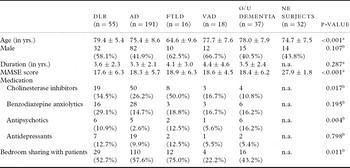
Table 2 presents the demographic and clinical indices of the subjects. Five dementia and NE subject groups were involved in the present study. There were significant differences in age, MMSE, and the ratio of bedroom sharing in six groups. As for medication, there was a significant difference in the frequency of cholinesterase inhibitors and antipsychotics prescription, but no significant differences were observed in the frequency of other drug usage in five dementia groups. Memantine or melatonin was not prescribed to any patient in this study.
Demographic and clinical valuables in five dementia groups and the NE subjects group

Notes: Values are n (%), or mean ± SD.
DLB: dementia with Lewy bodies; AD: Alzheimer's disease; FTLD: frontotemporal lobar degeneration; VaD: vascular dementia; O/U dementia: other/unspecified dementia; NE subjects: normal elderly subjects; MMSE: Mini-Mental State Examination; n.a.: not applicable.
Analysis by aone-way ANOVA or bFisher's exact probability test.
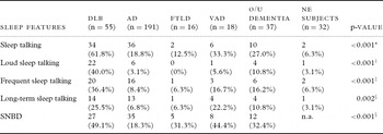
Table 3 shows the prevalence of sleep talking, three features of sleep talking, and SNBD in all the six groups. Sleep talking occurred most frequently in the DLB group, followed by the VaD group. It is noteworthy that only 6.3% of the NE subjects presented with sleep talking. There was a significant difference in the prevalence of sleep talking in these six groups. The prevalence of sleep talking in the DLB group was significantly higher than in other groups, except for the VaD group. Of the 34 DLB patients who had sleep talking, 22 patients (64.7%) showed loud sleep talking and the prevalence of loud sleep talking in the DLB group was significantly higher than that in the AD, O/U dementia, and NE subject groups. SNDB occurred most frequently in the DLB group and the prevalence of SNDB was significantly higher in the DLB group than in the AD group.
Frequency of sleep talking, loud sleep talking, frequent sleep talking, long-term sleep talking, and SNBD in five dementia groups and the NE subjects group

Notes: Values are n (%).
DLB: dementia with Lewy bodies; AD: Alzheimer's disease; FTLD: frontotemporal lobar degeneration; VaD: vascular dementia; O/U dementia: other/unspecified dementia; NE subjects: normal elderly subjects; SNBD: sleep and night-time behavior disorder; n.a.: not applicable.
Analysis by Fisher's exact probability test and Bonferroni Z-test.
*DLB significantly higher than AD, FTLD, O/U dementia, and NE subjects.
†DLB significantly higher than AD, O/U dementia, and NE subjects (FTLD was not compared with other groups).
‡DLB significantly higher than AD and NE subjects.
§DLB significantly higher than AD.
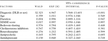
Multiple logistic regression analysis showed that a diagnosis of DLB was significantly associated with the presence of sleep talking (Table 4). There was no association between age, sex, duration of disease, MMSE score, bedroom sharing, or psychotropic drug usage and the presence of sleep talking.
Results of multiple logistic regression analysis associated with sleep talking

Note: DLB: dementia with Lewy bodies; MMSE: Mini-Mental State Examination.
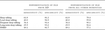
Table 5 shows the sensitivity and specificity of sleep talking, three features of sleep talking, and SNBD for the differential diagnosis of DLB from AD and that of DLB from all other types of dementia. The sleep talking yielded high specificity (81.2%) and some sensitivity (61.8%) for the differential diagnosis of DLB from AD. The loud sleep talking could improve the specificity (96.9%), but the sensitivity would decrease (40.0%). Even with the differentiation of DLB from all other dementias, the specificity of sleep talking and loud sleep talking did not change (79.4% and 95.8% respectively).
Sensitivity and specificity of sleep talking, three features of sleep talking, and SNBD for the differentiation of DLB from AD or all other dementias

Note: DLB: dementia with Lewy bodies; AD: Alzheimer's disease; SNBD: sleep and nighttime behavior disorder.
Discussion
Sleep talking is not necessarily a pathological symptom, and it often occurs in normal healthy people. Bjorvatn et al. (Reference Bjorvatn, Grønli and Pallesen2010) reported that sleep talking occurs at least once a week in 6.3% of adults in the general population. In the present study, 6.3% of NE subjects showed sleep talking, which is very similar to Bjorvatn et al.'s data. These findings indicate that the high prevalence of sleep talking in patients with dementia may be associated with some underlying pathological changes.
In this study, the prevalence of sleep talking differed according to the diagnostic group. In the DLB group, more than 60% of patients exhibited sleep talking, and the prevalence was significantly higher than in any other dementia types. Although the pathophysiology of sleep talking is not well understood, sleep talking is reported to be seen with high frequency in patients with RBD (Li et al., Reference Li, Wing, Lam, Zhang, Yu, Ho, Tsoh and Mok2010). Boeve (Reference Boeve2010) summarized demographics and clinical phenomenology of RBD, and demonstrated that abnormal vocalization is considered to characterize RBD, which is considered to be frequently associated with an underlying synucleinopathy, such as DLB (Boeve et al., Reference Boeve, Silber and Parisi2003a), and rarely with other neurodegenerative disorders. Ferman et al. (Reference Ferman, Boeve, Smith and Lin2011) reported that a history of RBD was present in 76% of autopsy-confirmed DLB patients. Therefore, RBD may be responsible for sleep talking in people with DLB.
We observed a high specificity of sleep talking (81.2%) for the differentiation of DLB from AD. In particular, the presence of loud sleep talking showed extremely high specificity (96.9%). Even in the differentiation of DLB from all other dementias, the specificity of loud sleep talking was kept high (95.8%). These findings suggest the usefulness of sleep talking to discriminate patients with DLB from those with other types of dementia. Although the utilization of SPECT and 123I-MIBG myocardial scintigraphy are limited to well-equipped hospitals, screening questions concerning sleep talking are easy to ask in clinical practice. Thus, patients suspected of having DLB should be questioned about the presence of sleep talking, in particular, the volume of sleep talking.
There was no significant difference between the DLB and VaD groups in the prevalence of sleep talking. This result may reflect low statistical power due to small sample size in the VaD group. However, the prevalence of sleep talking in the VaD group (33.3%) was higher than in NE subjects (6.3%), which did not reach statistical significance. To our knowledge, there have been no reports that investigated the prevalence of RBD in patients with VaD. Although RBD is frequently seen in patients with neurodegenerative diseases, RBD in patients with a pure pontine infarction has also been reported (Xi and Luning, Reference Xi and Luning2009). Some vascular damage around the pons may cause RBD in patients with VaD, and this might explain a certain prevalence of sleep talking in the VaD group.
Sleep disturbance occurs in many forms of dementia. Guarnieri et al. (Reference Guarnieri, Adorni and Musicco2012) reported that over 60% of persons with cognitive decline had one or more sleep disturbances. In this study, we also investigated the prevalence of sleep and nighttime behavior disorder. In the VaD and FTLD groups, the prevalence of SNBD was higher than that of sleep talking. On the other hand, in the DLB group, the prevalence of SNBD was lower than that of sleep talking. Severe daytime sleepiness predicts VaD (Guarnieri et al., Reference Guarnieri, Adorni and Musicco2012), and sleep-disordered breathing was frequent in VaD patients (Elwood et al., Reference Elwood, Bayer, Fish, Pickering, Mitchell and Gallacher2011). Anderson et al. (Reference Anderson, Hatfield, Kipps, Hastings and Hodges2009) have demonstrated sleep–wake disturbance in patients with FTD, who showed increased nocturnal activity and decreased morning activity. These findings suggest that each form of dementia may have a disease-specific sleep disturbance. Ferman and Boeve (Reference Ferman and Boeve2007) reported that sleep disturbance is helpful in differentiating DLB from AD early in the disease course. However, it may be more efficient to focus on sleep talking than to assess overall sleep disturbances for the differentiation of DLB from other dementias.
A recent review of RBD has described that the commonly used medications may induce or unmask latent RBD, and antidepressants are most commonly implicated in altering muscle control during REM and causing RBD (Trotti, Reference Trotti2010). On the other hand, based on a large case series and clinical experience, benzodiazepine clonazepam is considered the first-line treatment for RBD (Chenck and Mahowald, Reference Chenck and Mahowald1990). In the present study, no significant differences were observed in the frequency of benzodiazepine anxiolytics and antidepressant usage between the dementia groups. However, there were significant differences in the frequency of cholinesterase inhibitors and antipsychotic prescriptions. To our knowledge, there are no reports that indicate an association between antipsychotics and RBD. As for cholinesterase inhibitors, Boeve et al. (Reference Boeve, Silber and Ferman2003c) reported that among 50 patients with DLB and RBD who were treated with donepezil, none experienced significant benefit. Furthermore, multiple logistic regression analysis did not reveal any significant association between the use of these psychotropic drugs and the presence of sleep talking. Therefore, the difference in the frequency of sleep talking between different dementia types cannot be attributed to the effect of psychotropic drugs.
Several methodological issues limit the interpretation of results of this study. First, the diagnosis relied solely on clinical basis without histopathologic confirmation, with inevitably some uncertainty about the rate of misclassification. Although clinical studies are in fact influenced by the quality of clinical diagnosis, clinical studies with prospective clinical data collection can assess patients’ sleep disturbances more accurately than can autopsy studies with retrospective data review. Moreover, we supplemented clinical diagnosis with neuroimaging studies. Second, we obtained information about sleep talking from patients’ caregivers. However, nearly half of the caregivers did not share their bedroom with the patients. This condition may make it more difficult for the caregivers to note patients’ sleep talking and would increase the false-negative rate of sleep talking. In our study, approximately 60% of the patients with DLB had sleep talking, but this rate might be lower than the true rate. Third, although we considered that RBD was primarily responsible for sleep talking in the dementia patients, no patient with sleep talking was confirmed by PSG whether they had RBD or not. Therefore, the relationship between sleep talking and RBD can only be hypothesized. However, our main aim was to find an alternative to PSG, which can discriminate DLB from AD or all other dementias easily. In the future study, sleep talking in people with dementia need to be evaluated by PSG.
Despite these limitations, we believe that our findings are quite reliable because they are based on a prospective study design and on a consecutive patient series whose diagnosis was carefully made using widely accepted clinical criteria.
Conclusion
The questionnaire about sleep talking, especially the volume of sleep talking, may be useful in the clinical discrimination of DLB from not only AD but also from all other types of dementia.
Conflict of interest
None.
Description of authors’ role
Kazuki Honda designed this study, worked on data analysis, and wrote the paper. Yusuke Yatabe, Keiichiro Kaneda, Seiji Yuki, Yusuke Ogawa, Shiho Matuzaki, Atsuko Tsuyuguchi, Hibiki Tanaka, Hiroko Kashiwagi, Noriko Hasegawa, and Tomohisa Ishikawa helped in collecting the data. Mamoru Hashimoto supervised this study. Manabu Ikeda was responsible for the statistical design of the study.
Acknowledgments
The present study was undertaken with the support of grants provided by the Ministry of Health, Labour and Welfare (Research on dementia; H21-Dementia-General-005) for Manabu Ikeda and Mamoru Hashimoto. The authors gratefully acknowledge the assistance of staff of Department of Psychiatry and Neuropathobiology, Faculty of Life Sciences, Kumamoto University.