




No CrossRef data available.
Article contents
An Eimeria vaccine candidate appears to be lactate dehydrogenase; characterization and comparative analysis
Published online by Cambridge University Press: 13 May 2004
Abstract
An Eimeria acervulina protein fraction was identified which conferred partial protection against an E. acervulina challenge infection. From this fraction a 37 kDa protein was purified and its corresponding cDNA was cloned and shown to encode a lactate dehydrogenase (LDH). Full length cDNAs encoding LDH from two related species, E. tenella and E. maxima, were also cloned. The homology between the primary amino acid sequences of these three Eimeria LDH enzymes was rather low (66–80%), demonstrating an evolutionary divergence. The Plasmodium LDH crystal structure was used to generate a 3D-model structure of E. tenella LDH, which demonstrated that the many variations in the primary amino acid sequences (P. falciparum LDH and E. tenella LDH show only 47% identity) had not resulted in altered 3D-structures. Only a single LDH gene was identified in Eimeria, which was active as a homotetramer. The protein was present at similar levels throughout different parasitic stages (oocysts, sporozoites, schizonts and merozoites), but its corresponding RNA was only observed in the schizont stage, suggesting that its synthesis is restricted to the intracellular stage.
Information
- Type
- Research Article
- Information
- Copyright
- 2004 Cambridge University Press
INTRODUCTION
Eimeria species are the causative agents of intestinal coccidiosis, an enteritis that causes diarrhoea and reduces the productivity of many economically important animals, including chickens (reviewed by Reid, 1972). Presently, coccidiosis in chickens is primarily controlled by feed medication, using antibiotic drugs (Allen & Fetterer, 2002). In addition, live vaccines are available based on virulent strains or live attenuated strains (reviewed by Vermeulen, Schaap & Schetters, 2001). Analogous to a normal infection these vaccines induce a protective immunity that is species specific. Today's coccidiosis vaccines have the disadvantage that they are composed of multiple live Eimeria species, which makes them laborious to produce and relatively expensive. A subunit vaccine would not have these drawbacks and would have the added advantage that no live parasites are introduced. However, no subunit vaccine is available to control coccidiosis. In the search for candidate vaccine antigens, various groups have identified and cloned Eimeria antigens that should be immunogenic (Jenkins et al. 1989; Miller et al. 1989; Jenkins, Castle & Danforth, 1991; Crane et al. 1991; Tomley et al. 1991; Laurent et al. 1993; Vermeulen et al. 1993; Bumstead, Dunn & Tomley, 1995). Yet, none of the cloned Eimeria antigens have been developed into commercial products, underscoring their apparent limited value as potential vaccine antigens. These antigens were mostly sporozoite specific, and it can be disputed what role sporozoites without subsequent developmental stages may play to induce a protective immune response (Jeffers & Long, 1985). It appeared that intracellular sporozoite metabolism and/or limited schizont development were required for induction of a protective response (McDonald, Rose & Jeffers, 1986; McDonald et al. 1988; Jenkins et al. 1991).
Experiments in our own laboratory showed that curtailing Eimeria infections after 48 h with Toltrazuril (Baycox™) did not abrogate the immunizing effect of either E. tenella or E. acervulina infections, indicating that the first parasitic stages were sufficient to raise immunity (Panhuijzen & Vermeulen, unpublished results). Therefore, we focused in this study on the identification of protective parasitic antigens from the first intracellular schizont stages. A 37 kDa protein was identified that was partially protective and its corresponding cDNA identified it as a lactate dehydrogenase. The LDH sequence from three different Eimeria species is presented, the primary amino acid sequence is analysed in conjunction with a predicted 3D structure, and the expression pattern of Eimeria LDH is presented, demonstrating that Eimeria LDH is expressed in schizonts, but that its presence is not restricted to the schizont stage. Finally, the function of LDH is discussed in relation to the different parasitic life-cycle stages.
MATERIALS AND METHODS
Parasite isolation and purification
The Weybridge strain of E. tenella, the Houghton strain of E. acervulina and the Houghton strain of E. maxima were used to infect coccidia-free chickens (Shirley, 1986). Oocysts were purified, sporulated and stored according to the procedures of Long et al. (1976).
To obtain E. acervulina intracellular stages, 5-week-old chickens were infected with 108 sporulated oocysts. At 42 h post-inoculation the chickens were euthanized and the duodenum was removed. The tissue was washed in calcium/magnesium-free Hanks Balanced Salt Solution (Hanks BSS) containing 10 mg/ml glucose, cut into small pieces and suspended in the same buffer. Epithelial cells containing the parasites were released by incubation for 10 min in 2 mM EDTA in Hanks BSS. Cells were pelleted (400 g, 5 min) and intracellular parasites (trophozoite and schizont stages) were released from the host cells by saponin lysis (15 min in 0·1% saponin (w/v) in Hanks BSS, room temperature) and mechanical shearing (18 G needle). Schizonts (and trophozoites) were separated from host material by centrifugation through 45% (w/v) Percoll (Pharmacia) (700 g, 20 min, 4 °C). The pellet contained the schizont fraction.
Different sporulation stages of E. tenella oocysts were isolated as follows. Non-sporulated E. tenella oocysts were isolated from the caecal content of chickens, 7 days after infection. These oocysts were kept under anaerobic conditions during isolation and purification by adding sodium dithionite (0·01 M Na2S2O4) to prevent premature sporulation. Sporulation was initiated by removal of sodium dithionite (3 wash steps), followed by strong aeration in sodium bichromate solution (20% (w/v) in distilled water) at 28 °C for 0, 5.5, 24 or 72 h, i.e. samples were taken at different time-intervals. Microscopical examination showed no formation of sporocysts at 0 and 5.5 h sporulation, after 24 h sporocysts had formed in almost 100% of the oocysts but no sporozoites were present at that time, and at 72 h the sporocysts clearly contained developed sporozoites.
E. tenella merozoites were isolated from chickens according to a method described by Smith et al. (1995). Briefly, 10 chickens were each infected with 100000 sporulated oocysts and 96 h after infection the caeca were removed. These caeca were opened and washed with phosphate-buffered saline (PBS). The tissue was cut in small pieces and incubated for 30 min at 41 °C in Hanks BSS with 10 mM MgCl2, 0·25% trypsin and 1% taurocholic acid. Merozoites were filtered through gauze to remove debris. Merozoites were pelleted at 800 g for 10 min and resuspended in PBS, passed over a DE52 column, and centrifuged once more.
E. maxima gametocytes were isolated from chickens 6 days after infection, as previously described (Smith et al. 1995). Briefly, approximately 20 cm of intestine around the Meckels diverticulum was isolated and washed with ice-cold incubation buffer, (170 mM NaCl, 5 mM CaCl2, 10 mM glucose and 10 mM Tris–HCl, pH 7·0) with 1 mM phenylmethylsulfonyl fluoride (PMSF) and 1 mg/ml bovine serum albumin (BSA). The intestine was filled with incubation buffer (plus PMSF and BSA) containing 0·5 mg/ml hyaluronidase, sealed and incubated at 37 °C for 20 min in PBS while shaking. The intestine was subsequently opened lengthways and placed on a 20 μm nylon filter (Millipore, type NY20). Gametocytes were washed at room temperature with incubation buffer (plus PMSF and BSA) and filtered. The eluate was placed on an 11 μm filter (Millipore, type NY11), washed again and gametocytes were harvested from the filter. The gametocytes were subsequently centrifuged at 800 g for 5 min, resuspended and centrifuged again.
In vitro isolated E. tenella schizonts (and trophozoites) were cultured in Madin Darby Bovine Kidney (MDBK) cells, grown in Williams complete medium (Gibco Life technologies) with 10% foetal calf serum. Mitomycin-treated (80 μg/ml) MDBK cells were infected with purified E. tenella sporozoites (MOI=2) and cultured for 40 h at 41 °C, at which stage schizonts had formed. Prior to harvesting, cells were washed with PBS. Schizonts were subsequently analysed without any further purification.
Triton X–114 extraction and Prepcell fractionation
Triton X–114 extractions were carried out to obtain hydrophilic protein fractions of schizonts (and trophozoites) using a procedure described by Bordier (1981). Briefly, 108 to 109E. acervulina schizonts (and trophozoites) were sonicated and pre-condensed Triton X–114 was added to a final concentration of 10% (v/v). After thorough mixing the non-extractable material was pelleted, and the soluble fraction was layered over a 6% sucrose cushion, incubated for 10 min at 40 °C and centrifuged for 10 min at 400 g at room temperature. The hydrophilic proteins were collected and further separated on a 9% polyacrylamide Prepcell, under denaturing and reducing conditions (a 37 mm diameter Prepcell column was used, Bio-Rad Labs). Fractions were collected and analysed on SDS–PAGE, followed by in vivo protection studies.
Purification and analysis of RNA and DNA
Total RNA was isolated using Ultraspec TM RNA Isolation system (Campro Scientific BV). Genomic DNA was isolated according to Qiagen genomic DNA purification protocol. Northern blotting was performed with total RNA separated on a formaldehyde gel followed by capillary transfer using standard methods. Southern blotting was carried out with capillary transfer to Hybond-N+ (Amersham). Random primed probes were made from PCR products containing the complete open reading frames (ORFs). Non-translated sequences were omitted to prevent non-specific hybridization to repeated sequence elements. A 996 bp E. tenella LDH (EtLDH) PCR-product was generated with an EtLDH-forward primer (ATGGCGGTTTTCGAGAAGGT) and an EtLDH-reverse primer (TCACTCTGCAGCAGCGTCGG) and a 993 bp E. acervulina LDH (EaLDH) PCR-product was generated with an EaLDH-forward primer (ATGGCGGTCTTCGAGAAGAA) and an EaLDH-reverse primer (TTACTTGGATGCATCAAGAG). The PCR products were random-primed labelled with 32P-dATP, denatured and immediately hybridized to the blots. The blots were washed at moderately low stringency with 2×SSC/0·1% SDS (at 60 °C for 20 min), 1×SSC/0·1% SDS (at 60 °C for 20 min) and 0·5×SSC/0·1% SDS (at 60 °C for 20 min). cDNA libraries from E. acervulina oocysts and from E. tenella schizonts (with MDBK host cells) were prepared in lambda ZAPII phages (Stratagene) and screened with either antibodies or random primed labelled probes. The E. maxima LDH (EmLDH) complete ORF was cloned as follows: Two degenerate primers (TNGGNTCNGGNATGATHGG and AGNGCNGCNACCATNACRTC) (using standard IUB/IUPAC nucleic acid codes) were used in an RT–PCR reaction to amplify a 380 bp fragment from total E. maxima gametocyte mRNA. Based on the sequence of the 380 bp fragment, a specific forward primer (AGAATGGATTTATTACCAGT) was used in combination with an oligo-dT primer to amplify the 3′-part of the EmLDH transcript. The 5′-part was amplified with two nested reverse primers (TCCATCGCTTTTCCCATAGGCATATTAGGT and TAAGAATGCCATGGTGCCTCCGATCAT) using genome walking on E. maxima genomic DNA isolated from sporulated oocysts, according to the manufacturer (Universal Genomewalker™, Clontech, Palo Alto, USA). Using two specific primers at the start and stop codon of the EmLDH ORF (ATGGCGGTTTTTGAGCAA and TTACTTTCCTGCATCAAG resp.) the complete ORF was amplified with RT–PCR, cloned and sequenced. All molecular procedures were performed with standard protocols (Sambrook, Fritsch & Maniatis, 1989).
Antibodies, immunodetection and LDH assay
Polyclonal antibodies were raised in rabbits using standard procedures (Harlow & Lane, 1988). EaLDH antibodies were raised with Prepcell material containing the purified 37 kDa protein band. EtLDH antibodies were raised with a (His)6-tailed EtLDH fusion-protein, overexpressed in Salmonella and purified with Talon beads (Clontech).
Proteins were analysed on SDS–Tris–Glycine 4–20% polyacrylamide-gels (Novex), and either stained with Coomassie brilliant blue or Western blotted.
Lactate dehydrogenase isozymes were separated electrophoretically on native 5% Tris–acrylamide gels. The isozymes of LDH were visualized by staining of the enzyme activity with the nitro-blue tetrazolium method (Fine, Kaplan & Kuftinec, 1963). Enzyme activity in the gel was detected by formation of highly-coloured insoluble formazan. Procedures were according to the supplier's method (lactate dehydrogenase isoenzyme kit, Sigma).
3D modelling
EtLDH was modelled on the crystal structure of P. falciparum LDH (PfLDH), using Modeler 4.0 software as distributed with Quanta 98 (MSI, San Diego, USA). Standard settings were used for homology modelling and no further energy refinement of the resulting models was performed.
RESULTS
Selection of a partially protective hydrophilic early-schizont protein
First generation E. acervulina schizonts were obtained 42 h after infection of 5-week-old chickens. Intracellular parasites were isolated from duodenum epithelial cells and used to extract the hydrophilic proteins, which were further fractionated on an SDS–PAGE Prep-Cell (Fig. 1). A 37 kDa fraction, in particular, was shown in a preliminary trial to be partially protective (unpublished observations). The major 37 kDa protein was excised from the gel and used for amino acid sequencing and to prepare polyclonal antibodies in rabbits. Amino acid sequencing identified 1 major peptide: GWIKQEEVDDIVQK. The raised polyclonal antibodies specifically recognized a single protein of 37 kDa on Western blot with total E. acervulina extracts from either schizonts or sporozoites (results not shown).

Fig. 1. Purification of a 37 kDa protein from 42-h-old Eimeria acervulina schizonts. Shown is a Coomassie brilliant blue stained SDS–PAGE, with a sample of the hydrophilic fraction obtained after Triton X–114 extraction of purified schizonts. This material was further size fractionated over a Prepcell, and a sample of the pooled 37 kDa fractions is shown. Shown on the right is a molecular weight marker for reference.
Cloning and sequencing of the 37 kDa E. acervulina antigen
To identify the 37 kDa antigen, the monospecific antibody sera were used to clone its corresponding cDNA. Since the antigen was not only detected in schizonts but also in sporozoites it was decided to screen an E. acervulina sporozoite cDNA library, which was available. Six plaques were identified and partial sequence analysis showed that all clones contained homologous sequences. The full-length sequence from the longest clone was determined, having an insert of 1566 bps. The full-length cDNA encodes a 330 amino acid polypeptide, with a calculated molecular weight of 35857 Da, which is in close agreement with the size of the purified 37 kDa protein. Furthermore, this protein contained the deduced amino acid sequence from the analysed tryptic peptide (Fig. 2). Comparison of the deduced polypeptide against the NCBI-database showed highest homology with lactate dehydrogenase from Toxoplasma gondii (60% identity) and Plasmodium falciparum (50% identity). A lower but significant homology was seen with other LDH enzymes from vertebrates and plants (Table 1). Based on its homology, we named this protein E. acervulina LDH.

Fig. 2. Multiple alignment of Eimeria LDH enzymes with Apicomplexa LDH enzymes and vertebrate LDH enzymes. Shown are the aligned sequences of LDH from E. acervulina, E. maxima, E. tenella, Toxoplasma gondii LDH1 and LDH2, Plasmodium falciparum, human, chicken and dogfish. Amino acid identities are shown with grey and black shading. The conserved residues His195 and Arg171 are indicated with arrows (amino acid numbering is based on original LDH description from dogfish, White et al. 1976). A conserved Gln102 is replaced by Lys in Apicomplexa (also indicated with arrow). Shown in the 1st box is the extended Apicomplexan specific loop. Two highly conserved coccidia-specific regions are also boxed. Indicated boxes and amino acid residues are also depicted in the structural model (see Fig. 3). The determined amino acid sequence of a tryptic peptide from E. acervulina is indicated (overlined). Multiple alignments were made with ClustalX (Thompson et al. 1997) and edited with Genedoc (Nicholas, Nicholas & Deerfield, 1997). Nucleotide sequence data for Eimeria LDH was deposited in the GenBank™ under the Accession numbers AY143388 (EaLDH), AY143389 (EtLDH) and AY143390 (EmLDH). Other GenBank Accession numbers used were: T. gondii LDH1, AAC47443; T. gondii LDH2, AAC46863; PfLDH, AAA29633; Human LDH-A, CAA26088; Chicken LDH-A, CAA37824; Dogfish LDH-M, DEDFLMPID.
Table 1. Amino acid conservation of LDH between species (Shown is the percentage amino acid identity between species, based on the multiple alignment of Fig. 2 (abbreviations on the horizontal axis correspond with the species on the vertical axis).)

Cloning and sequencing of LDH from other Eimeria species
To determine if LDH is a conserved protein in different Eimeria species, and to define if LDH from other Eimeria species might also be immunogenic, the LDH homologues from E. tenella and E. maxima were also cloned.
The coding sequence of EaLDH was used as a probe to screen an E. tenella schizont cDNA library. Both strong and weak hybridizing plaques were purified, to identify potentially more than one LDH isoenzyme. Subsequent analysis showed that all clones encoded the same sequence (or parts thereof), with the exception of clone #18. The longest clone was sequenced and contained an 1876 bps long insert. The cDNA encoded a polypeptide of 331 amino acids, with a calculated molecular weight of 34965 Da. The deduced polypeptide was compared against the NCBI database and was most homologous to EaLDH (70% identity) (Table 1 and Fig. 2). Based on its homology this protein was named EtLDH. Sequence analysis of clone #18 showed that it did not encode another LDH cDNA. However, clone #18 confirmed that the screening had been performed at low stringency, since it contained low homology in its 3′-non coding region with the coding sequence from EaLDH. As the low stringency screening had specifically picked up a sequence non-related to LDH, it is unlikely that other LDH isoenzyme sequences were present in our E. tenella cDNA library.
To identify E. maxima LDH, degenerate primers were used in an RT–PCR on total RNA from E. maxima gametocytes. 3′-RACE–PCR was performed to obtain the complete 3′-part of EmLDH and 5′-genome walking was used to obtain the remaining 5′-sequence of EmLDH. Specific primers at the start and stop of the EmLDH coding sequence were used to amplify the full length product from first strand cDNA. EmLDH is a 330 amino acid polypeptide with a calculated molecular weight of 35951 Da. EmLDH is most homologous with EaLDH (80% identity) and less with EtLDH (66% identity) (Table 1 and Fig. 2).
Structure function analysis of Eimeria LDH
To characterize the molecular properties of Eimeria LDH, its primary structure was compared in a multiple alignment with related LDH molecules from T. gondii, P. falciparum, and three vertebrates (Fig. 2). Eimeria LDH enzymes had the closest relationship to each other (80, 70 and 66% identity), followed by T. gondii LDH enzymes (between 54 and 60%), P. falciparum LDH (47 to 50%) and finally vertebrate LDH enzymes (23–27%) (Table 1). The conservation follows the evolutionary divergence between these species: T. gondii being another member of the coccidia family, P. falciparum belonging to the same phylum of Apicomplexa, and the vertebrates being the least related. The multiple alignment shows that the various LDH enzymes have considerably diverged, but without dramatic changes, such as deletions or insertions. Furthermore, the Eimeria and Toxoplasma LDH enzymes clearly group together, having diverted from the P. falciparum LDH.
The primary structure of Eimeria LDH was analysed in more detail in conjunction with its predicted 3D structure. As will become clear from the analysis, a 3D model is much more informative to predict the enzymatic properties than a comparison based on primary structure. For this purpose EtLDH was used as an example and modelled on the crystal structure of PfLDH (Dunn et al. 1996). Despite the evolutionary divergence between both molecules (only 47% amino acid identity), EtLDH could be modelled on the PfLDH fold without major structural rearrangements. Figure 3 shows the predicted Cα trace of EtLDH superimposed on PfLDH. Two residues, His195 and Arg171 that are conserved between all LDH molecules, are indicated for reference in Figs 2 and 3 (amino acid numbering corresponds to the original numbering described for the dogfish sequence (White et al. 1976)). His195 is the active site residue, which donates a proton from the imidazolium moiety to pyruvate. Arg171 interacts via salt bridges with the carboxyl group of pyruvate, and helps to orient the substrate. Both residues are conserved in the Eimeria LDH molecules, and Fig. 3 shows that these residues are located at the same positions in the 3D structure in comparison with those of PfLDH.

Fig. 3. EtLDH structural model superimposed on PfLDH. Shown is the model of the Cα trace of EtLDH (orange and yellow) superimposed on the 3D crystal structure of PfLDH (purple and green). The NADH cofactor and the oxamate substrate analogue are inserted in the model. The side-chains of Lys102, Arg171 and His195 have been added for both structures. These three amino acids, NADH and oxamate are depicted with their atom colours (carbon=green, oxygen=red and nitrogen=blue). The coccidia-specific regions from Fig. 2 have been highlighted with green and yellow colours. The 3D structure of PfLDH was previously published (Dunn et al. 1996, PDB file 1LDG).
A third residue, Gln102, being conserved in vertebrate LDH molecules has changed into a lysine residue for Apicomplexa LDH enzymes in the primary alignment. Gln102 is involved in substrate-specific binding. Changing it into a positively charged residue has been shown to change the substrate specificity from pyruvate to oxaloacetate, thus changing the enzyme from a lactate dehydrogenase into a malate dehydrogenase (Wilks et al. 1988). However, in the 3D structures, Lys102 in both EtLDH and PfLDH is not localized at the same position as Gln102 in vertebrate LDH (data not shown). In fact, all Apicomplexa contain a unique extended substrate specificity loop, XTKXPGKSDKEW (residue 100–111), and Lys102 is located at the third position in this loop, having its side-chain facing away from the substrate site (the extended loop is shown in Fig. 3 on top of the green/yellow loop at the right site of the figure and lysine residue 102 of the loop is shown and labelled). In the 3D model Gln102 would co-localize with tryptophan, being the last residue from the extended loop. Thus the 3D model strongly suggests that EtLDH is not a malate but is indeed a lactate dehydrogenase.
The primary sequence comparison showed 2 regions of divergence between PfLDH with the group of Toxoplasma/Eimeria (Coccidia) LDH enzymes. The 1st region is located around 169–180 and the 2nd region is located around 246–259 (Fig. 2). Both regions are close to the substrate and nicotinamide ring of the cofactor. These regions are shown in the Cα trace with green/yellow. Although these conserved Coccidia boxes are substantially different in their primary sequence from PfLDH, their 3D structures are very similar. For example the first Coccidia box contains the previously mentioned Arg171, which is important for substrate positioning. Yet although the surrounding amino acids differ, Arg171 from EtLDH and PfLDH are located at identical positions in their 3D structures. Thus, despite the amino acid differences in these regions, it does not seem to change their function.
Finally, cofactor binding between PfLDH and EtLDH is structurally similar, showing many conserved hydrophobic residues that face the cofactor for both enzymes. It has been reported that the position of NADH with respect to the PfLDH protein is significantly altered compared to other LDH molecules (Dunn et al. 1996), and this is also observed for EtLDH. For example, the conserved Ser163 in other LDH molecules, which will hydrogen bond to NADH, is changed to a leucine in PfLDH and a conserved methionine for all Eimeria and Toxoplasma LDH molecules (located in the 1st green/yellow loop in Fig. 3).
In summary, although the primary sequence comparison of EtLDH with PfLDH only shows 47% identity, the structural model of EtLDH with its cofactor and substrate bound derived from PfLDH yields a properly folded protein structure with conserved active site features and would therefore be predicted to be a molecule with very similar properties.
Eimeria contains a single LDH enzyme
Since Toxoplasma contains 2 LDH enzymes (Yang & Parmley, 1997), we investigated whether Eimeria contains multiple LDH isoenzymes and/or LDH genes. Southern blots with both E. tenella and E. acervulina genomic DNA were hybridized in parallel with EaLDH and EtLDH probes to determine if Eimeria contains more than a single copy of the same LDH gene and/or if related isoenzymes might be present (Fig. 4). Genomic DNA was digested with 4 different restriction enzymes that recognize sites which are absent in the EaLDH and EtLDH cDNA sequence with the exception of Pst-I, that occurs once (10 bps before the 3′-end of the EtLDH ORF). The full-length open reading frame of either EaLDH or EtLDH were used as probes. The Southern blot only detected single and strongly hybridizing bands but no additional weakly hybridizing bands (with the exception of the first lane, containing 2 bands in the Pst-I digested E. acervulina DNA, which resulted from a Pst-I site in an EaLDH intron), thus showing no evidence for related isoenzymes. The positive hybridization of the cross-species LDH homologue (with only a 63% identity at the DNA level) confirmed that the screening had been performed at low stringency. In addition to the absence of related LDH isoenzymes, there was also no indication for multicopy and/or repeated LDH genes, because no additional bands were observed.

Fig. 4. Southern blot analysis of Eimeria LDH. Genomic DNA from E. acervulina and E. tenella was completely digested with either PstI, HindIII plus EcoRI, HindIII alone or EcoRI alone, separated on 0·7% agarose gel and blotted. The blot on the left was hybridized with a 993 bp EaLDH probe and the blot on the right with a 996 bp EtLDH probe, both probes comprise complete ORFs. Hybridizations were performed at moderately low stringency, showing strong hybridization with its corresponding gene product, and no additional bands. Note that the EaLDH probe weakly detects the EtLDH gene product (indicated with arrow). A sample of 10 μg digested genomic DNA was separated in each lane and a size marker (indicated in kbp) is shown at the right.
To corroborate the genomic data, in-gel enzymatic LDH-assays were performed to determine the migration pattern of active LDH. LDH is known to be functionally active as a tetramer, containing either identical subunits or different subunits (Markert, Shaklee & Whitt, 1975). If composed of different subunits, then they will migrate at multiple positions, as shown for bovine LDH (expressed in MDBK cells) where 5 bands were present (Fig. 5). These represent the tetrameric compositions A4, A3B1, A2B2, A1B4 and B4, A and B being two different LDH gene products, as previously described (Li, 1990). However, EtLDH was always present as a single band, and migrated at the same position for all the life-cycle stages tested: sporozoites, merozoites and schizonts (schizonts were difficult to unambiguously characterize since the 5 bovine bands might have obscured EtLDH activity). Thus during all the different parasitic stages tested only a single LDH enzyme was detected in E. tenella, which was functionally active as a homotetramer.

Fig. 5. LDH is active as a homo-tetramer in Eimeria tenella. LDH enzyme activity was measured after separation of total cell extracts on a native non-reducing polyacrylamide gel. LDH activity was visualized by formation of highly coloured insoluble formazan. Bovine LDH is present as 5 distinctive bands, representing different tetramer-combinations (as indicated). EtLDH is visible as a single band in the sporozoite, merozoite and merozoite plus chicken cells lanes. Chicken LDH is also clearly present as a single band. Different protein concentrations from total cell extracts were loaded in each lane: MDBK, 2·4 μg; MDBK+schizonts, 4·1 μg; sporozoites, 5·3 μg; merozoites, 5·5 μg; merozoites+chicken cells, 23 μg; chicken cells, 3·3 μg.
Expression of LDH during E. tenella life-cycle stages
Since Eimeria inhabits aerobic and anaerobic environments during its life-cycle and LDH is only required during anaerobic respiration, the expression pattern of LDH during the various life-cycle stages of Eimeria was studied. E. tenella was used as an example and material was isolated from different life-cycle stages and total RNA was isolated. Northern blot analysis with RNA from different E. tenella stages detected a single band of 1·8 kb in the schizont stage and not in oocysts or sporozoites (Fig. 6). The 1·8 kb transcript in schizonts was only weakly detected, but this is partially explained with the roughly 4-fold lower loading of specific E. tenella RNA due to an excess of MDBK RNA in this sample. Although the band was weak, the size of the 1·8 kb transcript did correlate with the 1876 nucleotides full length EtLDH cDNA. De novo LDH synthesis thus takes place in the schizont stage of E. tenella and not (or very little) in extracellular stages. It should be noted that gametocyte stages of E. tenella were not analysed, but that it is likely that EtLDH will be transcribed in gametocytes to some extend, because total RNA from E. maxima contained EmLDH transcripts (based on the RT–PCR results). Subsequently, the presence of LDH at the protein level was studied during the various life-cycle stages. Total protein from different parasitic stages was separated on denaturing polyacrylamide gel and analysed. Although a Coomassie brilliant blue-stained gel clearly indicated the appearance and disappearance of stage-specific E. tenella proteins (Fig. 7), a Western blot analysed with a polyclonal rabbit anti-EtLDH serum showed no variation in EtLDH levels between the different stages (Fig. 7). The in-gel LDH enzyme assay already had shown enzymatic activity in different parasitic stages, and in combination with the Western blot it demonstrated that EtLDH was present and functional throughout the different life-cycle stages. The invariable expression of EtLDH observed on Western blot was further substantiated in an immunohistochemical analysis of infected chicken caecal tissue, which identified EtLDH in first and second-generation trophozoite/schizont stages (Fig. 8). In conclusion, LDH is stage specifically transcribed in Eimeria, but its presence at the protein level remains constant during different life-cycle stages, suggesting that LDH must be a stable protein.

Fig. 6. Schizont-specific expression of EtLDH mRNA. A Northern blot is shown with total RNA from different E. tenella life-cycle stages. EtLDH mRNA was only detected in the schizont stage as a ~1·8 kb mRNA. mRNA of sporulated and non-sporulated oocysts showed no detectable EtLDH mRNA expression. A sample of 10 μg total RNA was loaded in each lane. Equal loading of total RNA was confirmed by comparing ribosomal RNA bands detected after ethidium bromide staining of the gel (not shown). T=0, contains non-sporulated oocysts; t=5.5 h contains non-sporulated oocysts; t=24 h contains fully sporulated oocysts with sporocysts; t=72 h contains fully sporulated oocysts with sporozoites; MDBK+Schizonts contains MDBK cells with 40-h-old E. tenella schizonts (E. tenella RNA was estimated to be 25% of the total RNA by comparing the 28S rRNA bovine band with the smaller 23S rRNA Eimeria band, not shown) and the last lane contains only MDBK cells. The blot was hybridized with a 996 bp EtLDH probe, comprising the complete ORF.

Fig. 7. LDH is present at equal levels during different life-cycle stages of Eimeria tenella. Total protein extracts from different E. tenella life-cycle stages were separated on denaturing and reducing polyacrylamide gels, and either stained with Coomassie brilliant blue or Western blotted. EtLDH was detected with an EtLDH specific polyclonal antiserum. Lanes contain total protein extracts from sporulating oocysts (0, 5.5, 24 and 72 h sporulation), purified sporozoites, purified merozoites, MDBK cells and MDBK cells 40 h after infection with E. tenella sporozoites. Each lane contains 27 μg total protein, with the exception of the MDBK and MDBK plus schizont lanes, which contain 108 μg total protein (to correct for the excess of MDBK cell material). Molecular weight markers are indicated.

Fig. 8. EtLDH expression in first and second-generation trophozoite/schizont stages. Caecal chicken tissue was analysed 48 h and 96 h after infection with sporulated Eimeria tenella oocysts. (A) A first generation schizont in a crypt at the bottom of a villus, 48 h after infection. (B and C) Second generation trophozoites and schizont stages in a heavily infected part of the caecum, 96 h after infection. (C) Higher magnification of second generation schizonts, which clearly stains all merozoites. EtLDH was visualized with a specific EtLDH polyclonal antibody, stained with peroxidase (haematoxylin blue was used for background staining).
DISCUSSION
Hydrophilic proteins from E. acervulina were fractionated and tested as protective antigens upon E. acervulina challenge. One fraction in particular could confer partial protection. Using amino acid sequence data and monospecific antibodies, the cDNA corresponding to the major protein from this fraction with a molecular weight of 37 kDa was cloned, and was found to encode lactate dehydrogenase, an enzyme which is conserved throughout evolution from bacteria to humans. Although the initial aim was to identify one or more protective schizont-specific antigens, Eimeria LDH was not confined to schizonts, but was also present in other parasitic life-cycle stages. Apart from E. acervulina, the corresponding LDH genes from E. tenella and E. maxima were also cloned, representing the 3 main pathogenic Eimeria species in chickens. In this paper the molecular and biochemical analysis of these Eimeria LDH enzymes has been described. The structure of Eimeria LDH was analysed with a 3D model, which was more informative than only a primary sequence comparison. Vaccination studies with these recombinantly expressed Eimeria enzymes will be described elsewhere (A. N. Vermeulen, manuscript in preparation). Alignment and 3D-modelling showed that Eimeria LDH enzymes were most homologous with each other, followed by other coccidial LDH enzymes. LDH homology between species correlated with taxonomic differentiation between species. The primary structure of Eimeria LDH was analysed together with a predicted 3D model superimposed on the crystal structure of PfLDH. These combined data clearly indicate first that Eimeria LDH functions as a lactate- and not a malate-dehydrogenase (not immediately evident from primary sequence alone, see Wilks et al. 1988), and secondly that its 3D structure is expected to be almost identical to PfLDH, yet its amino acid conservation is only 47%. Thirdly, the coccidia LDH enzymes contain highly conserved domains that have diverged from PfLDH, but without affecting the overall 3D structure. This could indicate that selective inhibitors of PfLDH, such as gossypol-related molecules (Gomez et al. 1997), might also be good inhibitors of Eimeria LDH, thus being potential anti-coccidials.
In contrast to Eimeria, T. gondii contains two LDH stage-regulated isozymes. Therefore we extensively searched for multiple LDH isoforms in Eimeria. However, both cloning and Southern blotting indicated that E. acervulina, E. tenella and E. maxima all contain only one LDH enzyme. The data were corroborated by Western blotting and in-gel LDH enzyme assays of different parasitic stages, which also identified only a single LDH enzyme. The presence of one LDH species in Eimeria agrees with earlier data from Shirley (Shirley, 1975; Shirley et al. 1989) describing only a single enzymatic LDH activity in oocyst stages of different Eimeria species and in various strains. Interestingly, the presence of a second LDH enzyme in Toxoplasma (LDH2) correlated with specific expression in the bradyzoite stage, which is lacking in Eimeria (Yang & Parmley, 1997). The latter authors suggested that the expression of a second LDH enzyme in bradyzoites might ensure that glycolysis continues in an anaerobic environment. Although the metabolically inactive oocyst is sometimes easily compared with bradyzoite cysts, the oocyst stages (from both Toxoplasma and Eimeria) are not strictly anaerobic stages (Coombs et al. 1997). Moreover, Eimeria oocysts need high oxygen levels for sporulation to occur. Thus oocyst stages may mostly utilize oxidative phosphorylation to generate energy and would not require LDH activity. In addition, oxidative phosphorylation would be the most economic usage of a limited energy supply, such as mannitol storage, allowing the oocyst to survive for as long as possible (Allocco et al. 1999). Therefore, we would speculate that LDH remains present in Eimeria oocysts, probably because it is a stable enzyme. We observed that after host cell invasion the parasite initiates new LDH mRNA transcription. During trophozoite/schizont stages the parasite is metabolically highly active and resides in a microenvironment with a low oxygen tension. It is likely that during the highly active metabolic stages aerobic energy supply is insufficient, thus requiring LDH to create an additional anaerobic metabolism. This would explain why LDH mRNA is synthesized in schizonts and would be in agreement with other reports describing that energy metabolism in oocysts is aerobic (in particular during sporulation) and is most likely anaerobic during intracellular stages (reviewed by Coombs et al. 1997).
Although apicomplexan LDH molecules appear structurally conserved, their primary amino acid sequences have extensively diverged, even within different Eimeria species (66–80% identity). This indicates that during evolution Eimeria LDH molecules were selected for having different primary sequences but being structurally and functionally unchanged. In comparison, other cloned Eimeria genes are often over 90% identical between different Eimeria species, such as an aspartyl proteinase (98% E. tenellavs. E. acervulina), a transhydrogenase (91% E. tenellavs. E. acervulina), a calcium/calmodulin protein kinase (91% E. tenellavs. E. maxima), a dnaK-type molecular chaperone HSP70 (92% E. maximavs. E. acervulina) and an immunoglobulin heavy chain binding protein (98% E. tenellavs. E. maxima) (Accession numbers respectively CAC20153, CAA80843, T18520, L08392, Z71757, CAA96438, S51682, S37165, CAA91253 and CAA91252). We hypothesize that immunological pressure may have selected for these altered primary LDH sequences. Although Eimeria LDH is a cytosolic enzyme and therefore not immediately presented to the immune system, one should realize that at the end of schizogony Eimeria generates a residual body containing cytosolic proteins (Pacheco, Vetterling & Doran, 1975). When the host cell ruptures, the residual body is deposited extracellularly and might trigger immune responses. If true, Eimeria LDH, which is highly expressed during schizogony, would be one of those extracellularly deposited schizont antigens that would be a potential target for the immune system to be recognized. However, the mechanisms involved need further study.
The authors are grateful to Paul van den Boogaart (Organon, Oss, the Netherlands) for assistance in the initial cloning of EaLDH, Corinne van Geffen for generating schizont cultures, Alida Boot-Groenink for performing initial LDH enzyme assays, John Janssen for performing the immunohistochemistry on Eimeria-infected chicken tissue and Theo Schetters for critical reading of the manuscript.
References
REFERENCES
ALLEN, P. C. &
FETTERER, R. H.
(
2002).
Recent advances in biology and immunobiology of Eimeria species and in diagnosis and control of infection with these coccidian parasites of poultry.
Clinical Microbiology Reviews 15, 58–65.CrossRefGoogle Scholar
ALLOCCO, J. J.,
PROFOUS-JUCHELKA, H.,
MYERS, R. W.,
NARE, B. &
SCHMATZ, D. M.
(
1999).
Biosynthesis and catabolism of mannitol is developmentally regulated in the protozoan parasite Eimeria tenella.
Journal of Parasitology 85, 167–173.CrossRefGoogle Scholar
BORDIER, C.
(
1981).
Phase separation of integral membrane proteins in Triton X-114 solution.
Journal of Biological Chemistry 256, 1604–1607.Google Scholar
BUMSTEAD, J. M.,
DUNN, P. P. &
TOMLEY, F. M.
(
1995).
Nitrocellulose immunoblotting for identification and molecular gene cloning of Eimeria maxima antigens that stimulate lymphocyte proliferation.
Clinical and Diagnostic Laboratory Immunology 2, 524–530.Google Scholar
CRANE, M. S.,
GOGGIN, B.,
PELLEGRINO, R. M.,
RAVINO, O. J.,
LANGE, C.,
KARKHANIS, Y. D.,
KIRK, K. E. &
CHAKRABORTY, P. R.
(
1991).
Cross-protection against four species of chicken coccidia with a single recombinant antigen.
Infection and Immunity 59, 1271–1277.Google Scholar
COOMBS, G. H.,
DENTON, H.,
BROWN, S. M. &
THONG, K. W.
(
1997).
Biochemistry of the coccidia.
Advances in Parasitology 39, 141–226.CrossRefGoogle Scholar
DUNN, C. R.,
BANFIELD, M. J.,
BARKER, J. J.,
HIGHAM, C. W.,
MORETON, K. M.,
TURGUT-BALIK, D.,
BRADY, R. L. &
HOLBROOK, J. J.
(
1996).
The structure of lactate dehydrogenase from Plasmodium falciparum reveals a new target for anti-malarial design.
Nature Structural Biology 3, 912–915.CrossRefGoogle Scholar
FINE, I. H.,
KAPLAN, N. O. &
KUFTINEC, D.
(
1963).
Developmental changes of mammalian lactic dehydrogenases.
Biochemistry 2, 116–121.CrossRefGoogle Scholar
GOMEZ, M. S.,
PIPER, R. C.,
HUNSAKER, L. A.,
ROYER, R. E.,
DECK, L. M.,
MAKLER, M. T. &
VANDER JAGT, D. L.
(
1997).
Substrate and cofactor specificity and selective inhibition of lactate dehydrogenase from the malarial parasite P. falciparum.
Molecular and Biochemical Parasitology 90, 235–246.CrossRefGoogle Scholar
HARLOW, E. &
LANE, D.
(
1988).
Antibodies, a Laboratory Manual.
Cold Spring Harbor Press,
NY, USA.
JEFFERS, T. K. &
LONG, P. L.
(
1985)
Eimeria tenella: immunogenicity of arrested sporozoites in chickens.
Experimental Parasitology 60, 175–180.CrossRefGoogle Scholar
JENKINS, M. C.,
AUGUSTINE, P. C.,
DANFORTH, H. D. & BARTA, J. R.
(
1991).
X-irradiation of Eimeria tenella oocysts provides direct evidence that sporozoite invasion and early schizont development induce a protective immune response(s).
Infection and Immunity 59, 4042–4048.Google Scholar
JENKINS, M. C.,
CASTLE, M. D. &
DANFORTH, H. D.
(
1991).
Protective immunization against the intestinal parasite Eimeria acervulina with recombinant coccidial antigen.
Poultry Science 70, 539–547.CrossRefGoogle Scholar
JENKINS, M. C.,
DANFORTH, H. D.,
LILLEHOJ, H. S. &
FETTERER, R. H.
(
1989).
cDNA encoding an immunogenic region of a 22 kilodalton surface protein of Eimeria acervulina sporozoites.
Molecular and Biochemical Parasitology 15, 153–161.CrossRefGoogle Scholar
LAURENT, F.,
BOURDIEU, C.,
KAGA, M.,
CHILMONCZYK, S.,
ZGRZEBSKI, G.,
YVORE, P. &
PERY, P.
(
1993).
Cloning and characterization of an Eimeria acervulina sporozoite gene homologous to aspartyl proteinases.
Molecular and Biochemical Parasitology 62, 303–312.CrossRefGoogle Scholar
LI, S. S.
(
1990).
Human and mouse lactate dehydrogenase genes A (muscle), B (heart), and C (testis): protein structure, genomic organization, regulation of expression, and molecular evolution.
Progress in Clinical Biological Research 344, 75–99.Google Scholar
LONG, P. L.,
MILLARD, B. J.,
JOYNER, L. P. &
NORTON, C. C.
(
1976).
A guide to laboratory techniques used in the study and diagnosis of avian coccidiosis.
Folia Veterinaria Latina 6, 201–217.Google Scholar
MARKERT, C. L.,
SHAKLEE, J. B. &
WHITT, G. S.
(
1975).
Evolution of a gene. Multiple genes for LDH isozymes provide a model of the evolution of gene structure, function and regulation.
Science 189, 102–114.Google Scholar
McDONALD, V.,
ROSE, M. E. &
JEFFERS, T. K. (
1986).
Eimeria tenella: immunogenicity of the first generation of schizogony.
Parasitology 93, 1–7.CrossRefGoogle Scholar
McDONALD, V.,
WISHER, M. H.,
ROSE, M. E. &
JEFFERS, T. K.
(
1988).
Eimeria tenella: immunological diversity between asexual generations.
Parasite Immunology 10, 649–660.CrossRefGoogle Scholar
MILLER, G. A.,
BHOGAL, B. S.,
McCANDLISS, R.,
STRAUSBERG, R. L.,
JESSEE, E. J.,
ANDERSON, A. C.,
FUCHS, C. K.,
NAGLE, J.,
LIKEL, M. H.,
STRASSER, J. M. &
STRAUSBERG, S.
(
1989).
Characterization and vaccine potential of a novel recombinant coccidial antigen.
Infection and Immunity 57, 2014–2020.Google Scholar
NICHOLAS, K. B.,
NICHOLAS, H. B. Jr. &
DEERFIELD, IID. W.
(
1997).
GeneDoc: analysis and visualization of genetic variation.
EMBNEW.News 4, 14.Google Scholar
PACHECO, N. D.,
VETTERLING, J. M. &
DORAN, D. J.
(
1975).
Ultrastructure of cytoplasmic and nuclear changes in Eimeria tenella during first-generation schizogony in cell culture.
Journal of Parasitology 61, 31–42.CrossRefGoogle Scholar
REID, W. M.
(
1972).
Coccidiosis. In
Diseases of Poultry, 6th Edn (
ed. Hofstad, M. S.
), pp. 945–977.
Iowa State University Press,
Iowa, USA.
SAMBROOK, J.,
FRITSCH, E. F. &
MANIATIS, T.
(
1989).
Molecular Cloning, a Laboratory Manual, 2nd Edn.
Cold Spring Harbor Press, NY, USA.
SHIRLEY, M. W.
(
1975).
Enzyme variation in Eimeria species of the chicken.
Parasitology 71, 369–376.CrossRefGoogle Scholar
SHIRLEY, M. W.
(
1986).
New methods for the identification of species and strains of Eimeria. In
Research in Avian Coccidiosis. Proceedings of the Georgia Coccidiosis Conference (ed. McDougald, L. R.,
Joyner, L. P. &
Long, P. L.), pp. 13–35.
University of Georgia,
Athens.
SHIRLEY, M. W.,
CHAPMAN, H. D.,
KUCERA, J.,
JEFFERS, T. K. &
BEDRNIR, P.
(
1989).
Enzyme variation and pathogenicity of recent field isolates of Eimeria tenella.
Research in Veterinary Science 46, 79–83.Google Scholar
SMITH, N.,
MILLER, C. M. D.,
PETRACCA, M. &
ECKERT, J.
(
1995).
Techniques for detecting immune responses of avian hosts.
COST 89/820, Guidelines on Techniques in Coccidiosis Research. European Commission (
ed. Eckert, J.,
Braun, R.,
Shirley, M. W. &
Coudert, P.), pp. 164–175. Office for Official Publications of the European Community, Luxemburg.
THOMPSON, J. D.,
GIBSON, T. J.,
PLEWNIAK, F.,
JEANMOUGIN, F. &
HIGGINS, D. G.
(
1997).
The ClustalX windows interface: flexible strategies for multiple sequence alignment aided by quality analysis tools.
Nucleic Acids Research 24, 4876–4882.CrossRefGoogle Scholar
TOMLEY, F. M.,
CLARKE, L. E.,
KAWAZOE, U.,
DIJKEMA, R. &
KOK, J. J.
(
1991).
Sequence of the gene encoding an immunodominant microneme protein of Eimeria tenella.
Molecular and Biochemical Parasitology 49, 277–288.CrossRefGoogle Scholar
VERMEULEN, A. N.,
KOK, J. J.,
VAN DEN BOOGAART, P.,
DIJKEMA, R. &
CLAESSENS, J. A.
(
1993).
Eimeria refractile body proteins contain two potentially functional characteristics: transhydrogenase and carbohydrate transport.
FEMS Microbiology Letters 110, 223–229.CrossRefGoogle Scholar
VERMEULEN, A. N.,
SCHAAP, D. &
SCHETTERS, T. P.
(
2001).
Control of coccidiosis in chickens by vaccination.
Veterinary Parasitology 100, 13–20.CrossRefGoogle Scholar
WHITE, J. L.,
HACKERT, M. L.,
BUEHNER, M.,
ADAMS, M. J.,
FORD, G. C.,
LENTZ, P. J. Jr.,
SMILEY, I. E.,
STEINDEL, S. J. &
ROSSMANN, M. G.
(
1976).
A comparison of the structures of apo dogfish M4 lactate dehydrogenase and its ternary complexes.
Journal of Molecular Biology 102, 759–779.CrossRefGoogle Scholar
WILKS, H. M.,
HART, K. W.,
FEENEY, R.,
DUNN, C. R.,
MUIRHEAD, H.,
CHIA, W. N.,
BARSTOW, D. A.,
ATKINSON, T.,
CLARKE, A. R. &
HOLBROOK, J. J.
(
1988).
A specific, highly active malate dehydrogenase by redesign of a lactate dehydrogenase framework.
Science 242, 1541–1544.CrossRefGoogle Scholar
YANG, S. &
PARMLEY, S. F.
(
1997).
Toxoplasma gondii expresses two distinct lactate dehydrogenase homologous genes during its life cycle in intermediate hosts.
Gene 184, 1–12.CrossRefGoogle Scholar

Fig. 1. Purification of a 37 kDa protein from 42-h-old Eimeria acervulina schizonts. Shown is a Coomassie brilliant blue stained SDS–PAGE, with a sample of the hydrophilic fraction obtained after Triton X–114 extraction of purified schizonts. This material was further size fractionated over a Prepcell, and a sample of the pooled 37 kDa fractions is shown. Shown on the right is a molecular weight marker for reference.
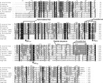
Fig. 2. Multiple alignment of Eimeria LDH enzymes with Apicomplexa LDH enzymes and vertebrate LDH enzymes. Shown are the aligned sequences of LDH from E. acervulina, E. maxima, E. tenella, Toxoplasma gondii LDH1 and LDH2, Plasmodium falciparum, human, chicken and dogfish. Amino acid identities are shown with grey and black shading. The conserved residues His195 and Arg171 are indicated with arrows (amino acid numbering is based on original LDH description from dogfish, White et al. 1976). A conserved Gln102 is replaced by Lys in Apicomplexa (also indicated with arrow). Shown in the 1st box is the extended Apicomplexan specific loop. Two highly conserved coccidia-specific regions are also boxed. Indicated boxes and amino acid residues are also depicted in the structural model (see Fig. 3). The determined amino acid sequence of a tryptic peptide from E. acervulina is indicated (overlined). Multiple alignments were made with ClustalX (Thompson et al. 1997) and edited with Genedoc (Nicholas, Nicholas & Deerfield, 1997). Nucleotide sequence data for Eimeria LDH was deposited in the GenBank™ under the Accession numbers AY143388 (EaLDH), AY143389 (EtLDH) and AY143390 (EmLDH). Other GenBank Accession numbers used were: T. gondii LDH1, AAC47443; T. gondii LDH2, AAC46863; PfLDH, AAA29633; Human LDH-A, CAA26088; Chicken LDH-A, CAA37824; Dogfish LDH-M, DEDFLMPID.

Table 1. Amino acid conservation of LDH between species
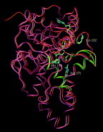
Fig. 3. EtLDH structural model superimposed on PfLDH. Shown is the model of the Cα trace of EtLDH (orange and yellow) superimposed on the 3D crystal structure of PfLDH (purple and green). The NADH cofactor and the oxamate substrate analogue are inserted in the model. The side-chains of Lys102, Arg171 and His195 have been added for both structures. These three amino acids, NADH and oxamate are depicted with their atom colours (carbon=green, oxygen=red and nitrogen=blue). The coccidia-specific regions from Fig. 2 have been highlighted with green and yellow colours. The 3D structure of PfLDH was previously published (Dunn et al. 1996, PDB file 1LDG).

Fig. 4. Southern blot analysis of Eimeria LDH. Genomic DNA from E. acervulina and E. tenella was completely digested with either PstI, HindIII plus EcoRI, HindIII alone or EcoRI alone, separated on 0·7% agarose gel and blotted. The blot on the left was hybridized with a 993 bp EaLDH probe and the blot on the right with a 996 bp EtLDH probe, both probes comprise complete ORFs. Hybridizations were performed at moderately low stringency, showing strong hybridization with its corresponding gene product, and no additional bands. Note that the EaLDH probe weakly detects the EtLDH gene product (indicated with arrow). A sample of 10 μg digested genomic DNA was separated in each lane and a size marker (indicated in kbp) is shown at the right.
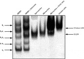
Fig. 5. LDH is active as a homo-tetramer in Eimeria tenella. LDH enzyme activity was measured after separation of total cell extracts on a native non-reducing polyacrylamide gel. LDH activity was visualized by formation of highly coloured insoluble formazan. Bovine LDH is present as 5 distinctive bands, representing different tetramer-combinations (as indicated). EtLDH is visible as a single band in the sporozoite, merozoite and merozoite plus chicken cells lanes. Chicken LDH is also clearly present as a single band. Different protein concentrations from total cell extracts were loaded in each lane: MDBK, 2·4 μg; MDBK+schizonts, 4·1 μg; sporozoites, 5·3 μg; merozoites, 5·5 μg; merozoites+chicken cells, 23 μg; chicken cells, 3·3 μg.
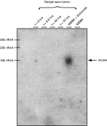
Fig. 6. Schizont-specific expression of EtLDH mRNA. A Northern blot is shown with total RNA from different E. tenella life-cycle stages. EtLDH mRNA was only detected in the schizont stage as a ~1·8 kb mRNA. mRNA of sporulated and non-sporulated oocysts showed no detectable EtLDH mRNA expression. A sample of 10 μg total RNA was loaded in each lane. Equal loading of total RNA was confirmed by comparing ribosomal RNA bands detected after ethidium bromide staining of the gel (not shown). T=0, contains non-sporulated oocysts; t=5.5 h contains non-sporulated oocysts; t=24 h contains fully sporulated oocysts with sporocysts; t=72 h contains fully sporulated oocysts with sporozoites; MDBK+Schizonts contains MDBK cells with 40-h-old E. tenella schizonts (E. tenella RNA was estimated to be 25% of the total RNA by comparing the 28S rRNA bovine band with the smaller 23S rRNA Eimeria band, not shown) and the last lane contains only MDBK cells. The blot was hybridized with a 996 bp EtLDH probe, comprising the complete ORF.
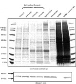
Fig. 7. LDH is present at equal levels during different life-cycle stages of Eimeria tenella. Total protein extracts from different E. tenella life-cycle stages were separated on denaturing and reducing polyacrylamide gels, and either stained with Coomassie brilliant blue or Western blotted. EtLDH was detected with an EtLDH specific polyclonal antiserum. Lanes contain total protein extracts from sporulating oocysts (0, 5.5, 24 and 72 h sporulation), purified sporozoites, purified merozoites, MDBK cells and MDBK cells 40 h after infection with E. tenella sporozoites. Each lane contains 27 μg total protein, with the exception of the MDBK and MDBK plus schizont lanes, which contain 108 μg total protein (to correct for the excess of MDBK cell material). Molecular weight markers are indicated.
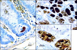
Fig. 8. EtLDH expression in first and second-generation trophozoite/schizont stages. Caecal chicken tissue was analysed 48 h and 96 h after infection with sporulated Eimeria tenella oocysts. (A) A first generation schizont in a crypt at the bottom of a villus, 48 h after infection. (B and C) Second generation trophozoites and schizont stages in a heavily infected part of the caecum, 96 h after infection. (C) Higher magnification of second generation schizonts, which clearly stains all merozoites. EtLDH was visualized with a specific EtLDH polyclonal antibody, stained with peroxidase (haematoxylin blue was used for background staining).
 You have
Access
- 21
- Cited by