Introduction
Ovarian functionality is a complex process controlled by the proper balance between follicular recruitment and atresia rates (Uri-Belapolsky et al., Reference Uri-Belapolsky, Shaish, Eliyahu, Grossman, Levi, Chuderland, Ninio-Many, Hasky, Shashar, Almog, Kandel-Kfir, Harats, Shalgi and Kamari2014), as well as by cellular interactions. At birth, mammalian ovaries contain thousands of follicles that host an oocyte in the earlier stages of development. These primordial follicles are quiescent and comprehend the ovarian follicular reserve pool. Follicles can be recruited and grow, encompassing primary and secondary follicles, which represent the preantral phase. The antral follicle is known as the latter stage of folliculogenesis and is the mature follicle containing the oocyte that can be ovulated (Piprek, Reference Piprek2016). Oocytes are surrounded by granulosa cells forming the follicle that is bordered by theca cells. The signalling and physical interactions among these cells play a key role in folliculogenesis. Mouse genetic models have provided important evidence of these cell interactions and their consequences to ovarian function (Monniaux et al., Reference Monniaux, Clément, Dalbiès-Tran, Estienne, Fabre, Mansanet and Monget2014).
Ligands produced by one cell type can bind to specific receptors present on another cell type and activate signalling pathways. Currently, the role of growth factors (Jones and Shikanov, Reference Jones and Shikanov2019) and hormones (Conforti et al., Reference Conforti, Esteves, Di Rella, Strina, De Rosa, Fiorenza, Zullo, De Placido and Alviggi2019) in follicular growth is being investigated, however there have been few studies about the role of inflammatory molecules on folliculogenesis. In this field, some studies have indicated that interleukin-1 deficiency prolongs ovarian lifespan in mice (Uri-Belapolsky et al., Reference Uri-Belapolsky, Shaish, Eliyahu, Grossman, Levi, Chuderland, Ninio-Many, Hasky, Shashar, Almog, Kandel-Kfir, Harats, Shalgi and Kamari2014; Yang, et al., Reference Yang, Pang and Miao2018) and more recently our group showed that CC-chemokine receptor 2 is expressed by oocytes included in growing follicles and is involved in the control of follicular mobilization and fertility lifespan (Santos et al., Reference Santos, Pereira, Viana, Russo and Campos-Junior2020).
Toll-like receptors (TLR) are transmembrane and intracellular proteins that participate in innate immune system development and activation (Gu et al., Reference Gu, Wang, Yin, Guo, Zhang, Zhang and Zhang2016; Liu et al., Reference Liu, Shimada and Richards2008; Atli et al., Reference Atli, Kose, Hitit, Kaya and Bozkaya2018; Vidya et al., Reference Vidya, Kumar, Sejian, Bagath, Krishnan and Bhatta2018; El-Zayat et al., Reference El-Zayat, Sibaii and Mannaa2019); in mice, there are 12 members (TLR1–TLR13), with TLR10 as a pseudogene (Jiang et al., Reference Jiang, Li, Hess, Guan and Tapping2016). Among these receptors, previous studies have demonstrated that TLR4 may be an important player in female fertility, as it is expressed by cumulus cells under homeostatic conditions (Shimada et al., Reference Shimada, Hernandez-Gonzalez, Gonzalez-Robanya and Richards2006, Reference Shimada, Yanai, Okazaki, Noma, Kawashima, Mori and Richards2008) and participates in ovulation and in cumulus–oocyte complex expansion (Hoshino et al., Reference Hoshino, Takeuchi, Kawai, Sanjo, Ogawa, Takeda, Takeda and Akira1999; Liu et al., Reference Liu, Shimada and Richards2008; Shimada et al., Reference Shimada, Yanai, Okazaki, Noma, Kawashima, Mori and Richards2008; Atli et al., Reference Atli, Kose, Hitit, Kaya and Bozkaya2018). Additionally, TLR4 is important in the female tract response to seminal fluid (Schjenken et al., Reference Schjenken, Glynn, Sharkey and Robertson2015), as well as for embryonic perinatal viability (Wahid et al., Reference Wahid, Dorian, Chin, Hutchinson, Rice, Olson, Moldenhauer and Robertson2015), as TLR4 blockade has been used to avoid preterm labour induced by lipopolysaccharide (LPS; a well characterized molecule associated with Gram-negative bacteria) (Wahid et al., Reference Wahid, Dorian, Chin, Hutchinson, Rice, Olson, Moldenhauer and Robertson2015). Despite this evidence, the role of TLR4 in ovarian physiology is not fully understood, therefore we hypothesized that TLR4 may be involved in folliculogenesis affecting mouse fertility. To explore these aspects, the aim of this study was to perform a quantitative analysis of reproductive performance, folliculogenesis and ovulatory responses in TLR4-deficient mice.
Materials and methods
Animals, facilities and experimental design
Adult mice C57BL/6 [wild type (WT), n = 102] and B6.129-Tlr4tm1Aki TLR4−/−; (Hoshino et al., Reference Hoshino, Takeuchi, Kawai, Sanjo, Ogawa, Takeda, Takeda and Akira1999, n = 94) used in this study were, respectively, obtained from the Universidade Federal de São João del Rei and Universidade Federal de Minas Gerais. These animals were genotyped and maintained isolated from other animals in a 12 h/12 h (light/dark) photoperiod, at a controlled temperature (21ºC) and received sterilized water and food ad libitum. Cages were filter topped and animals were inspected daily. Euthanasia was performed using high sedative doses (15.8 mg/kg xylazine and 139.6 mg/kg ketamine). Body and ovarian weights from all animals used in this study were measured, and the gonad somatic index (GSI; ovarian mass divided by bodyweight × 100) was also calculated. This study was approved by the Institutional Animal Care and Use Committee of the Universidade Federal University de São João del Rei (protocol number 001/2019 CEUA/FUSJ).
Reproductive performance and ovarian follicle quantification
To evaluate the effect of TLR4 deficiency on reproductive performance, 6-wk-old TLR4−/−♂ (n = 20) × TLR4−/−♀ (n = 20) and WT♂ (n = 20) × WT♀(n = 20) pairs were formed and maintained for 8 months. The following parameters were evaluated: number of pups per litter, birth spacing (inter-pregnancy interval), age at first birth and sex ratio. The evaluation of the estrous cycle was performed by vaginal cytology (Byers et al., Reference Byers, Wiles, Dunn and Taft2012). 6-wk-old TLR4−/− (n = 15) and WT (n = 15) females were evaluated during 8 weeks. The vaginal smears were collected every morning between 8 a.m. and 9 a.m., during all days of the experimental period. The estrous cycle length was determined by the time (in days) between two followed oestrus, and the frequency of the stages was calculated by the percentage of proestrus, estrum, metestrus and diestrus observed during the evaluation.
Ovaries were collected from WT (n = 11) and TLR4−/− (n = 11) adult female mice (in proestrus) at 6-wk-old and fixed in a 4% paraformaldehyde solution. Paraffin-embedded ovaries were serially sectioned (5 μm) and stained with haematoxylin and eosin. In every third ovarian section, the numbers of primordial, primary, secondary, antral and atretic follicles were counted. Follicles were classified as previously described (Campos-Junior et al., Reference Campos-Junior, Marinho Assuncao, Carvalho, Batista, Garcia and Viana2012; Pereira et al., Reference Pereira, Nascimento, Jorge, Segatelli, Coutinho, Viana and Campos-Junior2020; Santos et al., Reference Santos, Pereira, Viana, Russo and Campos-Junior2020) and follicles (from primordial to antral) showing morphological signs of death such as pyknosis, cellular fragmentation and disintegration were classified as atretic. Only follicles containing an oocyte with a visible nucleus were considered, to avoid double counting. Results are shown as the number of counted follicles per ovary (Campos-Junior et al., Reference Campos-Junior, Marinho Assuncao, Carvalho, Batista, Garcia and Viana2012; Santos et al., Reference Santos, Pereira, Viana, Russo and Campos-Junior2020).
Ovarian responsiveness to gonadotropins and female fertility evaluation
To evaluate the effects of TLR4 deficiency on ovarian responsiveness and ovulation rate, the number of ovulated oocytes was assessed after a superovulation protocol. Here, 20 IU of equine chorionic gonadotropin were intraperitoneally injected into 6-wk-old WT (n = 12) and TLR4−/− (n = 12) female mice. At 48 h later, 20 IU of human chorionic gonadotropin were also intraperitoneally injected, as previously described (Santos et al, Reference Santos, Pereira, Viana, Russo and Campos-Junior2020). Ovulated cumulus–oocyte complexes were isolated from the oviductal ampullae into PBS medium 14 h after the hCG pulse and counted.
Finally, to evaluate the effects of TLR4 deficiency on female fertility, 6-wk-old TLR4−/−♀ (n = 16) and WT♀(n = 16) were caged with a WT♂ (n = 8) of proven fertility for a mating period of 1 mo. Female mice were weighed at the beginning of the mating period and their body weight was evaluated daily. Vaginal plugs were observed and pregnant female mice were considered as those who had an increase of at least 3 g in 1 wk; they were euthanized close to delivery (19 days post coitum; dpc). Pregnancy was considered as having at least one fetus at laparotomy. The number of fetuses per uterus was calculated (Uri-Belapolsky et al., Reference Uri-Belapolsky, Shaish, Eliyahu, Grossman, Levi, Chuderland, Ninio-Many, Hasky, Shashar, Almog, Kandel-Kfir, Harats, Shalgi and Kamari2014).
Statistical analyses
Kolmogorov–Smirnov test was used to evaluate variables normality. Parametric data (body and ovarian weight, GSI, number of follicles, follicle activation and atresia rates, estrous cycle length, number of ovulated oocytes) were tested using unpaired Student’s t-test. Non-parametric endpoints (number of pups per litter, birth spacing, age at first birth, progeny sex ratios, frequency per stage, number of fetuses from heterozygous pairs) were compared using the Mann-Whitney test, and the pregnancy rate was compared using the chi-squared test. All analyses were performed by a biostatistician using GraphPad Prism v.8.4.2 software. Results are shown as mean ± standard error of the mean (SEM). A P-value < 0.05 was considered significant.
Results
TLR4 deficiency slightly altered reproductive performance
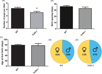
In the first analysis, when 6-wk-old TLR4−/−♂ × TLR4−/−♀ and WT♂ × WT♀ pairs were maintained for 8 months, it was observed that TLR4−/− pairs showed a reduced number of pups per litter 6.34 ± 0.32 (TLR4−/−♂ × TLR4−/−♀) vs 5 ± 0.3 (WT♂ × WT♀) (P = 0.03) (Fig. 1A). There was no significant difference (P > 0.05) in the birth spacing (inter-pregnancy interval) (Fig. 1B) or in the age at the time of their first birth (Fig. 1C). The pup sex ratio (TLR4−/− vs. WT) was also similar (P > 0.05) (Fig. 1D, E).
Reproductive performance of TLR4−/−♂ × TLR4−/−♀ and WT♂ × WT♀ pairs. (A) TLR4 deficiency significantly (P = 0.037) decreased the number of pups per litter. However, (B) birth spacing, (C) age at the time birth and (D, E) progeny sex ratios did not differ (P > 0.05) between TLR4−/−♂ × TLR4−/−♀ and WT♂ × WT♀ pairs.

TLR4 deficiency increased ovarian follicular reserve
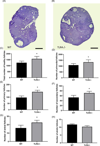
Body and ovarian weight, as well as GSI, were similar (P > 0.05) between WT and TLR4−/− mice (Fig. 2A–C). Histological analyses of a middle section of WT and TLR4−/− ovaries indicated that both were of similar size (Fig. 3A, B). The follicle counts demonstrated that adult TLR4−/− mice presented more (P < 0.001) healthy total follicles than in WT mice (Fig. 3C). Particularly, the number of primordial, primary, secondary and antral follicles was also increased (P < 0.05) in TLR4−/− females (P < 0.001) (Fig. 3D–F), however, the number of atretic follicles was similar (P = 0.1) between TLR4−/− and WT mice (Fig. 3H).
Biometric parameters of 6-wk-old TLR4−/− and WT female mice. (A) Body weight, (B) ovarian weight and (C) gonad somatic index (%) did not differ (P > 0.05) between WT and TLR4−/− mice.

TLR4 deficiency alters follicle endowment in adult female mice. A middle section of (A) WT and (B) TLR4−/− ovaries confirmed their similar sizes, and depicted the presence of all classes of follicles. In the follicular quantification analysis, (C) an increased (P < 0.001) total number of healthy follicles was observed in TLR4−/− females. In more detail, it was observed that TLR4 deficiency increased (P < 0.001) the number of (D) primordial, (E) primary, (F) secondary and (G) antral follicles. Finally, the number of (H) atretic follicles was not significantly different (P = 0.10) between TLR4−/− and WT mice. Bars, 4000 µm.

TLR4 deficiency altered the number of ovulated oocytes and fertility
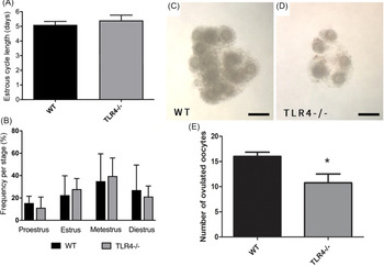
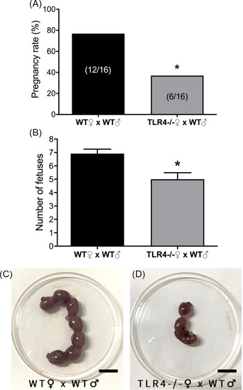
Interestingly, TLR4 ablation did not alter estrous cyclicity once there was no difference in cycle length (days) and its frequencies (proestrus, oestrus, metestrus, diestrus) between TLR4−/− and WT (P > 0.05; Fig. 4A, B). However, a lower (P = 0.006) number of cumulus–oocyte complexes was recovered from TLR4−/− oviducts, compared with WT (12.7 ± 2.3 vs. 16.4 ± 4; Fig. 4C–E). In heterozygous pairs, TLR4−/− females showed a reduced pregnancy rate compared with WT, 37.5% (6/16) vs. 75% (12/16), respectively (Fig. 5A). Moreover, at 19 dpc, a reduced number of fetuses was observed in the uterus of TLR4−/− compared with WT females (P = 0.007; Fig. 5B–D).
TLR4 deficiency effects on estrous cycle and ovulatory response. (A) Estrous cycle length (days) and (B) the frequency stages (proestrus, oestrus, metestrus, diestrus) were not altered in TLR4−/− (P > 0.05). Cumulus–oocyte complexes were recovered from the oviductal ampullae of (C) WT and (D) TLR4−/− mice, showing a reduced number of structures obtained from TLR4−/− females. (E) Number of oocytes harvested from TLR4−/− females was significantly lower (P = 0.006) compared with WT. Bars, 200 µm.

TLR4 deficiency alters female fertility. (A) TLR4−/− females showed a reduced pregnancy rate compared with WT, 37.5% (6/16) vs. 75% (12/16) (P = 0.002). (B) A reduced number of fetuses (P = 0.007) was observed in TLR4−/− mice uteri (19 dpc), as shown in (C) WT and (D) TLR4−/−. Bars, 10 mm.

Discussion
Ovarian follicular mobilization, gonadotropin responsiveness and embryonic developmental competence are important physiological mechanisms that act as decisive indicators of female fertility (Anderson et al. Reference Anderson, Newton, Anderson and Millar2018). This delicate balance can be altered by different hormones and growth factors and also by certain molecules that participate in different signalling pathways (Monniaux et al., Reference Monniaux, Clément, Dalbiès-Tran, Estienne, Fabre, Mansanet and Monget2014; Santos et al., Reference Santos, Pereira, Viana, Russo and Campos-Junior2020). To the best of our knowledge, studies on the potential roles of Toll-like receptor 4 (TLR4) on ovarian folliculogenesis and female fertility are non-existent. Therefore, in this study, we investigated these aspects using WT and knockout mice. Our main results showed that the lack of TLR4 led to an increased follicular reserve, contrasting with undesirable effects on female fertility, such as the reduction in: (i) the number of ovulated oocytes, (ii) the pregnancy rate and (iii) the number of pups per litter (in homozygous and heterozygous pairs).
TLRs are transmembrane proteins, widely recognized as key factors in the activation of immune and inflammatory responses (El-Zayat et al., Reference El-Zayat, Sibaii and Mannaa2019). TLR4 is best known for its involvement in bacteria-produced lipopolysaccharide recognition (Vidya et al., Reference Vidya, Kumar, Sejian, Bagath, Krishnan and Bhatta2018). Although its participation in ovarian cancer has been evaluated in recent years (Lupi et al., Reference Lupi, Cucielo, Silveira, Gaiotte, Cesário, Seiva and de Almeida Chuffa2020), there have been some reports in the published literature on the roles of TLR4 in ovulation and cumulus expansion (Liu et al., Reference Liu, Shimada and Richards2008; Shimada et al., Reference Shimada, Yanai, Okazaki, Noma, Kawashima, Mori and Richards2008; Vaure and Liu, Reference Vaure and Liu2014; Hosseini et al., Reference Hosseini, Dehghani-Mohammadabadi, Ghafarri Novin, Haji Molla Hoseini, Arefian, Mohammadi Yeganeh and Salehi2017; Atli et al., Reference Atli, Kose, Hitit, Kaya and Bozkaya2018), maternal–fetal interaction, preterm labour (Schjenken et al., Reference Schjenken, Glynn, Sharkey and Robertson2015; Wahid, et al., Reference Wahid, Dorian, Chin, Hutchinson, Rice, Olson, Moldenhauer and Robertson2015) and folliculogenesis (Gu et al., Reference Gu, Wang, Yin, Guo, Zhang, Zhang and Zhang2016). In the present study, there was no effect of TLR4 deficiency on the number of litters or on the time of the first birth. These findings may indicate that there was no alteration in puberty onset or in reproductive cycle length. However, TLR4 deficiency reduced the number of pups per litter in homozygous pairs, suggesting that this receptor may affect fertility. Similar to our findings, the depletion of some other proteins, such as Coilin (Walker et al., Reference Walker, Tian and Matera2009), Apc2 (Mohamed et al., Reference Mohamed, Hay, Reed, Smalley and Clarke2019), Fyn kinase (Luo et al., Reference Luo, McGinnis and Kinsey2010) and Aquaporin-4 (Sun et al., Reference Sun, Zhang, Fan, Ding, Sha and Hu2009) altered fertility; usually, this reduced fertility occurs due to changes in ovarian physiology and embryonic development.
Previous studies have demonstrated that TLR4 signalling is not only involved in inflammatory responses, but also regulates proliferation and apoptosis in several cell types (Ding et al., Reference Ding, Li, Zhou, Wang, Zhan and Zhou2010; Li et al., Reference Li, Che, Ji, Shi and Yu2017). Under specific circumstances, TLR4 signalling blocks cell proliferation in different tissues (Ji et al., Reference Ji, Sun, Shrestha, Darragh, Shirakawa, Xing, He, Carboneau, Kim, An, Ma, Oberholzer, Soleimanpour, Gannon, Liu, Naji, Kulkarni, Wang, Kersten and Qi2019; Kashani et al., Reference Kashani, Zandi, Bashash, Zaghal, Momeny, Poursani, Pourbagheri-Sigaroodi, Mousavi and Ghaffari2020). Nevertheless, different TLR4 signalling pathways include the activation of several intracellular ligands, culminating in NF-κB nuclear migration, which promotes the expression of cytokines, caspase-3, IFN-β and other factors that may promote apoptosis (Jung et al., Reference Jung, Lee, Jung, Ock, Lee, Lee and Suk2005). Therefore, it is reasonable to consider that TLR4 deficiency would prevent these signalling pathways. The larger follicular ovarian reserve here reported in TLR4-deficient mice would be a consequence of this atresia attenuation or due to an increase in germ cell proliferation. However, as in 6-wk-old mice, there was no difference in the number of atretic follicles, further studies must be performed in these models during ovarian development (in the pre-natal and perinatal periods), and also on the molecular mechanisms that control germ cell cycle.
TLR4 expression by granulosa cells is well documented in the published literature (Liu et al., Reference Liu, Shimada and Richards2008; Gu et al., Reference Gu, Wang, Yin, Guo, Zhang, Zhang and Zhang2016; Ernst et al., Reference Ernst, Amoushahi, Sørensen, Kragstrup, Ernst and Lykke-Hartmann2020). As follicle activation and growth are regulated by bidirectional communication between oocytes and follicular cells (Eppig et al., Reference Eppig, Wigglesworth and Pendola2002; Liu et al., Reference Liu, Shimada and Richards2008; Shimada et al., Reference Shimada, Yanai, Okazaki, Noma, Kawashima, Mori and Richards2008; Hosseini et al., Reference Hosseini, Dehghani-Mohammadabadi, Ghafarri Novin, Haji Molla Hoseini, Arefian, Mohammadi Yeganeh and Salehi2017), the lack of TLR4 would probably change this interaction. In addition, studies have shown that, although TLR4 is related to the recognition of pathogenic ligands that initiate innate immune responses, it can also be activated by non-pathogenic ligands, such as hyaluronan, which is found in the extracellular matrix around cumulus cells (Liu et al., Reference Liu, Shimada and Richards2008). Hyaluronan is synthesized by hyaluronic acid synthase that induces the formation of fragments and that, in turn, can interact with TLRs and promote the expression of pro-inflammatory cytokines and chemokines (Liu et al., Reference Liu, Shimada and Richards2008). Similarly, Adamts-1, a matrix metalloproteinase with disintegrin and thrombospondin motifs, has been suggested as a regulator of female fertility, primarily during the tissue remodelling process of ovulation (Brown et al., Reference Brown, Dunning, Robker, Boerboom, Pritchard, Lane and Russell2010). Therefore, TLR4 deficiency might have altered extracellular matrix, granulosa cells and oocyte dynamics, resulting in the reduced ovulatory rate and also would be related to a decreased pregnancy rate reported in this study for TLR4-deficient females. Additionally, other aspects such as granulosa cells responsiveness to gonadotropins and hormonal levels could be related to these contrasting findings (increased number of antral follicles and reduced number of ovulated oocytes), indicating that this article raises some questions that demand further investigations.
TLR ligands produced by the fetus interact with TLRs in fetal membranes and placental tissue and are important inflammatory regulators of normal on-time delivery (Wahid et al., Reference Wahid, Dorian, Chin, Hutchinson, Rice, Olson, Moldenhauer and Robertson2015). Specifically, TLR4 is involved in this process (Li et al., Reference Li, Block, Vierkant, Fogarty, Winham, Visscher, Kalli, Wang and Goode2016), and is expressed by fetal membranes; its pharmacological inhibition prevents preterm birth (Chin et al., Reference Chin, Dorian, Hutchinson, Olson, Rice, Moldenhauer and Robertson2016). Our previous data about the reduced number of pups per litter on TLR4−/−♂ × TLR4−/−♀ compared with WT♂ × WT♀ pairs confirmed those pharmacological data. Therefore, to exclude male influence, TLR4−/− and WT females were crossed with WT males. To avoid postnatal cannibalism, these heterozygous fetuses were recovered and counted just before delivery. In this experiment, reduced pregnancy rate and number of fetuses per uterus (in heterozygous pairs) were observed in TLR4-deficient females. Considering that the uterus undergoes immunological adaptation processes after copulation, TLR4 promotes the expression of inflammatory cytokines and chemokines in endometrium to improve receptivity to possible pregnancies (Schjenken et al., Reference Schjenken, Glynn, Sharkey and Robertson2015). Furthermore, there is an increase in TLR4 expression in the mouse uterus at the end of pregnancy (Wahid, et al., Reference Wahid, Dorian, Chin, Hutchinson, Rice, Olson, Moldenhauer and Robertson2015). Taken together, these data strongly suggested that TLR4 mediates inflammatory processes during pregnancy that would be important for proper embryonic development and fetal growth, consequently affecting successful birth and female fertility (Wahid, et al., Reference Wahid, Dorian, Chin, Hutchinson, Rice, Olson, Moldenhauer and Robertson2015).
Overall, this study reported for the first time in the published literature that, in mice, the lack of TLR4 increased ovarian follicular endowment, and represented a potential target for therapies to manipulate ovarian mobilization. However, TLR4 ablation also had some deleterious effects, such as reducing the number of ovulated oocytes, the pregnancy rate and the number of pups per litter. As the blockade of TLR4 signalling with anti-TLR4 monoclonal antibody and drugs (naloxone) has been tested to avoid the incidence of preterm labour induced by LPS (Wahid, et al., Reference Wahid, Dorian, Chin, Hutchinson, Rice, Olson, Moldenhauer and Robertson2015), further studies have to be performed to investigate the mechanism and causes of these reduced fertility parameters, as well as the potential side effects of these treatments on ovarian physiology.
In conclusion, TLR4 plays a contrasting role on murine female fertility. Contrastingly, while its deficiency increased the follicular endowment in adult ovaries, there was a reduction in the ovulation and pregnancy rates, potentially decreasing fertility.
Ethical approval
This study was approved by the Institutional Animal Care and Use Committee of the Universidade Federal University de São João del Rei (protocol number 001/2019 CEUA/FUSJ).
Financial support
This work was supported by the National Council for Scientific and Technological Development (CNPq, Brazil) and Minas Gerais State Research Foundation (FAPEMIG, Brazil). BAM received a scholarship from the Federal University of São João Del Rei.
Author contribution statement
PHACJ designed and supervised the experiments, and revised the manuscript. JPG and BAM performed the experiments and analyzed all data; JPG also prepared the manuscript.
Competing interests statement
The authors declare that there are no known conflicts of interest associated with this publication and there has been no significant financial support for this work that could have influenced its outcome.