Introduction
Parasite occurrence in conventional colonies of laboratory animals is very common. Infections can be problematic for animal breeding and experimentation due to developmental interference, and, consequently, biomedical research progress may be negatively impacted (Doyle et al., Reference Doyle, Monteiro, Graça, Santurio, Silva and Bertolin2006, Lytvynets et al., Reference Lytvynets, Langrova, Lachout and Vadlejch2013). Syphacia muris is a nematode that is frequently found in the intestinal tract of Wistar laboratory rats (Rattus norvegicus). Helminth eggs are very light and may be dispersed as aerosols, resulting in widespread environmental contamination (Baker, Reference Baker1998; Dix et al., Reference Dix, Astill and Whelan2004).
The life cycle of S. muris is direct. The female lays fertilized eggs in the colon and peri-anal regions of the rat (Lytvynets et al., Reference Lytvynets, Langrová, Lachout, Vadlejch, Fučíková and Jankovská2010). Eggs, which are infective within a few hours and contaminate other rodents, are highly resistant to the environment and can survive for extended periods of time at room temperature. The host becomes infected either by ingestion of eggs from the peri-anal region of an infected rat, or indirectly through food, water or other contaminated materials. Other possibilities include retro-infection, i.e. the hatching of eggs in the anus and subsequent movement of larvae into the colon. The pre-patent period of S. muris is 7–8 days, in which adult worms establish themselves in the caecum and ascending colon (Lewis & d'Silva, Reference Lewis and d'Silva1986; Baker, Reference Baker1998; Lytvynets et al., Reference Lytvynets, Langrová, Lachout, Vadlejch, Fučíková and Jankovská2010).
Several techniques have been described for diagnosing S. muris infections in laboratory rodents. The most common diagnostic techniques include taping, anal swab, spontaneous fluctuation, necropsy with microscopic examination of the caecal and colonic contents and histological examination (Pritchett & Johnston, Reference Pritchett and Johnston2002; Hill et al., Reference Hill, Randolph and Mandrell2009). The Hoffman, Pons and Janer (HPJ) technique is a spontaneous sedimentation method (Hoffman et al., Reference Hoffman, Pons and Janer1934) used to detect developmental forms of both protozoa and helminths, while the Willis technique (Willis, Reference Willis1921) is a spontaneous flotation method, used for detection of light structures such as protozoan cysts and light helminth eggs (Carvalho et al., Reference Carvalho, Moreira, Pena, Marinho, Bahia and Machado-Coelho2012). The Graham (Reference Graham1941) or taping method consists of making impressions of the peri-anal region of infected rodents using an adhesive tape (Hill et al., Reference Hill, Randolph and Mandrell2009).
Laboratory animals are of great importance to biological and biomedical experimentation, and sanitation insufficiencies can interfere with expected study results. As such, the aims of this study were to compare the three diagnostic methods for detection of S. muris in naturally infected rats and to qualify method appropriateness.
Materials and methods
Maintenance and peri-anal examination of rats
Thirty Wistar rats (R. norvegicus) with verified S. muris infection were chosen for the study. Male rats weighing in the range of 120–180 g and aged 6–8 weeks were used. Rats were bred in a conventional manner at the Centro de Experimentação Animal (CBEA) of Universidade Federal de Uberlândia (UFU). Rats were kept in cages with a maximum density of four rats per cage lined with bed shavings, with access to filtered water and fed with industrial feed. Colony room temperature was 22 ± 2°C, and artificial lighting consisted of a 12 h:12 h light–dark cycle. Rats were sacrificed following the method described by Luca et al. (Reference Luca, Alexandre, Marques, de Sousa, Merusse and Neves1996), referring to the study of intestinal parasites. The presence of S. muris infection was established by observation of any form of the helminth in the intestinal contents of rats at necropsy.
Rats were subjected to an in vivo taping examination using the Graham method (Graham, Reference Graham1941) according to Taffs (Reference Taffs1976), always in the afternoon. Transparent self-adhesive tape was placed in contact with the peri-anal region of each rat to collect S. muris eggs. The tape was then placed on a slide and examined using an optical microscope.
Collection of eggs from faeces
The spontaneous sedimentation method (Hoffman et al., Reference Hoffman, Pons and Janer1934) was performed by diluting collected faeces in distilled water and subjecting the sample to sedimentation over a period of 24 h. Samples were then stained with lugol and analysed using an optical microscope. To test the spontaneous flotation method (Willis, Reference Willis1921), samples were processed with a saturated solution of NaCl and transferred to a Borrel bottle, on top of which a slide was placed. After 5 min, slides were removed from the bottle and the material adhering to the slide was stained with lugol and analysed under a light microscope.
Data analysis
Statistical analysis was performed using GraphPad Prism 5.0 software (GraphPad Software Inc., San Diego, USA). The Fischer's exact test was used for comparison between methods of detection with levels of significant differences shown when P< 0.05.
Results and discussion
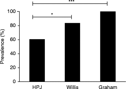
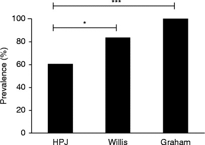
Among the methods used to detect S. muris eggs from 30 positive samples, the Graham method confirmed that 100% were positive compared with 83% and 60% using the Willis and HPJ methods, respectively (fig. 1). As all rats were confirmed positive for S. muris, the Willis and HPJ methods showed false-negative results. Using a Fisher's exact test, we attempted to compare which of the three methods would be more suitable for identification of pinworm eggs in the samples. A higher efficiency of egg detection was found using the Graham (P= 0.0001) compared with the Willis method (P= 0.0391), although these differences were not significant (P= 0.1124).
Three parasitological techniques – the Hoffman, Pons and Janer (HPJ), Willis and Graham methods – for detecting Syphacia muris eggs in the rat; level of significance: *P< 0.05; ***P< 0.0001.
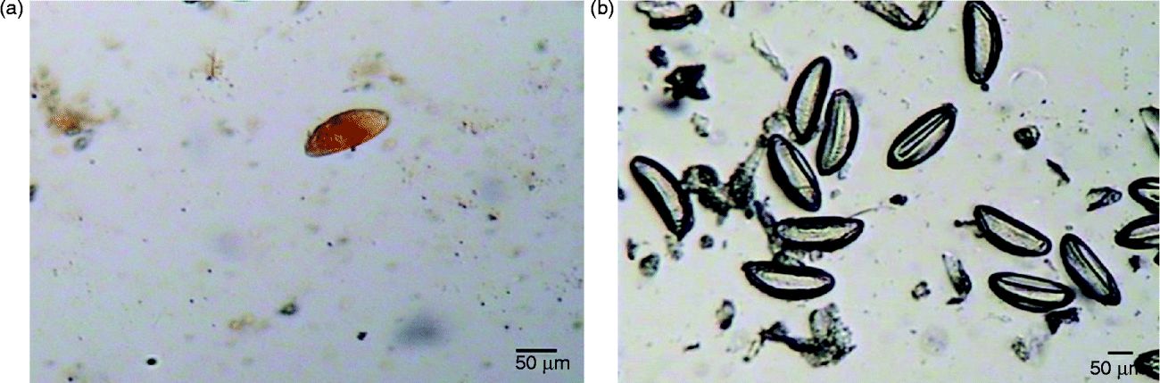
In the present study the Graham method was more effective in detecting eggs of S. muris when compared with the HPJ method, not only in providing a view of a large quantity of eggs in relation to field size (fig. 2) but also 100% detection with a relatively more rapid and simpler implementation compared with the other two methods.
Recovery of eggs of Syphacia muris from rats using the Hoffman, Pons and Janer (a) and Graham (b) methods, 100 × magnification. Images were captured with an optical microscope coupled to a Biocam system and PixelView software (XCapture PCI-PV-CX881P-F).

False-negative results (i.e. as those observed in the Willis and HPJ methods) have also been reported previously with use of tape. This may be related to factors such as worm burdens, sex and age of rodents, training, technical performance and test time (Taffs, Reference Taffs1976; Hill et al., Reference Hill, Randolph and Mandrell2009). Another important factor that should be taken into account for the diagnosis of S. muris infection is the frequency of egg production, as it can influence egg detection by the applied methods.
Pinworm species belonging to the genus Syphacia show high prevalence in laboratory rodents due to their short life cycles (Bicalho et al., Reference Bicalho, Araujo, Rocha and Carvalho2007). Under controlled conditions, in the afternoon hours female S. muris worms migrate from the intestine and caecum of the rat to deposit eggs around the peri-anal region (Taffs, Reference Taffs1976; Hill et al., Reference Hill, Randolph and Mandrell2009). Since most eggs are found in the peri-anal area and only a small portion will be eliminated with faeces, sedimentation and other traditional methods involving faecal collection, as well as egg quantification by floatation, are considered less effective, and are likely to underestimate Syphacia spp. infections. The Graham method is also the most suitable technique for detecting eggs of species such as Enterobius vermicularis (Jeandron et al., Reference Jeandron, Abdyldaieva, Usubalieva, Ensink, Cox, Matthys, Rinaldi, Cringoli and Utzinger2010), as fertilized females also lay eggs preferably around the peri-anal region of the host.
Establishing a reliable diagnostic method for S. muris is essential because this oxyurid is commonly found in laboratory rodent colonies, and infection can cause physiological changes in hosts that may compromise biomedical research data (Wagner, Reference Wagner1988; Gilioli et al., Reference Gilioli, Andrade, Passos, Silva, Rodrigues and Guaraldo2000; Lytvynets et al., Reference Lytvynets, Langrová, Lachout, Vadlejch, Fučíková and Jankovská2010; Tanideh et al., Reference Tanideh, Sadjjadi, Mohammadzadeh and Mehrabani2010). Besides S.muris, several other parasites have been detected in laboratory rodents, such as Hymenolepis nana, Hymenolepis diminuta, Syphacia obvelata, Aspiculuris tetraptera, Physaloptera, Paraspidodera uncinata and Trichinella spiralis; some of these have high zoonotic potential, making them difficult to control (Huq et al., Reference Huq, Karim and Sheikh1985; Tanideh et al., Reference Tanideh, Sadjjadi, Mohammadzadeh and Mehrabani2010; Hayashimoto et al., Reference Hayashimoto, Morita, Ishida, Yasuda, Kameda, Uchida, Tanaka, Ozawa, Sato, Takakura, Itoh and Kagiyama2013).
The use of laboratory animals in biomedical research has provided a better understanding of human and veterinary physiological and pathological processes (Casebolt et al., Reference Casebolt, Lindsey and Cassell1988; Bicalho et al., Reference Bicalho, Araujo, Rocha and Carvalho2007; Tanideh et al., Reference Tanideh, Sadjjadi, Mohammadzadeh and Mehrabani2010). Therefore, the quality, reliability and consequent standardization of these animals have a great impact on research, since only healthy and pathogen-free animals produce valid scientific data (Lytvynets et al., Reference Lytvynets, Langrová, Lachout, Vadlejch, Fučíková and Jankovská2010). Studies have demonstrated the importance of detection of infection in laboratory animals not only to ensure the reliability of scientific research but also to prevent the development of zoonoses, since approximately 150–200 diseases, including parasites, can be transferred from laboratory animals to healthy humans (Huq et al., Reference Huq, Karim and Sheikh1985; Tanideh et al., Reference Tanideh, Sadjjadi, Mohammadzadeh and Mehrabani2010).
Syphacia muris presents a great challenge in terms of control due to characteristics of their life cycle (i.e. as a retro-infection) and the ease of egg dispersion, resulting in contamination of other laboratory rodents and equipment (Dix et al., Reference Dix, Astill and Whelan2004). Resolving widespread contamination of laboratory animals becomes extremely laborious and costly, primarily due to interference with experimental data and the lengthy treatment period required. Thus, it is essential to establish effective methods for early detection of S. muris infection.
Choosing the most reliable method for screening of S. muris infection of laboratory rats also helps prevent future environmental contamination and infection. Considering the importance of the healthy condition of laboratory animals for both research and researchers, it is of fundamental importance to periodically monitor and evaluate these animals for parasites, as well as other pathogens such as viruses, bacteria and fungi.
Financial support
This study was supported by grants from the Coordenação de Aperfeiçoamento de Pessoal de Nível Superior (CAPES – Grant No. 3200601004M-8) (J.E.N.S and E.F.G.C.) and Fundação de Amparo à Pesquisa do Estado de Minas Gerais (FAPEMIG – Grant No. CBB-PPM-00 396-13).
Conflict of interest
None.
Ethical standards
Authors assert that all procedures contributing to this study comply with the ethical standards of laws of Brazil on the care and use of laboratory animals, in accordance with the ethical principles upon approval of the Comitê de Ética na Utilização de Animais of Universidade Federal de Uberlândia (protocol 112/12).