Introduction
Benign paroxysmal positional vertigo (BPPV) is classified into a lateral semicircular canal type and a posterior semicircular type, according to the features of positional nystagmus. Patients with the lateral canal type show horizontal nystagmus, and patients with the posterior canal type reveal torsional nystagmus. Positional nystagmus of the lateral canal type is classified as direction-changing geotropic nystagmus or direction-changing apogeotropic nystagmus. Geotropic nystagmus is classified into transient type or persistent type. Canalolithiasis (moving debris) accounts for transient geotropic nystagmus.Reference McClure1,Reference Baloh, Jacobson and Honrubia2 Persistent type, which lasts more than 1 minute, is caused by a light cupula.Reference Hiruma and Numata3–Reference Kim, Kim and Ban5 Apogeotropic nystagmus is persistent and explainable by a heavy cupula (cupulolithiasis).Reference Baloh, Yue, Jacobson and Honrubia6,Reference Bisdorff and Debatisse7 The pathophysiology of transient torsional nystagmus is a canalolithiasis in the posterior canal,Reference Epley8 while that of persistent torsional nystagmus is a heavy cupula in the posterior canal.Reference Imai, Takeda, Ito, Sekine, Sato and Midoh9,Reference Ichijo10
Few reports concerning the incidence of the subtypes have been reported. We performed a fixed-point observation study. Patients were prospectively aggregated for four years. The first aim of the study was to clarify whether the lateral canal type or posterior canal type was more common.
We observed that patients with BPPV felt vertigo at the time of awakening. Benign paroxysmal positional vertigo is associated with sleeping position, and there is a relationship between the affected side and head-lying side during sleep.Reference Korres, Papadakis, Riga, Balatsouras, Dikeos and Soldatos11 However, little information concerning the onset time is available. The second aim of the study was to assess the aetiology of BPPV by investigating the onset time of each subtype.
Materials and methods
Subjects
The subjects were 512 consecutive BPPV patients (374 females and 138 males, mean age of 61.4 years) who were diagnosed and prospectively aggregated between January 2016 and December 2019.
Inclusion and exclusion criteria
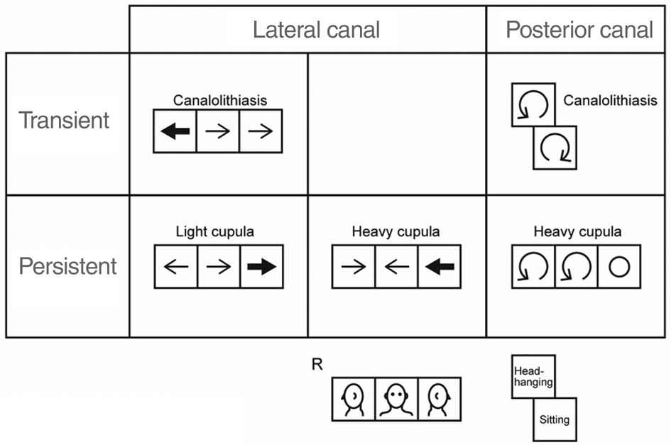
The inclusion criteria are shown in Figure 1. The exclusion criteria were: cochlear symptoms related to vertigo, central nervous system disorders and history of head trauma.
Five subtypes of benign paroxysmal positional vertigo. The affected side is the right side in this illustration. Arrows indicate the direction of nystagmus. Bold arrows represent stronger nystagmus. R = right

Nystagmus testing
We used an infrared charge-coupled device camera. First, the patient's eye movements were observed in the sitting position. Second, the seat was reclined slowly so that the lateral canal became earth-vertical. Next, the head was moved in yaw rotations leftward or rightward. If positive for nystagmus, head position was kept for 1 minute to determine whether the nystagmus was transient or persistent (the supine head roll test). When no nystagmus was elicited by the supine head roll test, the Dix–Hallpike testReference Dix and Hallpike12 was performed.
Benign paroxysmal positional vertigo subtypes
Lateral canalolithiasis
This was characterised by the following. There is no nystagmus in the sitting position. In the supine position, transient lying-down nystagmus frequently occurs. In the right-ear-down position, horizontal nystagmus toward the right occurs after a brief period, and then decays and stops within 1 minute. In the left-ear-down position, horizontal nystagmus toward the left occurs after a brief period, and then decays and stops within 1 minute.
Lateral light cupula
This was defined by the following. There is no nystagmus in the sitting position. In the right-ear-down position, horizontal nystagmus toward the right occurs and lasts for more than 1 minute. In the left-ear-down position, horizontal nystagmus toward the left occurs and lasts for more than 1 minute. In the supine position with the face straight up, persistent horizontal nystagmus occurs, and it ceases when the head is turned to the affected ear by 20–80° (neutral position). In the nose-down position with the face straight down, persistent horizontal nystagmus occurs, and its direction is opposite to that in the supine position.
Lateral heavy cupula
This was characterised by the following. There is no nystagmus in the sitting position. In the right-ear-down position, horizontal nystagmus toward the left occurs and lasts for more than 1 minute. In the left-ear-down position, horizontal nystagmus toward the right occurs and lasts for more than 1 minute. In the supine position with the face straight up, persistent horizontal nystagmus occurs and it ceases when the head is turned to the affected ear by 20–80° (neutral position). In the nose-down position with the face straight down, persistent horizontal nystagmus occurs, and its direction is opposite to that in the supine position.
Posterior canalolithiasis
This was defined by the following. The Dix–Hallpike test reveals upward vertical or torsional nystagmus, with some latency period in the head-hanging position. In the sitting position, downward vertical or torsional nystagmus occurs. In both positions, nystagmus decays and stops within 1 minute.
Posterior heavy cupula
This was characterised by the following. In the supine and affected-ear-down position, upward vertical or torsional nystagmus occurs and lasts for more than 1 minute. There is no nystagmus in the healthy-ear-down position. In the sitting position, no nystagmus or mild vertical (downward) nystagmus occurs. In the nose-down position, downward vertical or torsional nystagmus occurs and lasts for more than 1 minute.
Determining the affected side
In patients with lateral canalolithiasis, the direction of stronger nystagmus is the affected side. In patients with lateral light cupula and lateral heavy cupula, the side of the neutral position is the affected side. If the neutral position was 0°, we concluded that the affected side was unknown. In patients with posterior canal type, the direction of the torsional component in the supine or head-hanging position is the affected side.
Onset times assessment
We interviewed patients, and categorised the onset time into the following four groups: (1) during sleep; (2) at time of awakening in the morning; (3) morning (after awakening until noon); and (4) afternoon (from noon until going to bed). In patients with interrupted sleep, we categorised onset time as occurring during sleep.
Video-oculography
Nystagmus recordings were converted into digital data. Two-dimensional video-oculography was performed using ImageJ version 1.36 software (a public domain, Java-based image-processing program developed at the National Institutes of Health) and a Macintosh computer (operating system version 10.6.2). In order to analyse the horizontal and vertical components, the XY centre of the pupil was calculated.Reference Ikeda, Hashimoto, Horiike and Yamashita13 Upward deflection indicates rightward deflection of the eye.
Results
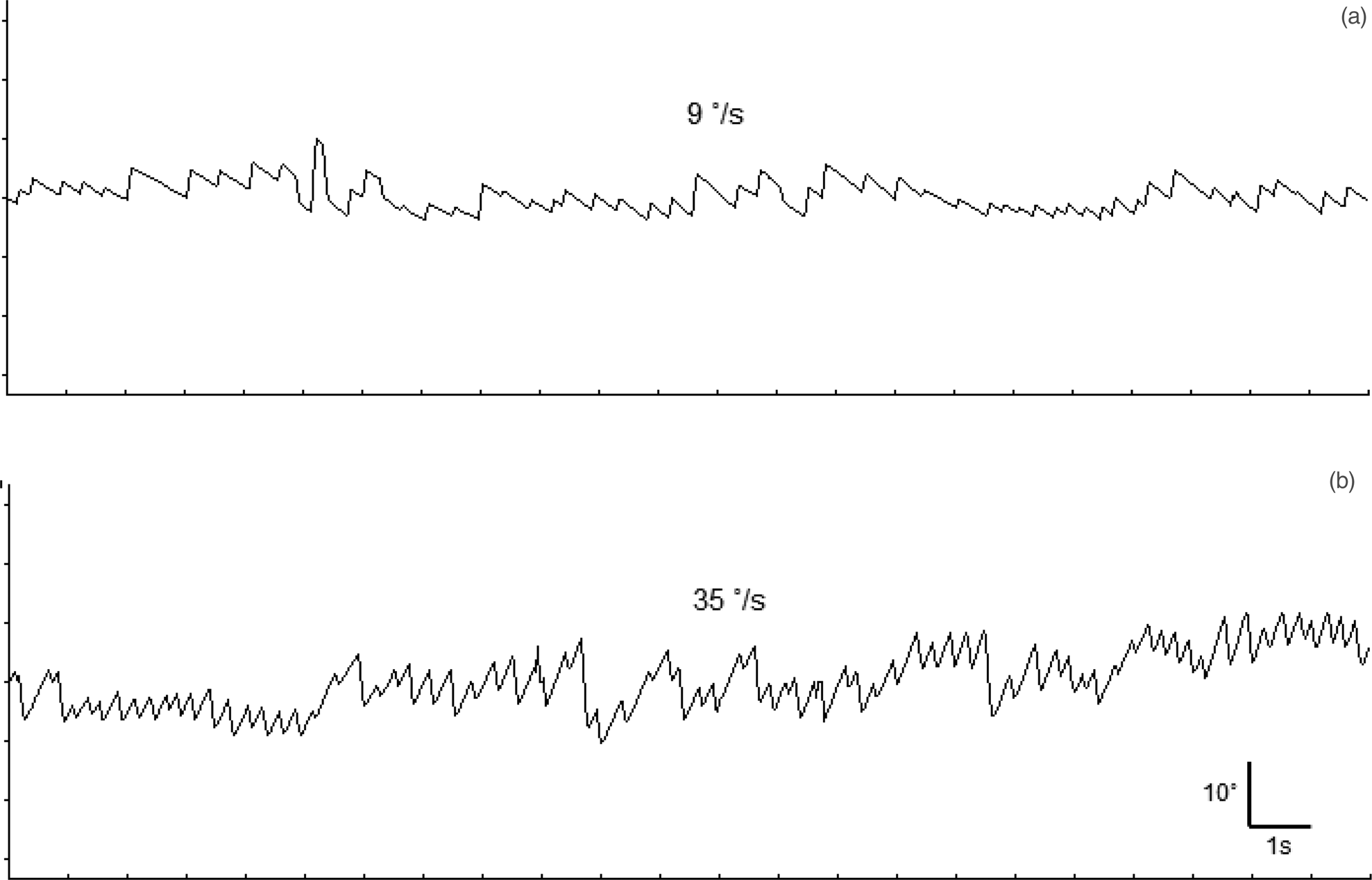
Video-oculography results of a patient with lateral light cupula are shown in Figure 2.
Video-oculography (position traces of the horizontal component) of persistent direction-changing geotropic positional nystagmus for (a) right-ear-down and (b) left-ear-down, in a patient with lateral light cupula. Note that the nystagmus does not fade. The numbers indicate maximum slow-phase velocity.

Age
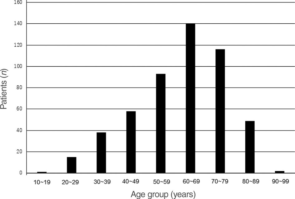
Age was not normally distributed (Figure 3). Most patients were in their sixties, followed by patients in their seventies.
Age distribution for all patients included in the analysis (n = 512). Most patients were in their sixties, followed by patients in their seventies.

Affected side
Table 1 shows the affected side of each subtype. In the lateral canal type, the left side was dominant. In the posterior canal type, the right side was dominant.
Affected side

Data represent numbers of cases
Subtypes
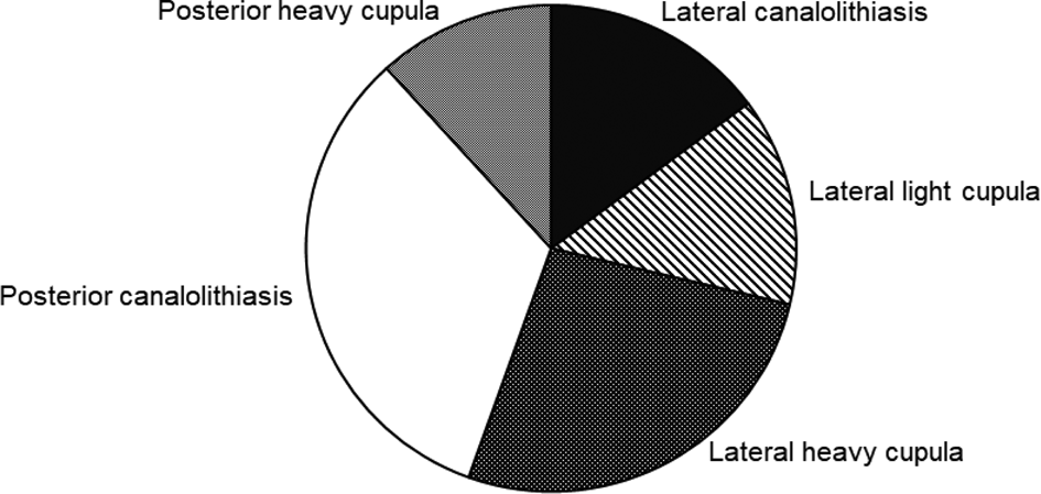
The proportions of the five subtypes are shown in Figure 4. Seventy-six patients (14.8 per cent) were found to have lateral canalolithiasis. Seventy-one patients (13.9 per cent) had lateral light cupula and 137 patients (26.8 per cent) had lateral heavy cupula. There were 168 patients (32.8 per cent) with posterior canalolithiasis and 60 patients (11.7 per cent) with posterior heavy cupula. Lateral canal type (55.5 per cent) was more common than posterior canal type (44.5 per cent).
Proportions of the five benign paroxysmal positional vertigo subtypes (n = 512). Lateral canal type was more common than posterior canal type.

Onset time
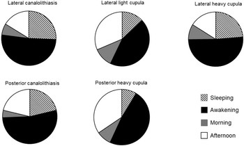
Figure 5 shows the onset time of the five subtypes. The time of awakening was the most common onset time in every subtype.
Onset times of the five benign paroxysmal positional vertigo subtypes. Time of awakening was the most common onset time in every subtype.

Lateral canalolithiasis
Vertigo occurred during sleep in 19 patients (25.7 per cent), at the time of awakening in 38 patients (51.4 per cent), in the morning in 5 patients (6.8 per cent) and in the afternoon in 12 patients (16.2 per cent).
Lateral light cupula
Vertigo occurred during sleep in 9 patients (12.9 per cent), at the time of awakening in 31 patients (44.3 per cent), in the morning in 8 patients (11.4 per cent) and in the afternoon in 22 patients (31.4 per cent).
Lateral heavy cupula
Vertigo occurred during sleep in 31 patients (23.7 per cent), at the time of awakening in 67 patients (51.1 per cent), in the morning in 12 patients (9.2 per cent) and in the afternoon in 21 patients (16.0 per cent).
Posterior canalolithiasis
Vertigo occurred during sleep in 34 patients (21.3 per cent), at the time of awakening in 85 patients (53.1 per cent), in the morning in 7 patients (4.4 per cent) and in the afternoon in 34 patients (21.3 per cent).
Posterior heavy cupula
Vertigo occurred during sleep in 5 patients (8.6 per cent), at the time of awakening in 28 patients (48.3 per cent), in the morning in 5 patients (8.6 per cent) and in the afternoon in 20 patients (34.5 per cent).
Discussion
Although several researchers have reported that the lateral canal type is rare,Reference Korres, Balatsouras, Kaberos, Economou, Kandiloros and Ferekidis14.Reference Cakir, Ercan, Cakir, Civelek, Sayin and Turgut15 we found the incidence of the lateral canal type to be higher than that of the posterior canal type. This discrepancy is attributed to the difference of the medical systems. Our hospital is a walk-in clinic; therefore, patients present with early-stage BPPV. In appointment-only clinics, patients need to wait for a longer time to visit. Consequently, the otologist cannot diagnose the lateral canal type accurately because the time from onset to remission of positional vertigo in patients with the lateral canal type is shorter than that in those with the posterior canal type.Reference Imai, Ito, Takeda, Uno, Matsunaga and Sekine16,Reference Shim, Ko, Lee and Park17
The second reason why the lateral canal type was more common than the posterior canal type is the order of examinations. We perform the supine head roll test first. If the Dix–Hallpike test is conducted first and the result is negative, the examiner sometimes neglects the supine head roll test.
• Benign paroxysmal positional vertigo (BPPV) is classified into five subtypes according to the features of positional nystagmus
• The lateral canal type (55.5 per cent) was more common than the posterior canal type (44.5 per cent)
• The time of awakening was the most common onset time in every subtype
• The aetiology of BPPV is closely related to sleep
The third reason reflects an awareness; some physicians do not recognise lateral canal BPPV, because it is a new concept and classification is not completely accepted. Therefore, some otologists might overlook the lateral canal type.
Patients with BPPV usually visit the internal medicine department because of a lack of cochlear symptoms. Recently, general physicians have begun to understand the pathophysiology of canalolithiasis, and they now refer dizzy patients to the otologist. Moreover, the internet has made it easier to access a specialist.
It was reported that more than 75 per cent of canalolithiasis patients experienced vertigo during sleep or at the time of awakening.Reference Ichijo18 The present study demonstrated similar results, even in patients with posterior heavy cupula and lateral light cupula. We found that time of awakening was the most common onset time in every subtype. These results suggest that the aetiology of BPPV is closely related to sleep. Ichijo proposed a micro-otoconia accumulation theory, wherein the pathological debris is an aggregate of micro-otoconia that developed over a long time period, which begins to slide by its own weight during sleep.Reference Ichijo18
Although the pathophysiology of light cupula is unclear, time of awakening is the most common time of onset, suggesting a light debris cupulolithiasis. Consider a hypothetical case of a female who likes to sleep in the right-ear-down position. While in bed, small light debris clings to the cupula of the left lateral canal. The attached tiny debris is not a cause of cupular deviation; however, if she keeps on sleeping in the same position, tiny light debris congregates on the left cupula by degrees. Finally, the buoyancy exerted on the cupula reaches a threshold and stimulates the hair cells.
The biggest problem is the identity of light debris. We speculate that the light debris is degenerated cells. In animal experiments, monocytes and lymphocytes were observed in the endolymphatic sac.Reference Yamane and Igarashi19 There are many macrophages in the human endolymphatic sac.Reference Kämpfe Nordström, Danckwardt-Lillieström, Laurell, Liu and Rask-Andersen20 If macrophages die and swell up, they become less dense.
Conclusion
The incidence of lateral canal type is higher than that of posterior canal type. Time of awakening is the most common onset time in every subtype; therefore, the aetiology of BPPV is strongly related to sleep.
Acknowledgement
We thank Dr Kazunori Futai for preparing some of the figures.
Competing interests
None declared