Introduction
The future of electrochemical energy storage depends on the concurrent advancement of constituent component materials and their satisfactory interaction with one another. We primarily look to increase the energy and power density of electrochemical cells through increasing electrode capacity, and remove the chemical energy available for release during device failure, as present in modern flammable organic liquid electrolytes used in lithium-ion batteries.Reference Dudney, West and Nanda1 Solid-state electrolytes (SSEs) may satisfy both these requirements, serving as a safe replacement for their organic liquid counterparts, while potentially enabling alkali metal anodes.
However, the introduction of SSEs into full cell batteries is accompanied by other constraints, both intrinsic and extrinsic.Reference Kerman, Luntz, Viswanathan, Chiang and Chen2 Intrinsically, though many SSEs exhibit high bulk ionic conductivity, grain boundaries may ultimately reduce the effective ionic conductivity to unusable levels, as in the case of solid-oxide perovskite lithium lanthanum titanate (LLTO),Reference Ban and Choi3 and may serve as regions of mechanical susceptibility, aiding lithium dendrite nucleation and penetration.Reference Varzi, Raccichini, Passerini and Scrosati4 Extrinsically, questions of electrolyte/electrode interfacial impedance, resulting from space chargeReference Maier5 and chemical and electrochemical compatibilityReference Richards, Miara, Wang, Kim and Ceder6,Reference Zhu, He and Mo7 of solid–solid interfaces, come into play. Despite the importance of these interfaces in the functionality of next-generation solid-state devices, there are surprisingly few studies focused on characterization of their interfaces, and even fewer providing in situ and operando insights.
In the past decade there have been significant efforts in applying in situ and operando measurements to electrochemical systems, though most work is limited to liquid-electrolyte systems. Development of intricate electrochemical testing cells for synchrotron end stations have enabled in situ and operando x-ray diffraction (XRD), x-ray absorption spectroscopy, and x-ray photoelectron spectroscopy (XPS).Reference Lin, Liu, Yu, Cheng, Singer, Shpyrko, Xin and Tamura8 In situ holders for electron microscopy have similarly progressed,Reference Mehdi, Gu, Parent, Xu, Nasybulin, Chen, Unocic, Xu, Welch, Abellan, Zhang, Liu, Wang, Arslan, Evans and Browning9–Reference Tripathi, Su and Hwang12 aided by the collaborative environment established by developers of transmission electron microscopy (TEM) holders. These methods have enabled dynamic observation of the complex changes within electrode materials during electrochemical cycling and have provided a variety of insights, including the dynamics of lithiation, phase transformations, and the elusive nature of the solid-electrolyte interface (SEI) formation. Such in situ electrochemical liquid testing methods are extensively described in previous review articles.Reference Lin, Liu, Yu, Cheng, Singer, Shpyrko, Xin and Tamura8–Reference Tripathi, Su and Hwang12
Attempts to observe similar processes in solid-state materials have added degrees of complexity due to their buried interfaces, often requiring the expansion of existing techniques and the development of entirely new methodologies. This perspective aims to review advanced characterization techniques applied to solid–solid interfaces, working toward in situ and operando characterization to elucidate the less-understood dynamic behavior and heterogeneous characteristics present at these interfaces.
Techniques to characterize buried solid–solid interfaces
Until recently, electrochemical techniques, primarily cyclic voltammetry and electrochemical impedance spectroscopy, were the main methods of characterizing buried interface resistances. These studies are prevalent in characterization of all-solid-state batteries, such as LiCoO2 (LCO)/lithium phosphorus oxynitride (LiPON)/LiReference Iriyama, Kako, Yada, Abe and Ogumi13 and LCO/Li1.5Al0.5Ti1.5(PO4)3 (LATP)Reference Amiki, Sagane, Yamamoto, Hirayama, Sudoh, Motoyama and Iriyama14 chemistries. Electrochemical testing of such cells suggested growing interfacial impedance, vaguely attributed to modified chemical bonding impacting charge-transfer characteristics.Reference Iriyama, Kako, Yada, Abe and Ogumi13 Thermal annealing studies showed reduced interfacial resistances and improved cyclability of the LCO/LiPON/Li cells, though physical interpretation was speculative, lacking further experimental evidence and simply attributing the change to modified bonding at the LCO/LiPON interface.Reference Iriyama, Kako, Yada, Abe and Ogumi13 The LCO/LATP cell utilized in situ formation of the anode, showing low charge-transfer resistance, though the nature of the anode was unknown at the time of the electrochemical testing.Reference Amiki, Sagane, Yamamoto, Hirayama, Sudoh, Motoyama and Iriyama14
Similar studies further evaluated the stability of an interface or interlayer, such as the insertion of a Nb interlayer between LCO and lithium lanthanum zirconium oxide (LLZO) electrolyte, observing reduced interfacial impedance without explicit mechanistic descriptions.Reference Kato, Hamanaka, Yamamoto, Hirayama, Sagane, Motoyama and Iriyama15 That is not to say that electrochemical methods are insufficient methods of probing such interfaces. In scenarios where constituents are expected to be kinetically stable, careful experimental design promotes isolation of such effects, as in the case of idealized solid-state interfaces such as some silver conductors.Reference Maier16 Control of materials selection can allow electrochemical methodologies to effectively isolate sources of charge-transfer resistance to the impact of lattice mismatch, defect chemistry, and equilibrium potentials.Reference Sata, Eberman, Eberl and Maier17 Indeed, an entire field has been based around this ideology—solid-state ionics/nanoionics—however, this will be not be covered in this article to focus on adapting advanced characterization techniques to more commercially relevant systems.
Until recently, the nature of stability of solid-state interfaces in applied lithium-ion battery materials was largely unknown, and questions of compatibility at interfaces was ascribed generally to issues of charge transfer across interfaces. Density functional theory (DFT) calculations were successfully employed to predict engineered interphases to counteract the effects of space charge, uncovering lithium niobate (LiNbO3) as a candidate to stabilize the cathode/sulfide–electrolyte interface.Reference Haruyama, Sodeyama, Han, Takada and Tateyama18 The insertion of a LiNbO3 interlayer at the LCO/LiPON interface was shown via XPS to modify lithium concentration, an effect that was attributed to non-faradaic Li migration.Reference Gittleson and El Gabaly19 A following body of computational research opened the question as to the thermodynamic stability of these interfaces—a fundamentally important step in interpreting the nature of these buried interfaces, providing potential explanations for the presence of interfacial resistances at solid−solid interfaces.Reference Richards, Miara, Wang, Kim and Ceder6,Reference Zhu, He and Mo7,Reference Zhu, He and Mo20 This work suggests that stable solid-state interphases effectively bridge the electrochemical window in much the same way the SEI does in their liquid electrolyte counterpart.
Thermodynamically driven decomposition is most clearly shown after applying high-temperature processing methods, as in the case of spinel cathodes interfaced with LATP; here, the increased temperature drives decomposition to a degree detectable by x-ray diffraction (XRD)—intuitively ruling out high-temperature co-sintering as a processing method.Reference Miara, Windmüller, Tsai, Richards, Ma, Uhlenbruck, Guillon and Ceder21 Combining results of electrochemical testing with ex situ evaluation has proven to be an effective technique for a more comprehensive understanding of the stability of solid–solid interfaces, and has yielded results consistent with computational predictions.
For the case of LCO/LiPON, LCO was predicted to decompose into a variety of constituents, dependent on the local bonding environment and lithiation state.Reference Zhu, He and Mo20 Experimentally, it was observed that cycling LCO/LiPON/Li thin-film batteries at elevated temperatures resulted in increasing interfacial impedance values. When extracted by a focused ion beam (FIB) system and observed by scanning transmission electron microscopy coupled with electron energy-loss spectroscopy (STEM/EELS), the increased interfacial impedance was correlated with the growth of a structurally decomposed LCO interlayer, exhibiting chemical signals consistent with Li2O and disordered rock-salt Co3O4.Reference Wang, Lee, Xin, Han, Grillon, Guy-Bouyssou, Bouyssou, Proust and Meng22 X-ray photoelectron spectroscopy (XPS) was similarly applied to explain growing impedance in Na-metal/Na-sulfide symmetric cells; as predicted by computational modeling, Cl-doping was observed to promote the formation of NaCl, which stabilized the interface, preventing further electrolyte decomposition.Reference Wu, Kompella, Zhu, Lee, Lee, Chu, Nguyen, Ong, Banerjee and Meng23 Similar to the case of LCO/LiPON, DFT modeling of phase diagrams is partially correct, yet it generally fails to differentiate the kinetics of phase formation.
Though ex situ methods have proven convenient for interpreting interfacial behavior, they are ineffective for certain material classes. For example, despite the importance Li-metal anodes play in increasing energy densities of next-generation electrochemical cells, methods for structural characterization have been limited until recently. There are many reasons for this, including Li-metal’s high reactivity and soft mechanical properties, forming native oxides under environments controlled to below ppm of oxygen and moisture, and its susceptibility to melting/vaporization under exposure to high-energy probes, as in FIB preparation methods.Reference Lee, Wynn, Schroeder, Alvarado, Wang, Xu and Meng24
Cryogenic electron microscopy (cryo-EM), a tool commonplace to the biological field, has recently secured its place as a necessary tool in the characterization of lithium metal. With cryogenic TEM (cryo-TEM) the sample temperature is maintained low enough to counteract local heating during exposure to an electron beam. Such recent work has elucidated the amorphous nature of electrodeposited lithium under certain plating conditions,Reference Wang, Zhang, Alvarado, Wang, Sina, Lu, Bouwer, Xu, Xiao, Zhang, Liu and Meng25 demonstrating the stability of lithium while imaging at atomic resolution.Reference Li, Li, Pei, Yan, Sun, Wu, Joubert, Chin, Koh, Yu, Perrino, Butz, Chu and Cui26 Similarly, recent work has shown the necessity of cryogenic temperatures during FIB milling (cryo-FIB) of alkali metals. Room-temperature melting has been shown to drastically alter lithium morphology, obscuring the nature of lithium stripping and plating, and rendering the Li-metal/SSE interface unstable. Indeed, there is further demonstration of the effectiveness of cryo-FIB in maintaining morphological features present in stripping/plating experiments under a variety of electrolytes.Reference Lee, Wynn, Schroeder, Alvarado, Wang, Xu and Meng24 Having demonstrated the relative importance of maintaining cryogenic temperatures while exposing lithium metal to electron beams, a next step would be to apply such cryo-EM analyses to the Li/SSE interfaces. This would require complete environmental isolation of the FIB-prepared sample from milling to insertion into the TEM—a markedly difficult task.
In situ observation of thermodynamic interface modification
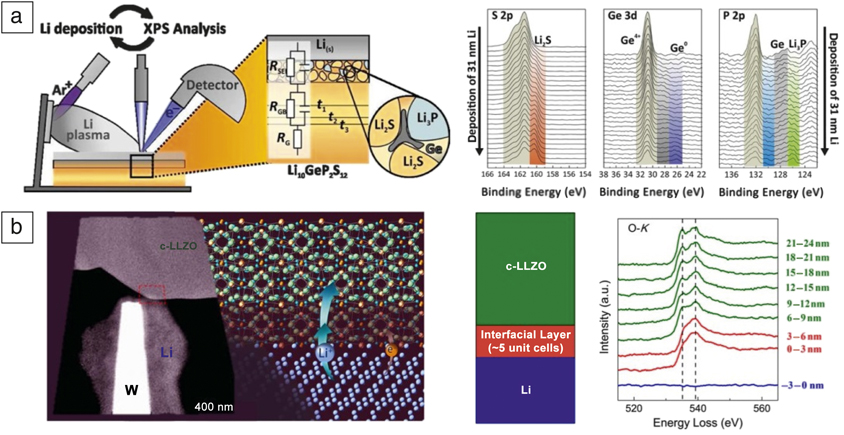
Furthering the notion of thermodynamic incompatibility/decomposition, a variety of in situ observations of solid–solid interface reactions have been pursued through XPS and TEM. Utilizing high-vacuum-transfer systems, a number of researchers have taken surface-science approaches, combining surface-sensitive XPS with intermittent growth of various materials to measure modification in chemical bonds as materials are grown upon one another (Figure 1a). Such results have been useful in characterizing the chemical bonding and intrinsic band-structure modification, as well as observing chemical decomposition at solid–solid interfaces. Examples include the deposition of Li metal on LiPON, suggesting the chemical formation of computationally predicted species (Li2O, Li3PO4, and Li3N),Reference Schwöbel, Hausbrand and Jaegermann27 direct observation of titanium reduction at the LLTO/Li interface,Reference Wenzel, Leichtweiss, Krüger, Sann and Janek28 and similarly the decomposition of L10GeP2S12 into Li3P, Li2S, and a Li-Ge alloy upon deposition of Li;Reference Wenzel, Randau, Leichtweiß, Weber, Sann, Zeier and Janek29 these techniques have provided valuable explanations of increased interfacial impedance and validated existing thermodynamic models, though such methods lack the crucial effect of the electrochemical probe present in actual devices.
In situ techniques have been developed to characterize the thermodynamic and kinetic stability of solid-state interphases as formed in controlled environments. (a) In situ deposition of lithium upon solid electrolytes coupled with x-ray photoelectron spectroscopy (XPS) has been applied to a number of systems, here showing decomposition of L10GeP2S12 when coated with Li. Adapted with permission from Reference Reference Wenzel, Randau, Leichtweiß, Weber, Sann, Zeier and Janek29. © 2016 American Chemical Society. (b) Physical contact between Li and solid electrolyte LLZO in a transmission electron microscope with electron energy-loss spectrometer, showing fine structure modification commensurate with structural modification. Adapted with permission from Reference Reference Ma, Cheng, Yin, Luo, Sharafi, Sakamoto, Li, More, Dudney and Chi30. © 2016 American Chemical Society.

Monitoring in situ reactions within a STEM was performed using a lithium-coated probe, made to contact aluminum-doped garnet LLZO (Li7−3xAlxLa3Zr2O12), shown in Figure 1b. STEM coupled with EELS provides the capability to characterize a material structurally, probing with atomic-scale resolution, and adds the advantage of mapping detailed chemical states. In this work, when the Li probe was brought into contact with the LLZO, a distinct chemical change was observed, showing a modified oxygen K-edge, suggesting the loss of the cubic phase, Li K-edge changes, consistent with the presence of excess Li, and Zr L-edge modification consistent with Zr reduction. Interestingly, these results do not show decomposition of the electrolyte, but rather the modification to tetragonal LLZO, a less conductive interphase, but negligibly so, given the thickness of the interlayer.Reference Ma, Cheng, Yin, Luo, Sharafi, Sakamoto, Li, More, Dudney and Chi30 The results emphasize the importance of kinetically limited reactions at these interfaces, and suggest that while more reactive materials will thermodynamically decompose in the presence of highly reducing lithium metal, certain electrolytes possess some degree of stability and will at least require external sources of energy to drive decomposition.
In situ observation of electrochemically driven interface modification
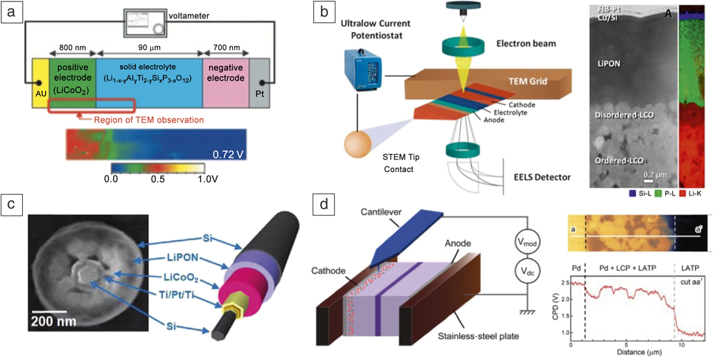
Electrochemical in situ experiments involve the design and fabrication of electrochemically active devices, with an ultimate goal of elucidating links between state of charge and the nature of the interface, yet few techniques have been successfully developed with this capability. The first demonstration of an electrochemically active device in TEM involved an LiCoO2 cathode and Li1+x+yAlyTi2–ySixP3–xO12 electrolyte, forming the anode in situ during charge (Figure 2a). A cross section of the cathode/electrolyte interface was thinned via FIB, allowing a range of TEM characterization to be performed. Primary findings via electron holography allowed mapping of the electric potential as a function of state of charge. The potential distribution showed a sharp potential drop at the cathode/electrolyte interface, with linear decreases through each component.Reference Yamamoto, Iriyama, Asaka, Hirayama, Fujita, Nonaka, Miyahara, Sugita and Ogumi31,Reference Yamamoto, Iriyama, Asaka, Hirayama, Fujita, Fisher, Nonaka, Sugita and Ogumi32 This technique was more recently applied to observe the potential distribution as a function of voltage in LiPON solid electrolyte.Reference Aizawa, Yamamoto, Sato, Murata, Yoshida, Fisher, Kato, Iriyama and Hirayama33 Beyond the exposure of only one interface in the full cell device, the bulk of the device likely dominates the electrochemical performance, leaving to question the magnitude of the field distribution within the thinned region. Nevertheless, an electron-transparent region in an electrochemically active battery was an important step in the development of TEM-based in situ techniques.
Advanced methods for local in situ interface characterization: (a) Electron holography allows dynamic visualization of electric-field distribution. Reprinted with permission from Reference Reference Yamamoto, Iriyama, Asaka, Hirayama, Fujita, Fisher, Nonaka, Sugita and Ogumi32. © 2010 Wiley. (b) Scanning transmission electron microscopy (STEM)/electron energy-loss spectroscopy (EELS) enables structural and chemical mapping, uncovering interface decomposition.Reference Wang, Santhanagopalan, Zhang, Wang, Xin, He, Li, Dudney and Meng35 (c) Transmission electron microscope (TEM) image of 3D nanowire-based batteries shows dynamic behavior of the solid electrolyte as a function of thickness. Adapted with permission from Reference Reference Ruzmetov, Oleshko, Haney, Lezec, Karki, Baloch, Agrawal, Davydov, Krylyuk, Liu, Huang, Tanase, Cumings and Talin36. © 2012 American Chemical Society. (d) Kelvin probe force microscopy uncovers electric potential distribution across all-solid-state battery cross sections. Reprinted with permission from Reference Reference Masuda, Ishida, Ogata, Ito and Fujita37. © 2017 Royal Society of Chemistry. Note: LCO, LiCoO2; CPD, contact potential difference; LCP, LiCoPO4; LATP, Li1.5Al0.5Ti1.5(PO4)3; LiPON, lithium phosphorus oxynitride.

The first true electrochemically active nanobattery to expose all active interfaces was prepared by extracting a cross section of a full cell thin-film battery via FIB (Figure 2b). Work by Santhanagopalan demonstrated electrochemical performance of a cross section of an LCO/LiPON/amorphous-Si full cell first within the FIB, demonstrating the importance of the dwell time during preparation in preventing damage and decomposition of the LiPON solid electrolyte.Reference Santhanagopalan, Qian, McGilvray, Wang, Wang, Camino, Graetz, Dudney and Meng34 A similar wedge-shaped nanobattery was then transferred to a scanning tunneling microscope holder, allowing precise manipulation of an electrical contact, used for in situ biasing within the TEM. This probe, coupled with a low-current potentiostat, allowed the first demonstration of potentiostatic in situ cycling of a solid-state device in the TEM, while maintaining current densities consistent with functioning devices. Using STEM coupled with EELS, an interphase of decomposed LCO was observed, shown to consist of lithium oxides and lithium peroxides with highly oxidized cobalt ion species. This work not only uncovered the interfacial phenomena responsible for the increasing charge-transfer resistance at the LCO/LiPON interface, it also demonstrated the importance in in situ methodologies to observe potential metastable states while mitigating the effects of sample exposure to ambient conditions.Reference Wang, Santhanagopalan, Zhang, Wang, Xin, He, Li, Dudney and Meng35
An alternative design, fabricating the nanobattery in a core–shell configuration around a conductive nanowire is shown in Figure 2c.Reference Masuda, Ishida, Ogata, Ito and Fujita37 After isolating the electrochemically active material from the base of the nanowire, the nanowire is biased with a similar mechanically manipulated probe tip, as mentioned previously. Using this technique, it was determined that the critical component to prevent shorting is the thickness of the LiPON layer. When too thin, the layer was observed to self-discharge, and the electrochemically driven formation of voids within LiPON was observed via bright-field TEM. The three-dimensional (3D) configuration of the nanobattery makes chemical evaluation of the interfaces ambiguous, though it enabled explanation of the shorting phenomena in these 3D devices.
In situ Kelvin probe force microscopy (KPFM) has been employed to explore the electric potential distribution across cross sections of solid-state batteries,Reference Masuda, Ishida, Ogata, Ito and Fujita37 similar to that observed by electron holography (Figure 2d).Reference Yamamoto, Iriyama, Asaka, Hirayama, Fujita, Fisher, Nonaka, Sugita and Ogumi32 Here, a potential is measured through the scanning probe tip across the battery cross section, effectively measuring the electrical potential distribution across the battery, and showing regions of lithium depletion.Reference Masuda, Ishida, Ogata, Ito and Fujita37 This technique offers the advantage of imaging more area of larger devices than is possible in TEM, although this technique requires complex preparation using a cross-sectional ion mill with environmental transfer capabilities, as well as a scanning probe within an inert environment for testing. While previous measurements were performed on composite electrodes, such a test could be applied to parallel cells to allow deconvolution of space-charge effects.
Other efforts to characterize solid-state interfaces with nonlocal probes have been made via solid-state nuclear magnetic resonance (NMR) and magnetic resonance imaging (MRI) of LiReference Zhu, He and Mo7 ions, as in the case of the Li/Li10GeP2S12/Li symmetric configuration.Reference Chien, Feng, Tang, Rosenberg, O’Neill, Zheng, Grant and Hu38 In situ MRI provided the ability to map Li distribution through the thickness of the electrolyte as a function of state of charge, and correlated this distribution to an inhomogeneous strain state through the thickness of the electrolyte. With continuous cycling, Li distribution clearly correlates with cracking within the solid-state electrolyte.
Remaining challenges and the future of in situ and operando techniques
While the techniques described here have effectively been employed to characterize interfaces, there is ample room for improvement, with the goal of simultaneously extracting chemical, structural, and mechanical information as a function of state of charge. It is worth noting that synchrotron x-ray methods have not played a big role in the characterization of solid-state interfaces, likely due to the complexities in data convolution in buried interfaces of complex devices. Electron microscopy at the present time is the only method capable of collecting structural and chemical information under electrochemical stimulus for solid state interfaces. Advancement of in situ and operando electron microscopy methodologies requires further development in the current state of the art, reducing beam–material interaction induced damage while improving spatial and temporal resolution during data acquisition; an ideal characterization technique would meet most of the requirements outlined in Figure 3.
In situ probing of solid-state interfaces requires development of a range of technologies, enabling increased temporal and spatial resolution, low current measurements with minimal noise, environmental transfer capabilities, and application of cryogenic temperatures.

Use of high-energy probes is accompanied by many practical considerations, primarily beam damage effects. High-energy electrons damage materials through radiolysis (decomposition due to ionizing radiation), primary knock-on damage, and local heating effects.Reference Williams and Carter39 Solid electrolytes are electronically insulating out of necessity and are often amorphous, and as such are generally susceptible to thermal degradation. Traditional methods of combating heating effects, such as carbon coating, are incompatible with in situ biasing (i.e., device shorting), but rather require fine control of the experimental design by limiting electron dose and selecting more robust electrolytes. It is noted in works of SanthanagopalanReference Santhanagopalan, Qian, McGilvray, Wang, Wang, Camino, Graetz, Dudney and Meng34 and Wang,Reference Wang, Santhanagopalan, Zhang, Wang, Xin, He, Li, Dudney and Meng35 and similarly by the community of electron microscopists,Reference Taheri, Stach, Arslan, Crozier, Kabius, LaGrange, Minor, Takeda, Tanase, Wagner and Sharma10 that the electron dose is of critical importance both for the stability of the materials being probed and in the interpretation of electronic probe outputs, and should be recorded and reported carefully. Selection of electrolytes that are more robust to beam exposure, such as oxides LLTO or LLZO, will enable quantitative observation of dynamic cathodic/anodic behaviors.
Beyond damaging effects, electron probes also have the potential to alter the total current injected into the battery. Revisiting the length scale of nanobatteries, the necessarily small cross section required for TEM analysis demands similarly low currents to promote reasonable electrochemical behavior. For example, a dense LiCoO2 nanobattery with a cross section of 10 μm2 and cathode thickness of 2 μm demands a current on the order of 10 pA to achieve a cycling rate of 1 C, though lower charge rates are required for systems with sluggish kinetics or large interfacial transfer impedance. While this current may be on the order of magnitude of STEM probe currents, the likelihood of the electron probe interacting with the electrochemical probe is minimal due to the low degree of internal scattering; however, the degree of scattering is dependent on the material density and the sample geometry. Currents below the pA range may be impacted by electromagnetic noise within instruments, and reduced current testing within a TEM is currently being evaluated.
Improved detector acquisition rates have the potential to capture reconstruction in solid-state interphases, as in the case of atomically resolved grain-boundary dynamics reconstructions.Reference Radetic, Gautam, Ophus, Czarnik and Dahmen40 Direct-detection cameras have further enabled reduced-dose image acquisition, as effectively demonstrated in biological sciences,Reference Milazzo, Cheng, Moeller, Lyumkis, Jacovetty, Polukas, Ellisman, Xuong, Carragher and Potter41 and will likely be useful in capturing dynamic phenomena in sensitive battery materials. However, such high-speed comes with the downside of yielding overwhelmingly large data sets. Direct detection cameras capable of capturing 24 megapixel images at a rate of 1500 frames per s, producing datasets on the order of terabytes, are nearing the limit of modern hard drive technology. Efficient storage protocol and automated large-data set analysis methods are areas of great interest to the field.
When characterizing nanoscale devices, it behooves the researcher to keep a realistic picture of the nature of the modifications to their device. For example, the reduced in-plane length scale of a nanobattery used in the in situ TEM methodologies is notably different from that of its parent device. Fabrication of the nanobattery increases the edge/interface ratio otherwise negligible in a thin-film device. Arguments as to field distribution should incorporate models with geometric considerations, as free surfaces likely alter the equilibrium field distribution through the bulk of the nanobattery.Reference Dudney, West and Nanda1 Similarly, kinetic limitations of the electrodes place an upper bound on the applied current densities. Analogous to alkali metal ions’ propensity to form dendrites in liquid cells, the presence of an exposed interface may provide a low energy site for nucleation should the conditions present itself, ultimately resulting in “dead” metal extruding outside of the device; such material may be rendered largely inactive due to its position outside of the electric field potential. Attempts to bypass the complexities associated with full cell devices include an FIB-assembled nanobattery, with an FIB-thinned LiCoO2 particle as the cathode, FIB lamella of LLZO as electrolyte, and Au anode, all affixed together using FIB-deposited Pt.Reference Gong, Zhang, Jiang, Shi, Zhang, Yang, Zou, Wang, Yu, Xiao, Hu, Gu, Li and Chen42 While such fabrication methods may provide insight into the dynamic nature of cathode materials under extreme conditions, such construction requires careful evaluation of the input current to make any link between the electrochemical state of a device and the phenomena being observed. In such work, with a cross section required to achieve atomic-scale resolution via conventional TEM, structural change was observed with the application of current on the order of 1 mA;Reference Gong, Zhang, Jiang, Shi, Zhang, Yang, Zou, Wang, Yu, Xiao, Hu, Gu, Li and Chen42 considering the size of the cross section for imaging, this translates to a current density on the order of 108 mA/cm2—in excess of current densities present in conventional devices. Such approximations are necessary, and as long as they are addressed appropriately, they do not limit the impact of the results presented.
Further, studying electrochemical evolution of alkali-metal anodes will in itself require a host of engineering accommodations. Environmental isolation is paramount in the case of alkali metal anode-based batteries, as the reduced dimensions greatly increase the surface-to-volume ratio. Even if environmental isolation can be accomplished, there is the question of stability under the electron beam, which has been demonstrated to be mitigated by cryo-EM techniques. Finally, to apply electrochemical methods requires the in situ biasing capabilities demonstrated in past work.
However, one may note an inherent incompatibility between cryogenic temperatures and the reduced kinetics of interfacial decomposition at reduced temperatures. While cryo-EM has proven to be useful, further work is needed to study variability in holder temperature with respect to lithium stability under the electron beam. A researcher could imagine tuning temperatures for lithium/interface stability, interface kinetics, and lithium transport within the device. Conversely, in situ heating holders, coupled with in situ biasing holders, would promote thermodynamic decomposition, emulating the effect of cycling batteries at elevated temperatures.
Summary and outlook
In spite of fundamental limitations, the outlined techniques are currently the promising methodologies capable of locally probing buried solid-state interfaces, enabling simultaneous structural and chemical characterization as a function of state of charge. The most striking aspect of the development of solid-state electrolytes, as evident from this analysis or from a similar literature search, is the limited number of studies devoted to explaining poor materials compatibility. Understandably, the degree of incompatibility was only recently called into question in a host of DFT studies, and similarly due to the complexity of analysis of these systems. Even in the limit of ex situ analyses, these materials are inherently reactive under ambient conditions, often shrouding the true electrochemical modifications present. While thermodynamic stability is in question in many compounds, kinetics of decomposition serves as a further variable, likely resulting in discrepancies between theory and experiment. Only through the application of in situ methodologies can we capture the dynamic and heterogeneous nature of interfaces, preventing environmental effects from taking hold, and further preventing electrochemical relaxation when the device is stopped from biasing.
Acknowledgments
The authors acknowledge funding support for the development of all-solid-state batteries and in situ focused ion beam and transmission electron microscope holder development by the US Department of Energy, Office of Basic Energy Sciences, under Award No. DE-SC0002357.

Thomas A. Wynn is a doctoral candidate in the Materials Science Program at the University of California, San Diego. He received his BS degree in materials science and engineering from The Ohio State University and his MS degree in chemical engineering and materials science from the University of California, Davis. Prior to graduate research, he worked as a research technologist at Los Alamos National Laboratory. His research interests include solid-state interface characterization and anionic redox-active materials in secondary batteries. Wynn can be reached by email at twynn@eng.ucsd.edu.

Jungwoo Z. Lee is a doctoral candidate in the Department of Nanoengineering at the University of California, San Diego. She received her BA degree in physics from Wellesley College. She was a research engineer at the Panasonic Boston Laboratory and the Massachusetts Institute of Technology Photovoltaic Research Laboratory, developing manufacturing systems and imaging techniques. Her research focuses on thin-film solid-state battery materials and devices, along with cryogenic microscopy techniques, to elucidate structural and chemical mechanisms in sensitive Li-ion battery materials. Lee can be reached by email at jzlee@eng.ucsd.edu.

Abhik Banerjee is a postdoctoral scholar in the Department of Nanoengineering at the University of California, San Diego. He received his BSc and MSc degrees in chemistry from the University of Calcutta, India. He earned his PhD degree in 2015 from National Chemical Laboratory, India. He was then a postdoctoral scholar at Ulsan National Institute of Science and Technology, South Korea. His research interests include the design principles and interfacial charge-transfer limitations of solid-state electrolytes for Li and Na-ion batteries. Banerjee can be reached by email at a7banerjee@eng.ucsd.edu.

Y. Shirley Meng holds the Zable Endowed Chair in Energy Technologies and is professor of nanoengineering and materials science at the University of California, San Diego. She is the founding director of the Sustainable Power and Energy Center. She received her PhD degree in advanced materials for micro- and nanosystems from the Singapore–Massachusetts Institute of Technology (MIT) Alliance in 2005, after which she worked as a postdoctoral research fellow and research scientist at MIT. Her research group, Laboratory for Energy Storage and Conversion, focuses on functional nano- and microscale materials for energy storage and conversion. Meng can be reached by email at shirleymeng@ucsd.edu.