Introduction
Entomopathogenic fungi have been used widely to control insect pest because they are considered environmentally friendly alternatives to synthetic pesticides. Fungi are one of the most studied insect antagonists for their pest management benefits (Marcelino et al. Reference Marcelino, Giordano, Gouli, Gouli, Parker, Skinner, TeBeest and Cesnik2008; Tanyeli et al. Reference Tanyeli, Sevim, Demirbag, Eroglu and Demir2010; Kocacevik et al. Reference Kocacevik, Sevim, Eroglu, Demirbag and Demir2015; Yucel et al. Reference Yucel, Gozuacik, Gencer, Demir and Demirbag2018; Goffre et al. Reference Goffre, Jensen, Lopez Lastra, Humber and Folgarait2021). Several isolates of fungi have been commercialised as biopesticides for the control of various insect pests (Faria and Wraight Reference Faria and Wraight2007; Zimmermann Reference Zimmermann2007). Entomopathogenic fungi are well known for losing virulence when repeatedly subcultured on artificial media, but virulence can be restored when the pathogen is passed through the original or a heterologous insect host (Butt and Goettel Reference Butt, Goettel, Navon and Ascher2000; Butt et al. Reference Butt, Wang, Shah, Hall, Eilenberg and Hokkanen2006). However, isolates differ in their stability when cultured on artificial media, with some isolates clearly losing stability more rapidly than others (Hutwimmer et al. Reference Hutwimmer, Wagner, Affenzeller, Burgstaller and Strasser2008). Stable isolates can retain their virulence when subcultured on artificial media for multiple generations, whereas unstable isolates usually lose virulence after a few cycles of subculturing (Butt et al. Reference Butt, Wang, Shah, Hall, Eilenberg and Hokkanen2006; Loesch et al. Reference Loesch, Hutwimmer and Strasser2010). Stability of isolates during repeated in vitro cultivation is essential for the successful production of fungal biopesticides.
When an isolate is subcultured on artificial media, phenotypic changes, including changes in colour, growth rates, and form, production of sectors, and a decline in spore production are observed. In addition, the production of the cuticle-degrading enzyme, subtilisin-like Pr1, which is important for insect penetration and subsequent pathogenicity, can decline (Leger et al. Reference Leger, Charnley and Cooper1986; Small and Bidochka Reference Small and Bidochka2005; Perinotto et al. Reference Perinotto, Angelo, Golo, Camargo, Quinelato, Santi and Bittencourt2014). Repeated subculturing commonly results in low levels of Pr1 production, and monitoring production of this enzyme has been shown to be an excellent tool to monitor the quality of a fungus during mass production (Shah et al. Reference Shah, Wang and Butt2005; Safavi et al. Reference Safavi, Shah, Pakdel, Rasoulian, Bandani and Butt2007).
In addition to the stability of the entomopathogenic fungal isolates, their tolerance to the adverse effects of environmental conditions must also be retained or enhanced during production and formulation. Abiotic factors limit the development of Metarhizium spp. (Clavicipitaceae) and reduce effectiveness, with virulence against insect pests and the persistence of conidia in the field being directly affected by high temperature (Zimmermann Reference Zimmermann1982; Dimbi et al. Reference Dimbi, Maniania, Lux and Mueke2004; Barreto et al. Reference Barreto, Luz, Mascarin, Roberts, Arruda and Fernandes2016), low relative humidity (Arthurs and Thomas Reference Arthurs and Thomas2001; Ment et al. Reference Ment, Gindin, Glazer, Perl, Elad and Samish2010; Athanassiou et al. Reference Athanassiou, Kavallieratos, Rumbos and Kontodimas2017), and ultraviolet radiation (Braga et al. Reference Braga, Flint, Messias, Anderson and Roberts2001; Fernandes et al. Reference Fernandes, Rangel, Braga and Roberts2015; Acheampong et al. Reference Acheampong, Hill, Moore and Coombes2020). Studies to overcome these adverse effects have focused on formulating conidia. For example, mixing conidia with oil provides protection against high temperatures, ultraviolet radiation, and rapid dehydration in low-humidity environments (Xavier-Santos et al. Reference Xavier-Santos, Lopes and Faria2011; Paixao et al. Reference Paixao, Muniz, Barreto, Bernardo, Mascarin, Luz and Fernandes2017; Muniz et al. Reference Muniz, Paixão, Barreto, Luz, Arruda, Angelo and Fernandes2020).
The objectives of the present study were (1) to determine the stability of Metarhizium isolates, in terms of morphological, genotypic, and biochemical properties and (2) to formulate conidiospores as an oil-in-water emulsion to extend isolate survival and pathogenicity in nature.
Materials and methods
Insect collection
Third-instar larvae of Melolontha melolontha (Coleoptera: Scarabaeidae) were collected from naturally infested plantation fields by digging up soil with a pickaxe in the vicinity of Trabzon, Turkey. After collection, larvae were put into plastic boxes (20 × 20 cm) containing sufficient soil and plant roots as food until bioassays were performed.
Fungal cultures
Nine Metarhizium anisopliae isolates were provided from the microbiology laboratory of the Department of Biology, Karadeniz Technical University, Trabzon, Turkey (Table 1). The isolates were selected because they possessed high virulence against insect pests.
The indigenous Metarhizium anisopliae isolates used in stability tests and their origin.

Fungi were initially cultured on Sabouraud dextrose–agar (Merck, Darmstadt, Germany) at 28 ± 1 °C for three days. After incubation, a single colony of each isolate was transferred to fresh Sabouraud dextrose–agar plates and incubated at 28 ± 1 °C until fully grown (∼2 weeks). Fully sporulating cultures were used for bioassays.
Screening tests
Two-week-old fungus cultures were used in pathogenicity tests. Fungal spores were harvested from the agar plates using a sterile scalpel and transferred to 10 mL of sterile distilled water containing 0.01% Tween 80 (Thermo Fisher Scientific Ltd., Waltham, Massachusetts, United States of America). The conidial suspensions were passed through sterile muslin to remove agar pieces and hyphal debris. Conidia were counted using a Neubauer hemocytometer (Sigma-Aldrich, St. Louis, Missouri, United States of America), and the concentration was adjusted to 1 × 10⁷ conidia/mL. The efficacy of fungi was tested on third-instar Me. melolontha larvae. After application of 1 mL of fungal suspensions to larvae with a hand-operated sprayer, larvae were placed in plastic boxes (20 × 20 cm) containing sterile damp sand and plant roots. Assessments were performed with 10 larvae for each fungus and repeated three times. Control larvae were treated with 0.01% aqueous Tween 80. Assessments were conducted at 28 ± 2 °C and 16:8-hour light:dark photoperiod for 15 days (Biryol et al. Reference Biryol, Efe, Eski, Demirbag and Demir2020). Dead insects were removed and placed in Petri dishes with moist filter paper to allow mycosis to occur. Mortality and mycosis rates were corrected for control mortality using Abbott’s formula (Abbott Reference Abbott1925).
Repeated subculturing
Initially, four Metarhizium isolates (KTU-2, KTU-51, KTU-60, and Gg-12) that had high insecticidal activity according to screening tests were passaged through Galleria mellonella (Lepidoptera: Pyralidae) larvae to ensure their virulence. Ten larvae were dipped into conidial suspension (1 × 10⁷ conidia/mL) for three to five seconds each and then were incubated at 28 °C until mycosis. Conidia collected from cadavers were used to re-isolate the fungus onto Sabouraud dextrose–agar–yeast medium (2% sucrose, 0.25% yeast extract, 1% peptone, and 0.02% chloramphenicol) incubated in the dark at 28 °C for 15 days. This was designated as the first subculture. Subculturing was then repeated 12 times. Each cycle of subculture included 10 replicates. The first, sixth, and twelfth subcultures were evaluated in the following experiments, per Ansari and Butt (Reference Ansari and Butt2011).
Effect of repeated subculture on isolate phenotype
The Metarhizium isolates were visually examined after one, six, and twelve subcultures (15 days old) for changes in colony colour and shape and infertile field formation. Suspensions of 1 × 10⁶ conidia/mL were prepared from these subcultures, spread on Sabouraud dextrose–agar–yeast medium with a glass rod and immediately exposed to ultraviolet radiation (306 nm) for 0, 15, and 30 minutes to determine the effects of repeated subculture on ultraviolet-radiation resistance. The treatments were performed in an ultraviolet-radiation chamber with UV-B fluorescent lamps (TL20W/12RS; Philips, Amsterdam, The Netherlands), and each treatment was repeated three times. After treatment, the Petri dishes were incubated at 28 °C for 24 hours, and germination rates were determined in a Neubauer hemocytometer (Braga et al. Reference Braga, Flint, Messias, Anderson and Roberts2001). At least 300 conidia per plate were examined under a light microscope, and conidia were considered germinated when the germ tube extended at least to a length equal to one-half of the diameter of the spores.
Effects of repeated subculture on isolate genotype
Random amplified polymorphic DNA polymerase chain reaction analyses and restriction profiles of the pr1 gene were performed on the Metarhizium isolates after one, six, and twelve subcultures. Genomic DNA was extracted from each isolate using the Quick-DNA™ Fungal/Bacterial MiniPrep Kit (Zymo Research, Irvine, California, United States of America) following the manufacturer’s instructions. Random amplified polymorphic DNA polymerase chain reaction analyses were performed using five primers: OPA-03, AGTCAGCCAC; OPA-08, GTGACGTAGG; OPB-04, GGACTGGAGT; OPB-07, GGTGACGCAG; and OPE-01, CCCAAGGTCC (Wang et al. Reference Wang, Types and Butt2002). Polymerase chain reaction was performed in a 50-µL reaction volume containing 5 µL of 10 × Taq DNA polymerase reaction buffer, 200 µM of each deoxynucleoside triphosphate synthesis, 50 pmoL of each primer, 2.5 units of Taq DNA polymerase, and 50 ng of genomic DNA. Polymerase chain reaction conditions were as follows: initial denaturation at 95 °C for five minutes; 35 cycles at 95 °C for one minute, at 55 °C for 55 seconds, and at 72 °C for two minutes, followed by a final extension at 72 °C for 10 minutes. The polymerase chain reaction products were separated on 1.0% agarose gel that was stained with ethidium bromide. Evidence for genotypic changes was determined by examining the polymerase chain reaction products.
In addition, a segment (∼1200 bp) of the pr1 gene was amplified by nested polymerase chain reaction using two primer pairs (METPR1, 5′-CACTCTTCTCCCAGCCGTTC-3′ and METPR2, 5′-AGGTAGGCAGCCAGACCGGC-3′; and METPR4, 5′-GTAGCTCAACTTCTGCACTC-3′ and METPR5, 5′-TGCCACTATTGGCCGGCGCG-3′). The first polymerase chain reaction was performed using the extracted Metarhizium DNA as a template and the outer pair of primers (METPR1 and METPR4). An aliquot of the first polymerase chain reaction, as a template, and the inner pair of primers (METPR2 and METPR5) were used in the second polymerase chain reaction. Polymerase chain reaction conditions were adapted from Leal et al. (Reference Leal, Bertioli, Butt, Carder, Burrows and Peberdy1997). The polymerase chain reaction products were loaded on 1.0% agarose gels that contained ethidium bromide, scanned under ultraviolet light for visualisation, and sent to Macrogen Inc. (Amsterdam, The Netherlands) for direct sequencing. To determine the polymorphism in the pr1 gene, the fragment of pr1 gene amplified by nested polymerase chain reaction was treated with DdeI, HpaII, and RsaI restriction endonucleases, successfully revealing any polymorphisms (Leal et al. Reference Leal, Bertioli, Butt, Carder, Burrows and Peberdy1997). The digest mixture included 5 µL of polymerase chain reaction product, 0.5 µL of restriction enzyme, and 2 µL of 10 × restriction enzyme buffer in a total volume of 20 µL. Digestions were performed at 37 °C for four hours. Restriction fragments were separated by electrophoresis in 1.0% agarose gels (containing 1 µL/mL ethidium bromide) at 80 mA in Tris-acetate-EDTA buffer.
Effect of repeated subculture on isolate sporebound Pr1 activity
The activity of Metarhizium Pr1 enzyme after one, six, and twelve subcultures was determined after growth in minimal medium containing 0.2% (weight/volume) glucose, 0.6% (weight/volume) NaNO3, 0.2% (weight/volume) peptone, 0.05% (weight/volume) yeast extract, and 0.8% chitin (supplementary carbon source). One millilitre of 1 × 10⁶ conidia/mL fungus suspension was inoculated into minimal medium (20 mL) in an Erlenmeyer flask, incubated on an orbital shaker (150 rpm) at 28 °C for 96 hours. After incubation, the medium was filtered through Whatman No. 1 filter paper, and the total protein concentration of the filtrate was determined using bovine serum albumin as a standard, according to Bradford (Reference Bradford1976). The concentrations of the proteins were equalised, and 5 μL of the supernatant was transferred to 96-well dishes for each isolate. Five microlitres of N-succinyl-Ala-Ala-Pro-Phe-pNA substrate (10 mM) and 90 μL of Tris-hydrochloride buffer (0.1 M, pH 0.8) were added into the supernatant in each well and incubated at 37 °C for 30 minutes (Perinotto et al. Reference Perinotto, Angelo, Golo, Camargo, Quinelato, Santi and Bittencourt2014). After incubation, the absorbance was measured at 405 nm with a spectrophotometer, and the Pr1 enzyme activity of the isolates was determined. Assessments were performed in triplicate for each isolate.
Effect of repeated subculture on isolate virulence
A single replicate plate after one, six, and twelve subcultures of each isolate was used to assess the stability of virulence. Fungal suspensions were prepared as above. Ten G. mellonella third-instar larvae were inoculated with suspensions containing 1 × 10⁶ conidia/mL by hand-operated spray. Inoculated larvae were transferred into plastic containers with artificial diet and observed for 15 days. To determine the mycosis rate, surface-sterilised dead larvae were transferred to Petri dishes and left to incubate at 28 °C in 60% relative humidity in a moisture chamber. The mortality and mycosis values were corrected using Abbott’s formula (Abbott Reference Abbott1925).
Mycoinsecticide production
Based on the results of the assessments described above, the isolate KTU-2 was selected for development of a prototype mycoinsecticide. Mass production was performed by biphasic fermentation. For production of blastospores in the liquid fermentation, Sabouraud dextrose–yeast broth was inoculated with the conidiospores (2 × 10⁶ conidia/mL) and incubated at 28 °C on an orbital shaker (150 rpm) for four days. The production of conidia then occurred on a solid substrate (cracked rice). Polyethylene bags containing 200 g parboiled cracked rice were autoclaved at 121 °C for 30 minutes. The bags with moistened media were then cooled, and 10 mL of the fungal suspension (1 × 10⁸ blastospores/mL) were added to each bag. The contents of the bags were gently mixed at 15-minute intervals for one hour to ensure homogeneity of the inoculum, and the bags were kept at 28 °C for one month. The rice colonised by the fungus was transferred to Kraft paper bags (20 × 30 cm) and allowed to dry for 10 days. The spores then were collected by sifting the substrate through a sieve (45 mesh), and the moisture content was decreased to less than 5% using a vacuum desiccator. To determine the production level, 1 g of spore powder was suspended in 99 mL of 0.01% aqueous Tween 80, and the concentration of spores was determined using a Neubauer hemocytometer.
The KTU-2 spore powder was formulated to protect its physical and chemical properties and to extend its survival and pathogenicity in nature. An oil-in-water emulsion was prepared by mixing 10 g of conidia, 1 mL of vegetable oil, 1 mL of Triton X-100, 0.5 mL of silicone, 1 mL of Na2CO3, and 96.5 mL of sterile distilled water. The mixture was homogenised at 4 °C using a magnetic stirrer for one hour to give a stable emulsion (Ummidi and Vadlamani Reference Ummidi and Vadlamani2014). The spore concentration was determined with a Neubauer hemocytometer.
Insecticidal activity of an oil-in-water emulsion
The efficacy of oil-in-water mycoinsecticide was tested on the larvae of Me. melolontha by pot culture assay. Five different concentrations of formulation (from 109 to 105 conidia/mL) were prepared by serial dilution. Ten larvae were released onto strawberry plants grown in pots, and 10 mL of serially diluted formulations were applied to the soil surface and mixed into the upper 15 cm of the soil using a small rake. Distilled water was used as a control, and the whole experiment was repeated five times. Experiments were performed at 28 ± 2 °C, 60% relative humidity, and 12:12-hour light:dark photoperiod. The development of strawberry plants was examined for 15 days, and then soil was examined for dead insects, which were transferred to a humid chamber to stimulate fungal sporulation on the cadaver surface. Concentration-response mortality data were subjected to probit regression analysis (Finney Reference Finney1971) using SPSS 25.0 statistical software (IBM, Armonk, New York, United States of America), and lethal concentrations (LC50 and LC95) were estimated.
Results
Effect of fungal isolates on Melolontha melolontha larvae
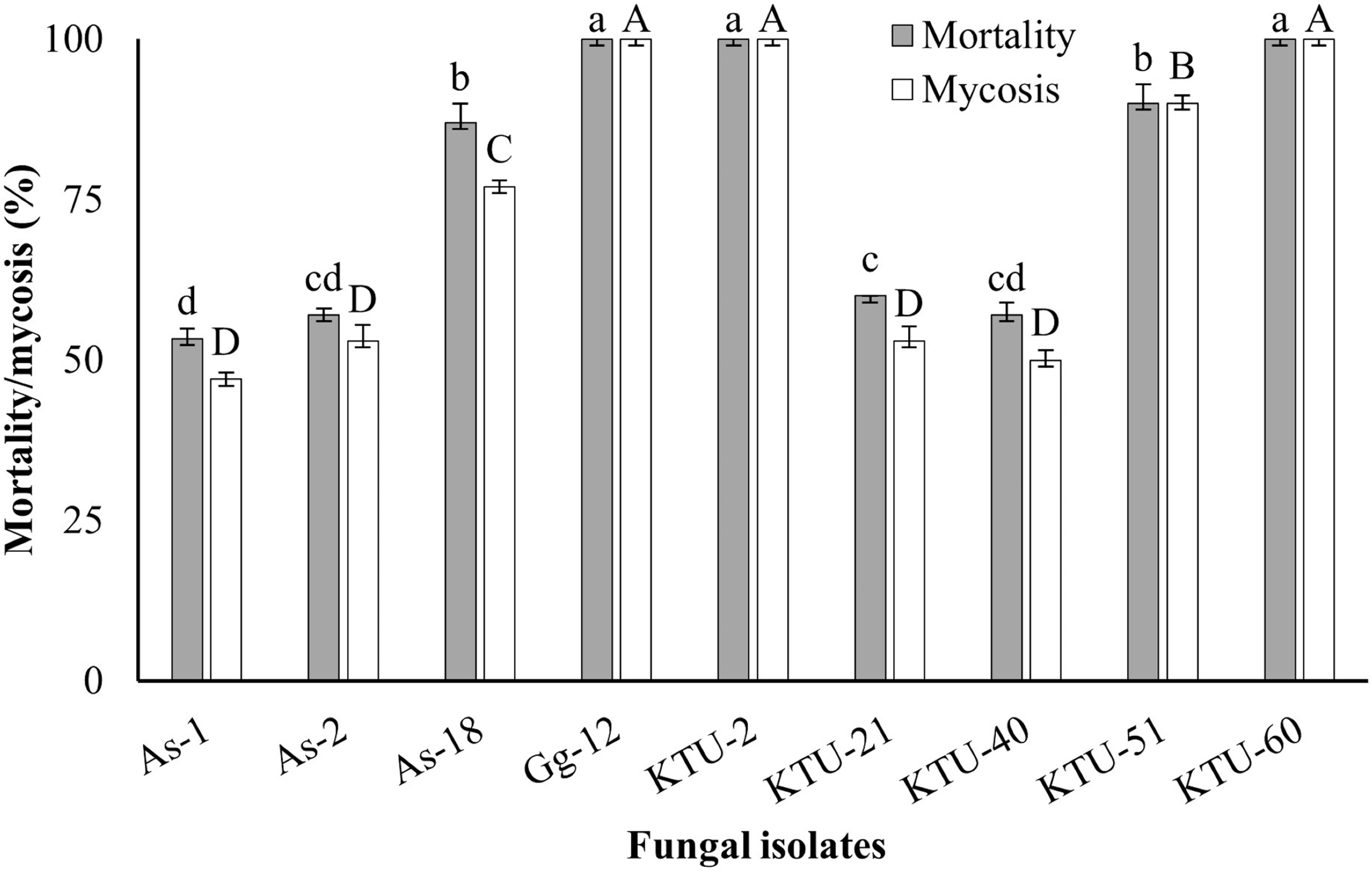
The virulence of nine Metarhizium isolates on Me. melolontha larvae was determined, and the isolates that had high virulence were selected for use in further assessment. All Metarhizium isolates showed virulence on the larvae of Me. melolontha. Isolates caused mortality ranging from 53% to 100% (F = 41.6; df = 9; P < 0.05). The isolates Gg-12, KTU-2, and KTU-60 were more virulent, causing complete mortality within 15 days (Fig. 1).
Corrected mortality and mycosis rate of Metarhizium anisopliae isolates on the larvae of Melolontha melolontha 15 days after application of 1 × 10⁷ conidia/mL fungal suspension. Mycosis indicates the sporulation rate in the moisture chamber after death. Mortality indicates the mean of three replications. Bars show the standard deviation of the means.

Effects of repeated subculture on isolate phenotype
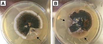
Repeated subculture affected the isolates differently. For the isolate Gg-12, the colony margin initially was circular and entire, but by the sixth subculture, it became uneven with infertile-sector formation at the sides of the colonies. After 12 subcultures, only a small fertile region remained in the centre of the colony. For the isolate KTU-2, colony morphology did not change with repeated subculture, and no colour change occurred in the spores. Sporulation occurred across the entire colony after six subcultures, and all spores were fertile. However, by the twelfth subculture, infertile areas were present. For the isolate KTU-51, although no change in colony diameter occurred with repeated subculture, the sporulation rate decreased and the spore colour changed from green to pale yellow. Also, infertile areas appeared after six subcultures, with the frequency increasing through to the twelfth subculture. Large triangular sectors containing no growth occurred in the sixth and twelfth subcultures (Fig. 2). For the isolate KTU-60, the size of the colonies decreased by the twelfth subculture, but no colour change was observed. However, infertile areas were observed in the centres of the colonies, which began to shrink beginning with the sixth subculture.
Sector formation in Metarhizium anisopliae isolate KTU-51 after repeated subculture on artificial medium. Arrows indicate the triangular sectorisation. A, six subcultures, and B, 12 subcultures.

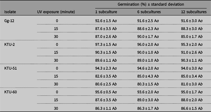
The germination rates of spores exposed to ultraviolet radiation for different durations differed among the isolates (P < 0.05) but not between number of subcultures (P > 0.05; Table 2).
Means (± standard deviation) of germination rates for conidia of four Metarhizium anisopliae isolates, exposed to ultraviolet radiation after one, six, and twelve subcultures.

Means followed by the different uppercase letters within rows indicate the differences amongst the subcultures. Means followed by the different lowercase letters within columns indicate the differences amongst the ultraviolet-radiation treatments (P < 0.05).
Effect of repeated subculture on isolate genotype
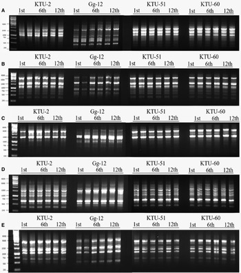
The DNA of the four isolates was successfully amplified using the OPA, OPB, and OPE primers and gave the same banding pattern on agarose gel, with no genotypic change observed (Fig. 3).
Random amplified polymorphic DNA polymerase chain reaction profile of repeated subcultures of Metarhizium anisopliae isolates amplified using A, OPA-03, B, OPA-08, C, OPB-04, D, OPB-07, and E, OPE-01 primers. First line of each gel is GeneRuler 1-kb DNA ladder (Thermo Scientific, Waltham, Massachusetts, United States of America).

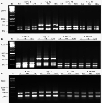
When the pr1 gene products were digested with the RsaI restriction endonuclease, two fragments were obtained for the KTU-2, KTU-51, and KTU-60 isolates, and these were the same lengths (∼320 and 430 bp) across the subculture cycles tested. For isolate Gg-12, two fragments (∼320 and 620 bp) were obtained and were also stable with repeated subculture. When the gene products were digested with the HpaII restriction endonuclease, the KTU-51, KTU-60, and KTU-2 isolates yielded the same fragments (∼210 and 290 bp), whereas the Gg-12 isolate gave four fragments (200, 280, 330, and 500 bp); this was stable with repeated subcultures. With the DdeI restriction endonuclease, two fragments (300 and 500 bp) were obtained for all isolates for all subculture cycles tested (Fig. 4).
Digestion of pr1 gene region amplified from different subcultures by restriction enzymes A, RsaI, B, HpaII, and C, DdeI. M is GeneRuler 1 kb DNA ladder (Thermo Scientific).

Effects of repeated subculture on isolated sporebound Pr1 activity
No significant difference was observed between the Pr1 activity between isolates or subculture cycle. The Pr1 enzyme activity ranged from 1.10 to 1.18 nmol of released p-nitroanilide/mol/minute (P > 0.05).
Effects of repeated subculture on isolate virulence
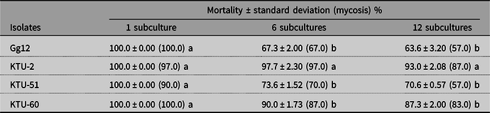
All isolates led to complete mortality of G. mellonella larvae at the first subculture. However, the virulence of isolates Gg-12, KTU-51, and KTU-60 significantly declines by their sixth subcultures (P < 0.05). By the twelfth subcultures, the highest virulence was obtained with isolate KTU-2, at 93%, whereas isolate Gg-12 had declined to 64% (Table 3).
Effect of repeated subculture on Galleria mellonella larvae on isolate efficacy.

Means followed by the same letters within rows are not significantly different (P > 0.05). Mycosis indicates the sporulation rate in the moisture chamber after death.
Mycoinsecticide production and insecticidal activity
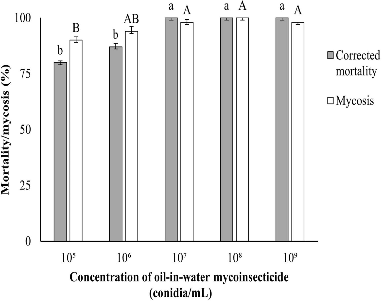
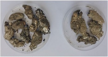
Mass production of conidia from isolate KTU-2 yielded a product with 1 × 10¹⁰ conidia/g and the oil-in-water emulsion with 1010 conidia/mL. Different concentrations of this formulation that were tested on Me. melolontha larvae with pot assays caused 80% mortality at even the lowest concentration (105 conidia/mL). However, full mortality was obtained with the 1 × 10⁷, 1 × 10⁸, and 1 × 10⁹ conidia/mL concentrations (Fig. 5). The mycosis on the dead larvae confirmed that deaths were due to fungal infection (Fig. 6). The lethal concentrations LC50 and LC95 of this mycoinsecticide were estimated at 9.29 × 10³ and 2.1 × 10⁶ conidia/mL, respectively (df = 3; χ2 = 1.047).
Means (± standard deviation) of the concentration-response experiment for oil-in-water mycoinsecticide on the larvae of Melolontha melolontha. Mortality indicates the mean of five replications. Bars show the standard deviation of the means. Different lower case and upper case letters represent statistically significant differences amongst mortalities and mycosis, respectively (P < 0.05). Mycosis indicates the sporulation rate in the moisture chamber after death.

Mycoses of cadavers of Melolontha melolontha larvae infected with oil-in-water emulsion of KTU-2 in the pot experiments. The larvae infected with M. anisopliae KTU-2 were covered with mycelia, and dark green colour of conidia was observed.

Discussion
Stability of entomopathogenic fungi through repeated in vitro subculturing is desirable for large-scale production of mycoinsecticides. However, degradation has been observed in nearly all the taxa of entomopathogenic fungi with in vitro subculturing (Butt et al. Reference Butt, Wang, Shah, Hall, Eilenberg and Hokkanen2006; Ansari and Butt Reference Ansari and Butt2011). However, this degradation varies considerably between entomopathogenic fungal isolates. A key outcome of the present study was the selection and production of a stable isolate of M. anisopliae that has potential as a mycoinsecticide.
The present study demonstrated that morphology and virulence of Metarhizium isolates can change with repeated subculture. Although morphological properties of the KTU-51, KTU-60, and Gg-12 isolates changed, the KTU-2 isolate remained relatively stable. Infertile areas were observed by the sixth subculture in the other isolates. Shah and Butt (Reference Shah and Butt2005) observed the formation of morphological variants or sectors was often observed in fungal cultures that are maintained on artificial media. Those authors reported that when M. anisopliae isolate V245 was subcultured on media with a carbon:nitrogen ratio of 35:1, sterile white mycelium growth was observed, and the formation of triangular infertile sectors was observed when the isolate was subcultured on potato–dextrose–agar medium. In our study, triangular infertile sectors were observed at the sixth subculture of the isolate KTU-51 on Sabouraud dextrose–agar medium. Sterile white mycelium growth was observed in the centre of isolate KTU-60, which began to become smaller after the sixth subculture. Similarly, Safavi (Reference Safavi2011) observed that Beauveria bassiana (Cordycipitaceae) DEBI008 and DEBI007 isolates showed fluffy hyphal growth and lower radial growth with 15 subcultures on Sabouraud dextrose–agar.
In the present study, the germination of spores of the subcultures of Metarhizium test by exposure to ultraviolet radiation for 15 and 30 minutes did not differ with subculture cycle. However, although a number of studies have tested the effects of ultraviolet radiation on the germination of Metarhizium, none have considered the effect of repeated subculture. Santoro et al. (Reference Santoro, Zorzetti, Constanski and Neves2014) subcultured B. bassiana 17 times on potato–dextrose–agar and then tested the effect of ultraviolet-radiation exposure. Ultraviolet irradiation was found to reduce spore viability, but this decrease was unaffected by repeated subculture.
Metarhizium anisopliae is known to be prone to genotypic change during repeated subculture on artificial media, and random amplified polymorphic DNA polymerase chain reaction is a potentially useful method to detect these genetic changes (Cobb and Clarkson Reference Cobb and Clarkson1993; Leal et al. Reference Leal, Bertioli, Butt and Peberdy1994, Reference Leal, Butt, Peberdy and Bertioli2000; Tigano-Milani et al. Reference Tigano-Milani, Gomes and Sobral1995). Vandenberg and Cantone (Reference Vandenberg and Cantone2004) observed no change in banding pattern for any isolate using arbitrary primers for random amplified polymorphic DNA polymerase chain reaction when Paecilomyces fumosoroseus (Cordycipitaceae) isolates ARSEF 4461, 4481, and 4491 were repeatedly subcultured on Sabouraud dextrose–agar medium. Also, Ansari and Butt (Reference Ansari and Butt2011) subcultured M. anisopliae and M. brunneum through G. mellonella larvae and did not observe genetic change with repeated subculture. These reports indicate that some isolates of entomopathogenic fungi can be genetically stable when subcultured either in vitro or in vivo. Similarly, in the present study, when subcultures of Metarhizium (Gg-12, KTU-2, KTU-51, and KTU-60) isolates were amplified by random amplified polymorphic DNA polymerase chain reaction, no variation was observed (Fig. 3). In contrast, Nahar et al. (Reference Nahar, Kulkarni, Kulye, Chavan, Kulkarni and Rajendran2008) found that repeated subculture of M. anisopliae M34412 on potato–dextrose–agar medium resulted in significant genetic changes that were revealed by random amplified polymorphic DNA analysis.
The sporebound Pr1 enzyme of M. anisopliae is important in the initial stage of fungal spore penetration through the insect cuticle. Some Metarhizium isolates that are spontaneous pr1-deficient mutants have low or no virulence. Wang et al. (Reference Wang, Types and Butt2002) reported that although the M. anisopliae wild-type isolate V275 yielded more than 90% mortality on Tenebrio molitor (Coleoptera: Tenebrionidae) larvae, three genetically identical spontaneous pr1-deficient mutants showed reduced lethal activity (20%). The presence of the pr1 gene in all subcultures of isolates examined in the present study was determined by nested polymerase chain reaction, and no difference was observed between subculture cycles for each isolate using restriction enzymes (Fig. 4). Subculturing may also cause a decline in virulence of fungal entomopathogens by switching off the genes and affecting the sporebound Pr1 activity. Safavi (Reference Safavi2011) found that when the B. bassiana isolate DEBI008 was subcultured on T. molitor larvae, Pr1 activity was lower after 15 subcultures (0.92 µmol NA/minute) than it was in the control (1.61 µmol NA/minute). Also, Ansari and Butt (Reference Ansari and Butt2011) subcultured two M. anisopliae isolates on Sabouraud dextrose–agar, and the Pr1 activity of conidia of the M. anisopliae isolate V275 decreased from the first subculture (0.57 µmol NA/min) to the sixth (0.34 µmol NA/min) and twelfth subcultures (0.30 µmol NA/min). However, another isolate, ARSEF 4556, was unaffected by the same subculturing. Similarly, in the present study, no significant difference in Pr1 activity between isolates and subcultures (P > 0.05) was observed.
Phenotypic and genotypic changes caused by subculturing mostly result in attenuation of the virulence. This attenuation varies according to genus, species, or even isolate. Early studies by Hall (Reference Hall1980) reported that subculturing of Verticillium lecanii (Cordycipitaceae) on Sabouraud dextrose–agar up to 50 times resulted in phenotypic change but no significant decrease in virulence in Macrosiphoniella sanborni (Hemiptera: Aphididae). Also, Vandenberg and Cantone (Reference Vandenberg and Cantone2004) reported that the virulence of the P. fumosoroseus isolate 4491 in Plutella xylostella (Lepidoptera: Plutellidae) larvae did not alter even after 30 in vitro subcultures on Sabouraud dextrose–agar–yeast. Subculturing on Sabouraud dextrose–agar medium up to 12 times did not reduce the virulence of M. anisopliae V275 and M. brunneum ARSEF 3297 isolates in Tenebrio molitor larvae, although a decline in Pr1 activity was observed (Ansari and Butt Reference Ansari and Butt2011). Similarly, in the present study, stable virulence was found in the Metarhizium isolates tested, which maintained their virulence in G. mellonella larvae after 12 subcultures.
In the present study, the M. anisopliae isolate KTU-2 retained the highest virulence after 12 subcultures. Therefore, this stable isolate was selected for mycoinsecticide production. Conidia of KTU-2 were produced on cracked rice because they can expand their extracellular enzymes and conidiophores to larger surface areas on solid substrates (Lee et al. Reference Lee, Song and Hwang2003; Arzumanov et al. Reference Arzumanov, Jenkins and Roussos2005; Guijarro et al. Reference Guijarro, Melgarejo and Cal2007; Verma et al. Reference Verma, Brar, Tyagi, Surampalli and Valero2007). Although conidia of entomopathogenic fungi show high virulence in various pest insects under laboratory conditions, their virulence in the field is adversely affected by abiotic factors such as ultraviolet radiation, low humidity, and nonoptimal temperatures. To overcome these limitations, conidia of KTU-2 were formulated in an oil-in-water emulsion. Oil-based formulations not only protect the conidia from detrimental abiotic factors but also increase the performance of entomopathogenic fungi by favouring the spread and adhesion of conidia onto the hydrophobic cuticle of the target pests (Perinotto et al. Reference Perinotto, Angelo, Golo, Camargo, Quinelato, Santi and Bittencourt2014; Paixao et al. Reference Paixao, Muniz, Barreto, Bernardo, Mascarin, Luz and Fernandes2017; Oliveira et al. Reference Oliveira, Lopes, Rezende and Delalibera2018). In the present study, an oil-based formulation of KTU-2 showed 80% mortality even at the lowest dose (105 conidia/mL) within 15 days. In addition, our formulation caused complete mortality on Me. melolontha at 1 × 10⁷ conidia/mL, with a lethal concentration (LC50) of about 9.29 × 10³ conidia/mL. Similarly, Hernandez-Velazquez et al. (Reference Hernandez-Velazquez, Berlanga-Padilla and Barrientos-Lozano2000) found that an oil emulsion with M. anisopliae MaPL40 resulted in complete mortality on Schistocerca piceifrons (Orthoptera: Acrididae) after eight days at the concentration of 1 × 10⁶ conidia/mL. Although several studies have investigated the efficacy of formulated conidia against coleopteran pests, including scarab beetles under laboratory or field conditions (Chelvi et al. Reference Chelvi, Thilagaraj and Nalini2011; Erler and Ates Reference Erler and Ates2015; Ibrahim Reference Ibrahim2017), limited studies are available about the use of mycoinsecticide on Me. melolontha. Melocont, a wettable powder formulation of B. brongniartii, was used to control Me. melolontha population in a potato field, achieving a 64% decrease in population density (Laengle et al. Reference Laengle, Pernfuss, Seger and Strasser2005), but M. anisopliae isolate NCAIM 362 as a wettable powder tested against Me. melolontha larvae in potato fields had no effect (Putnoky-Csicsó et al. Reference Putnoky-Csicsó, Tonk, Szabó, Márton, Bogdányi and Tóth2020). Importantly, the oil formulation used in the present study showed a significant effect on Me. melolontha. Also, in the pot experiments, untreated strawberry plants were severely damaged by larval feeding but remained healthy, flowered, and fruited when the formulation was applied.
In conclusion, the stability of fungi during repeated subculture on artificial media is crucial for the production of mycoinsecticides. The present study shows that the stability of Metarhizium isolates can vary, but the M. anisopliae isolate KTU-2 proved to be effective and stable. An oil-in-water formulation of this isolate was developed as a prototype, and its insecticidal activity was determined. Future research that looks into the large-scale production and field evaluation of this formulation is warranted.
Acknowledgements
The authors thank the Karadeniz Technical University (Project number: FBA-6915) for financial support and Dr. I.T. Riley for advice during preparation of the manuscript.
Conflict of interest
The authors declare no conflict of interest.