Implications
The ATP-binding cassette transporter G2 (ABCG2) is an efflux drug transporter expressed in biological barriers that transports xenobiotics into milk. The bovine ABCG2 Y581S genetic change is clinically relevant in the excretion of fluoroquinolones into milk, with important implications for the control of drug residues. Our results showed that this polymorphism also affects the accumulation of antioxidants and fundamental nutrients. Thus, bovine ABCG2 and its genetic variant could act as a key participant in the development of new strategies affecting nutritional quality of the milk and its antioxidant capacity by altering the content of uric acid, the enterolactone and vitamin B2.
Introduction
ATP-binding cassette transporter G2 (ABCG2), the G2 member of the ABC protein superfamily, is a transporter that is highly expressed in the lactating mammary gland and is therefore considered to be largely responsible for the active secretion of some endogenous and exogenous compounds, including drugs, into milk (Jonker et al., Reference Jonker, Merino, Musters, van Herwaarden, Bolscher, Wagenaar, Mesman, Dale and Schinkel2005; van Herwaarden et al., Reference van Herwaarden, Wagenaar, Merino, Jonker, Rosing, Beijnen and Schinkel2007). Recently, ovine and bovine variants of ABCG2 have been cloned to facilitate the understanding of the interspecies variations of the transporter function (Real et al., Reference Real, Gonzalez-Lobato, Baro, Valbuena, de la Fuente, Prieto, Alvarez, Marques and Merino2011; Gonzalez-Lobato et al., Reference Gonzalez-Lobato, Real, Herrero, de la Fuente, Prieto, Marques, Alvarez and Merino2014). In these terms, Mealey (Reference Mealey2012) reviewed the relevance of the interaction between drugs and ABC transporters in the veterinary field with a special mention to the reported polymorphisms. Cohen-Zinder et al. (Reference Cohen-Zinder, Seroussi, Larkin, Loor, Everts-van der Wind, Lee, Drackley, Band, Hernandez, Shani, Lewin, Weller and Ron2005) described a non-synonymous single-nucleotide polymorphism (SNP; A/C, rs43702337) encoding a substitution of a Ser by a Tyr at aminoacidic position 581 (Y581S) of bovine ABCG2. Based on the two-dimensional topology model by Wassermann et al. (Reference Wassermann, Halwachs, Lindner, Honscha and Honscha2013), the Y581S polymorphism of the bovine ABCG2 is located in the extracellular loop between transmembrane domains 5 and 6 of the transporter. This genetic change was reported to alter milk yield and composition (Cohen-Zinder et al., Reference Cohen-Zinder, Seroussi, Larkin, Loor, Everts-van der Wind, Lee, Drackley, Band, Hernandez, Shani, Lewin, Weller and Ron2005; Olsen et al., Reference Olsen, Nilsen, Hayes, Berg, Svendsen, Lien and Meuwissen2007). Y581S SNP frequency can reach 20% in some Holstein populations (Ron et al., Reference Ron, Cohen-Zinder, Peter, Weller and Erhardt2006). Moreover, this SNP has been described in vitro as a gain-of-function polymorphism revealing a higher capacity of the Y581S variant (Real et al., Reference Real, Gonzalez-Lobato, Baro, Valbuena, de la Fuente, Prieto, Alvarez, Marques and Merino2011) that produces a higher in vivo transport of antimicrobials such as danofloxacin into milk (Otero et al., Reference Otero, Barrera, de la Fuente, Prieto, Marques, Alvarez and Merino2013 and Reference Otero, Real, de la Fuente, Prieto, Marques, Alvarez and Merino2015). The involvement of ABCG2 in milk secretion and plasma levels of endogenous compounds has been extensively studied in humans. Some of these studies include the effect of ABCG2 and its human SNPs on the distribution of vitamins such as folic acid and vitamin K3 and uric acid (Jani and Krajcsi, Reference Jani and Krajcsi2014). Regarding the Dairy Industry, interest in the study of the excretion of uric acid in ruminant milk is based on the fact that uric acid is a strong antioxidant similar to α-tocopherol, carotenoids and ascorbic acid, which may affect milk quality due to their antioxidant properties (Larsen and Moyes, Reference Larsen and Moyes2010). Diet composition is another factor that alters milk nutritive capacity and its properties. A significant concentration of enterolactone (a microbiota-derived metabolite from the dietary lignan secoisolariciresinol) has been reported in bovine milk (Antignac et al., Reference Antignac, Cariou, Le-Bizec and André2004). Milk from cows fed with whole flaxseed and flaxseed meal supplements or white clover grass silages have been reported to have greater concentrations of enterolactone (Steinshamn et al., Reference Steinshamn, Purup, Thuen and Hansen-Moller2008; Petit et al., Reference Petit, Gagnon, Mir, Cao and Cui2009). Enterolactone possesses antioxidant activity and dietary intake of lignans, and their derivatives, as part of a healthy diet, have been associated with protection against cardiovascular disease, diabetes and metabolic syndrome, cancer, oxidative stress and inflammation (Adolphe et al., Reference Adolphe, Whiting, Juurlink, Thorpe and Alcorn2010). Recently, enterolactone has been identified as a substrate of the human and murine abcg2 (Miguel et al., Reference Miguel, Otero, Garcia-Villalba, Tomas-Barberan, Espin, Merino and Alvarez2014). Riboflavin deficiency can occur in populations fed with diets poor in dairy products. Milk and other dairy products are considered as riboflavin-rich foods and make the greatest contributions of vitamin B2 intake. Riboflavin has been previously described as murine and human ABCG2 substrate in vitro (van Herwaarden et al., Reference van Herwaarden, Wagenaar, Merino, Jonker, Rosing, Beijnen and Schinkel2007).
The effect of ABC transporters and their polymorphisms on drug pharmacokinetics has been previously reported, but their role in the transport of endogenous substrates, which could be directly involved in milk quality, adds intriguing new features to this research. The main aim of this work therefore was to test the effect of the Y581S SNP of the bovine ABCG2 transporter on secretion into milk of enterolactone, uric acid and riboflavin. The present study compares bovine ABCG2-mediated transport of these compounds in cell cultures transduced with bovine ABCG2 and its Y581S variant, and their levels in plasma and milk of Y/Y 581 homozygous and Y/S 581 heterozygous lactating cows.
Material and methods
Chemicals
Mitoxantrone, uric acid and riboflavin were obtained from Sigma-Aldrich (St. Louis, MO, USA). Enterolactone was purchased from Toronto Research Chemicals (Toronto, ON, Canada). Ko143 was purchased from Tocris (Bristol, UK). Ultrapure Millipore water was used for all the experiments. All the other chemicals were of analytical grade and obtained from commercial sources.
Cell cultures
MDCK-II (Madin–Darby canine kidney epithelial cell) parental cells were provided by Dr A. H. Schinkel (Netherlands Cancer Institute). MDCK-II subclones stably transduced with the bovine ABCG2 (WT-bABCG2) and its Y581S variant (Y581S-bABCG2) were generated by our group (Real et al., Reference Real, Gonzalez-Lobato, Baro, Valbuena, de la Fuente, Prieto, Alvarez, Marques and Merino2011). These transduced cell lines express bABCG2 protein at similar levels. The cells were cultured in Dulbecco’s modified eagle medium (DMEM) with GlutaMAX (LifeTechnologies Inc., Carlsbad, CA, USA), supplemented with 10% (v/v) fetal calf serum (MP Biomedicals, Solon,OH, USA), penicillin (50 U/ml) and streptomycin (50 μg/ml; Life Technologies), at 37°C and pH 7.4 in an atmosphere with 5% CO2 (Gonzalez-Lobato et al., Reference Gonzalez-Lobato, Real, Herrero, de la Fuente, Prieto, Marques, Alvarez and Merino2014).
Transport studies
Transport assays using Transwell plates (3.0 μm pore size, 24 mm diameter; Transwell 3414; Costar, Corning Inc., Corning, NY, USA ) at a density of 1.0×10⁶ cells per well were carried out as previously described by Pulido et al. (Reference Pulido, Molina, Merino, Mendoza, Prieto and Alvarez2006) with parental MDCK-II cells and their transduced subclones stably expressing the bovine ABCG2 transporter (WT-bABCG2) and its Y581S variant (Y581S-bABCG2). The experiments were performed in triplicate. Two hours before the start of the experiment, medium at both the apical (AP) and basolateral (BL) sides of the monolayer was replaced with 2 ml of transport medium without serum, and either with or without the specific ABCG2 inhibitor Ko143 (1 μM). The experiment started (t=0) when the medium in either the AP or BL compartment was replaced with fresh medium containing physiological concentrations of the compounds studied (100 µM of uric acid; 10 µM of enterolactone; 0.1 µM of riboflavin; Larson and Moyes Reference Larsen and Moyes2010; Koop et al., Reference Koop, Monschein, Pauline Macheroux, Knaus and Macheroux2014; Miguel et al., Reference Miguel, Otero, Garcia-Villalba, Tomas-Barberan, Espin, Merino and Alvarez2014). In the case of enterolactone and uric acid, transport medium used was Opti-MEM I Reduced Serum medium (Life Technologies-Gibco 31985-047). In the case of riboflavin transport assays, transport medium consisted of Hanks’ Balanced Salt solution (Sigma-Aldrich) supplemented with 4-(2-hydroxyethyl)-1-piperazineethanesulfonic acid, HEPES (25 mM), sodium bicarbonate (4 mM) and glucose (19 mM). Cells were incubated at 37°C in 5% CO2 and 100 μl aliquots of culture media were taken at t=2 and 4 h and stored at −20°C. Concentrations of studied compounds were subsequently determined by HPLC and mass spectrometry. The ratio BL-AP/AP-BL represents the relative transport ratio (i.e. the apically directed translocation divided by the basolaterally directed translocation) at 4 h.
Accumulation assays
In vitro accumulation assays were carried out as described previously (Pavek et al., Reference Pavek, Merino, Wagenaar, Bolscher, Novotna, Jonker and Schinkel2005). Mitoxantrone (MXR, 10 µM) was used as fluorescence substrate of ABCG2, and enterolactone (100 and 200 µM) was used to inhibit the transporter (Miguel et al., Reference Miguel, Otero, Garcia-Villalba, Tomas-Barberan, Espin, Merino and Alvarez2014). Cells were cultured in 12-well plates (20×103 cells/well) in complete medium for 36 h to subconfluence. Medium was aspirated, and cells were preincubated in prewarmed Opti-MEM medium with or without enterolactone for 60 min before adding mitoxantrone. The mixture was incubated for 1 h at 37°C. Cells were washed in ice-cold PBS and trypsinized. Collected cells were sedimented by centrifugation and resuspended in PBS with 2.5% of fetal calf serum. Relative cellular accumulation of MXR was determined by flow cytometry using a CyAn ADP cytometer (Beckman Coulter, Fullerton, CA, USA). Excitation and emission wavelengths for mitoxantrone were 635 and 650 nm. Fluorescence of accumulated substrate in populations of at least 5000 cells was quantified from histogram plots using the median of fluorescence (MF) as direct and indirect values relativized to inhibition of the specific ABCG2 inhibitor Ko143 (100%). ABCG2 inhibition increases accumulation of mitoxantrone in transduced cells and thus increases MF. Possible background fluorescence from enterolactone was checked in appropriate channels, but was found to be negligible in all cases. Flow cytometry data were processed and analyzed using WinMDI software (The Scripps Institute, West Lafayette, IN, USA).
Animals
Animals were housed and handled according to institutional guidelines complying with European legislation (2010⁄63⁄EU). Lactating Holstein-breed cows aged between 2 and 5 years and weighing 600–800 kg were used. There were no differences in age, weight or milk yield between both sets of cows. The daily milk yield averaged 41±7 kg. The overall lactation period was mid-late. Animals were genotyped for the Y581S polymorphism of the bovine ABCG2 transporter (Otero et al., Reference Otero, Real, de la Fuente, Prieto, Marques, Alvarez and Merino2015) and grouped into five Y/S heterozygous cows and six Y/Y homozygous cows. Milk and plasma sampling was performed on the private Garfi SAT farm located at Santa María del Monte del Condado (Leon, Spain). Sample collection (one per animal) was performed early in the morning. Blood samples were collected from the tail vein. Milk samples were manually collected. Blood samples were centrifuged at 1000 g for 15 min and plasma was collected. Plasma and milk samples were stored at −20°C until analysis.
Analysis by UPLC-ESI-QTOF-MS/MS of enterolactone
Plasma and milk samples (100 µl) were mixed with sodium acetate buffer and treated with hydrochloric acid in methanol, following the method of Bolca et al. (Reference Bolca, Urpi-Sarda, Blondeel, Roche, Vanhaecke, Possemiers, Al-Maharik, Botting, De Keukeleire, Bracke, Heyerick, Manach and Depypere2010), re-dissolved in methanol and filtered (0.22 µm) before injection into the LC-MS system.
Qualitative and quantitative analyses were developed using an Agilent 1290 Infinity LC system coupled to the 6550 Accurate-Mass Quadrupole time-of-flight (QTOF; Agilent Technologies, Waldbronn, Germany) using an electrospray interface with jet stream technology. Chromatographic and mass spectrometry conditions were the same as those previously published for the analyses and quantification of enterolactone (Miguel et al., Reference Miguel, Otero, Garcia-Villalba, Tomas-Barberan, Espin, Merino and Alvarez2014). Enterolactone was identified in plasma and/or milk samples based on the accurate mass and isotopic pattern given in QTOF-MS acquisition mode, fragmentation information offered by targeted MS/MS experiments and direct comparison with an authentic standard. Enterolactone was quantified with the calibration curve obtained with its own standard, linear from 2.6 to 2000 nM with R 2 ⩾0.997. Limit of detection and quantification were 0.8 and 2.6 nM, respectively. Recovery percentages were 60% in plasma and 70% in milk. Method repeatability was ⩽5% for intra-day repeatability and ⩽10% for inter-day repeatability.
Analysis by HPLC for uric acid and riboflavin
The HPLC system consisted of a Waters 600 pump, a Waters 717 plus autosampler and a Waters 2487 UV detector for uric acid analysis and a Waters 486 fluorescence detector for riboflavin analyses. Integration was performed using Millenium 32 Chromatography manager software (Waters Corporation, Milford, MA, USA).
Chromatographic conditions for uric acid analyses were based on those described by Safranow et al. (Reference Safranow, Machoy and Ciechanowski2000) with minor modifications. Briefly, a Reverse Phase Phenomenex Synergy 4.6 µm 250×4.6 mm column was used and mobile phases consisted of trifluoroacetic acid (TFA) 0.1% and methanol (99 : 1) and the flow rate was set to 1 ml/min. UV absorbance was measured at 292 nm. Experiments for uric acid transport were analyzed by directly injecting 50 µl of medium into the HPLC system. Standard samples in Opti-MEM medium ranged from 0.5 to 100 µg/ml. Uric acid from plasma and milk samples was extracted according to the method published by Liu et al. (Reference Liu, Lin, Yan, Chen, Hoidal and Xu2003) consisting of precipitating proteins from 100 µl of milk or plasma with 200 µl of perchloric acid (10%). After sample vortexing, they were centrifuged and supernatants were evaporated under nitrogen stream. Samples were reconstituted in 100 µl of methanol and immediately injected. Caffeine 50 µg/ml was used as internal standard for both plasma and milk. Standard samples ranged from 1.56 to 25 µg/ml in plasma and from 6.25 to 100 µg/ml in milk.
For riboflavin analysis, HPLC conditions were based on a previously published method (van Herwaarden et al., Reference van Herwaarden, Wagenaar, Merino, Jonker, Rosing, Beijnen and Schinkel2007). The mobile phase consisted of 50 mM ammonium acetate (pH 5)/methanol (60 : 40), the flow rate was set to 1 ml/min and a Reverse Phase Phenomenex Synergy 4.6 µm 250×4.6 mm column was used. Fluorescence was measured with excitation and emission wavelengths set at 372 and 520 nm, respectively. Samples were thawed and kept protected from light in brown Eppendorf tubes, and a sample of 50 µl of culture media was directly injected into the HPLC system. Standard samples in transport solution were prepared, yielding a concentration range from 0.0006 to 0.3 µg/ml.
Statistical analysis
To compare levels of enterolactone or uric acid between Y/Y 581 homozygous and Y/S 581 heterozygous cows, a two-sided unpaired Student’s t-test was used throughout to assess the statistical significance of differences between the two sets of data as well as to compare WT-bABCG2 and Y581S-bABCG2 in accumulation assays. Differences were considered to be statistically significant when P<0.05.
To compare between the six experimental conditions of transepithelial transport experiments of enterolactone, uric acid or riboflavin, one-way ANOVA was used to test the differences in the mean of the ratios of transport using a post hoc Bonferroni test (P<0.05). Statistical analyses were performed with the SPSS statistics 21 software.
Results
Enterolactone
Transport and inhibitory activity of enterolactone by bovine ABCG2-transduced subclones cells. Relation with in vivo results (see Figures 1 and 2 and Table 1).
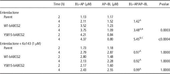
We used transduced WT-bABCG2 cells and their variant Y581S-bABCG2 to study enterolactone transport mediated by bovine ABCG2. Our results demonstrated that the efflux ratio (BL–AP/AP–BL at 4 h) that measures preferential AP transport of enterolactone (10 µM; Table 1) in both transduced subclones was significantly increased with respect to MDCK-II parental cells. When the selective ABCG2 inhibitor Ko143 was used, transport was completely inhibited resulting in an efflux ratio (BL–AP/AP–BL at 4 h) equal to that of the MDCK-II parental cell line. Of the transduced subclones, the apically directed translocation of enterolactone was higher for Y581S-bABCG2 cells (around 20%) compared with WT. These results confirm that enterolactone is transported by bovine ABCG2 with significant differences in the efflux ratio between two variants, the Y581S being more active than the WT variant.
Concentration of enterolactone (10 μM) permeated towards apical (BL–AP transport) or basal (AP–BL transport) compartments in cell cultures in the absence and in the presence of inhibitor

ABCG2=ATP-binding cassette transporter G2.
Results are expressed as mean (n=3).
Residual error term resulting from the ANOVA=0.216.
A,B,CMean ratio values within a column not sharing the same superscripts differ significantly at P<0.01.
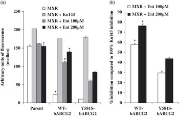
Evaluation of the inhibitory activity of enterolactone was measured in accumulation assays with flow cytometry experiments. ABCG2 expression prevents MXR accumulation and the inhibition of ABCG2 will result in an accumulation of MXR in MDCK-II cells. The ability of enterolactone to reverse the reduced MXR accumulation in cells transduced with the transporter was tested in ABCG2 bovine subclones (Figure 1a). The percentage of inhibition of the different concentrations of enterolactone for bovine wild type/Y581S-transduced cells was related to the effect of the reference inhibitor Ko143 (set at 100% inhibition of ABCG2, Figure 1b). Our results showed that enterolactone was able to inhibit the transporter increasing the accumulation of MXR, in a dose-dependent manner, showing high percentages of inhibition, especially in the bovine wild-type transduced cells (60% to 80% inhibition at 100 to 200 µM; Figure 1b). This result supported the dual interaction of enterolactone, as substrate and inhibitor, with the bovine ABCG2 transporter.
Effect of the enterolignan enterolactone on the accumulation of mitoxantrone (at 10 µM) in parent MDCK-II cells and in their WT bABCG2 and Y581S bABCG2-transduced subclones. Cells were pre-incubated with or without the specific ABCG2 inhibitor Ko143 (1 µM) or enterolactone at the indicated concentrations. (a) Fluorescence units, (median). (b) Percentage of inhibition of the different concentrations of enterolactone. Data are expressed as mean±SEM (n=3, *P<0.05). Percentage of inhibition was related to the effect of the reference inhibitor Ko143 (set at 100% inhibition of ABCG2).

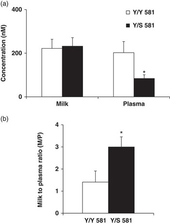
The relation between in vitro and in vivo results was established by analyses of milk and plasma enterolactone concentration in non-carriers (Y/Y) and animals carrying the polymorphism (Y/S) were performed. We first determined the levels of enterolactone in plasma and milk (Figure 2a), and then compared the milk/plasma ratio between both groups of cows (Figure 2b). The results showed a significant decrease in plasma enterolactone concentration in Y/S animals (84.1±17 v. 202±51 nM in Y/S and Y/Y cows, respectively). In contrast, enterolactone levels in milk were higher than those obtained in plasma but similar in both set of cows, indicating a higher secretion capacity of enterolactone by the Y/S group as shown by the milk/plasma ratios (2.99±0.46 for the Y/S cows as compared with 1.44±0.50 in the Y/Y cows). These results support a stronger secretion of enterolactone into milk by the Y/S animals.
Milk and plasma concentration (a) and milk/plasma ratio (b) of enterolactone in Y/Y 581 and Y/S 581 lactating cows. Results are expressed as mean±SEM (n=5 to 6; *P<0.05).

Uric acid
Transport of uric acid by bovine ABCG2-transduced subclones. Milk/plasma ratio in cows (see Table 2 and Figure 3).
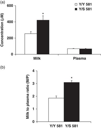
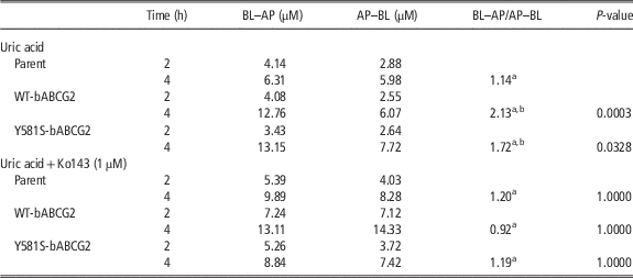
Parental and bovine transduced cell lines were grown to confluent polarized monolayers on porous membrane filters, and vectorial transport of the uric acid (100 μM) across the monolayers was determined. The vectorial efflux transport, BL–AP/AP–BL at 4 h, of uric acid showed a significant increase in the apically directed translocation in bovine subclones with significant differences with respect to the MDCK-II parental cell line in the efflux ratios (Table 2), although no differences were obtained when both variants were compared. The AP transport was abolished in the presence of Ko143, thus corroborating that uric acid transport was mediated by bovine ABCG2 in a specific manner. Figure 3 shows the uric acid concentration in milk and plasma obtained in both sets of cows. No differences were observed for uric acid levels in plasma. Conversely, significant differences were obtained in uric acid milk levels (421.1±54.2 µM for Y/S cows v. 254.4±26.3 µM for the Y/Y cows). Thus, the milk/plasma ratios observed for uric acid also indicate a higher secretory activity for this compound in the Y/S cows.
Milk and plasma concentration (a) and milk/plasma ratio (b) of uric acid in Y/Y 581 and Y/S 581 lactating cows. Results are expressed as mean±SEM (n=5 to 6; *P<0.05).

Concentration of uric acid (100 μM) permeated towards apical (BL–AP transport) or basal (AP–BL transport) compartments in cell cultures in the absence and in the presence of inhibitor

ABCG2=ATP-binding cassette transporter G2.
Results are expressed as mean (n=3).
Residual error term resulting from the ANOVA=0.326.
a,bMean ratio values within a column not sharing the same superscripts differ significantly at P<0.05.
Riboflavin transport (see Figure 4)
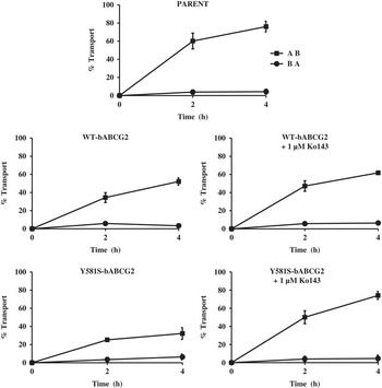
Vitamin B2 may represent an endogenous ligand for ABCG2 in the mammary gland. Our results on vectorial transport of vitamin B2 in bovine-transduced cells show that parental MDCK-II cells displayed a high BL-directed translocation (AP–BL, influx flow), whereas AP-directed translocation was very low (BL–AP, efflux flow) (Figure 4). In addition to the presence of an active transepithelial absorptive riboflavin transport in the basolateral membrane, our results support the presence of another riboflavin transport mechanism mediated by ABCG2 possibly counteracting the endogenous absorptive process. Indeed, in MDCK-II cells overexpressing bovine ABCG2, the percentage of basolateral translocation or influx flow at 4 h was markedly decreased from 76±0.34 in MDCK-II parent cells to 52.1±2 in WT-bABCG2 cells and 32.2±3.3 in Y581S-bABCG2 cells. This transport was completely reversed by Ko143, thus corroborating riboflavin transport mediated by both bovine ABCG2 variants. The results obtained show a significant increase in the ratio BA–AP/AP–BA from 0.060±0.024 in MDCK-II parent cells v. 0.120±0.024 in WT-bABCG2 and 0.223±0.016 Y581S-bABCG2, resulting in a higher transport of riboflavin in the Y581S cells.
Transepithelial transport of riboflavin (0.1 µM) in polarized parent MDCK-II and in their WT bABCG2 and Y581S bABCG2-transduced subclones in the absence or in the presence of Ko143 (1 µM). ●, Translocation from the basolateral to the apical compartment. ■, Translocation from the apical to the basolateral compartment. The fraction of riboflavin transported to the acceptor compartment was presented as a percentage of the total riboflavin added to the donor compartment at the beginning of the experiment. Results are expressed as mean±SEM (n=3).

Despite these convincing in vitro results, no differences in riboflavin levels in milk were observed between both sets of cows (3.17 nM±0.17 for Y/Y cows v. 3.61 nM±0.16 for Y/S cows), which suggests that the Y581S polymorphism does not affect riboflavin levels in milk.
Discussion
The study of the association between in vitro and in vivo results is difficult because of the complex interaction of genomic, proteomic and metabolomic factors and the important role of animal characteristics such as age, nutrition and stage of lactation in cows. In this study, we examine the relevance of the Y581S polymorphism on the transport of endogenous and dietary compounds present in cow milk as previously reported for milk secretion of fluoroquinolone drugs such as danofloxacin (Otero et al., Reference Otero, Barrera, de la Fuente, Prieto, Marques, Alvarez and Merino2013 and Reference Otero, Real, de la Fuente, Prieto, Marques, Alvarez and Merino2015).
Our results show that the in vitro models can act as a valuable tool to test the potential role of the Y581S bovine polymorphism in the secretion of drugs (Gonzalez-Lobato et al., Reference Gonzalez-Lobato, Real, Herrero, de la Fuente, Prieto, Marques, Alvarez and Merino2014) and dietary-derived compounds such as enterolactone (Miguel et al., Reference Miguel, Otero, Garcia-Villalba, Tomas-Barberan, Espin, Merino and Alvarez2014) into milk. The role of enterolactone as an inhibitor of ABCG2 could have a significant impact on milk quality and the control of milk residues.
Uric acid
The presence of some key compounds such as antioxidants and vitamins could affect milk properties with effects in the Dairy Industry. At the moment, several tools have been developed to predict milk accumulation of some of these compounds, which might help to elucidate possible mechanisms behind the interindividual differences in milk quality. Nonetheless, the effect of the alteration of specific compounds on milk quality-related parameters has not yet been clearly established. This is the case of uric acid, a purine metabolite able to act as a potent-free radical scavenger and thereby provide protection against oxidative damage (Larsen and Moyes, Reference Larsen and Moyes2010). Therefore, the altered secretion of uric acid might contribute to the overall redox potential of milk together with vitamins such ascorbic acid or α-tocopherol (Aycicek et al., Reference Aycicek, Erel, Kocyigit, Selek and Demirkol2006). The milk uric acid concentration in the Y/S 581 cows was significantly higher compared with the Y/Y 581 cows (Figure 3). This result supports the function of mammary gland ABCG2/BCRP transporter in uric acid transport in milk as well as the differential role of the ABCG2 polymorphism. The uric acid concentration could increase the potential redox in milk as suggested by preliminary physico-chemical analyses of milk from both cow groups (milk redox value was significantly increased in Y/S 581 polymorphic cows; JA Otero et al., unpublished results). On the other hand, the role of ABCG2 in the extrarenal transport of uric acid is well established (Hosomi et al., Reference Hosomi, Nakanishi, Fujita and Tamai2012), as is the strong interaction between uric acid and the transporter as evidenced by the inhibition of ABCG2 by uric acid at clinically relevant concentrations in humans (IC50 value=365 µM; Dankers et al., Reference Dankers, Mutsaers, Dijkman, van den Heuvel, Hoenderop, Sweep, Russel and Masereeuw2013).
Enterolactone
The enterolactone concentration in cows’ milk agrees with the average content reported in previous studies (Antignac et al., Reference Antignac, Cariou, Le-Bizec and André2004; Gagnon et al., Reference Gagnon, Cortes, da Silva, Kazama, Benchaar, dos Santos, Zeoula and Petit2009). Flax hulls increase concentration of enterolactone in bovine milk and the abundance of some antioxidant genes in the mammary gland, which can promote an antioxidant environment and protect against oxidative stress damage in the mammary gland (Cortes et al., Reference Cortes, Palin, Gagnon, Benchaar, Lacasse and Petit2012; Schogor et al., Reference Schogor, Palin, Santos, Benchaar, Lacasse and Petit2013). Our in vitro results presented enterolactone not only as a specific and potent ABCG2 substrate but also as a compound more actively transported by the Y581S variant of the bovine ABCG2 transport compared with the WT variant. This is the first time that such an effect of the Y581S SNP is described for a natural component derived from silage intake. In dairy cows, lignans are metabolized in the rumen and are available for absorption in the small intestine as enterolignans (Petit et al., Reference Petit, Gagnon, Mir, Cao and Cui2009). Some authors have pointed out the high concentration of enterolactone in milk rather than its low uptake (Steinshamn et al., Reference Steinshamn, Purup, Thuen and Hansen-Moller2008; Petit et al., Reference Petit, Gagnon, Mir, Cao and Cui2009). Njastad et al. (2014) have recently reported that factors such as reconjugation and secretion into the rumen and intestinal wall as well as in cells from the udder could be limiting factors in the content of dietary isoflavones and lignans into milk. ABCG2 has been previously reported to be involved in intestinal conjugation and hepatic metabolism of isoflavones (Alvarez et al., Reference Alvarez, Real, Perez, Mendoza, Prieto and Merino2010). Furthermore, ABCG2 expression in bovine rumen, intestine, liver and mammary gland has recently been reported (Lindner et al., Reference Lindner, Halwachs, Wassermann and Honscha2013; Haslam and Simmons, Reference Haslam and Simmons2014). Plasma levels of enterolactone in Y/S 581S cows (84.10±17.00 nM) were lower than those obtained for Y/Y 581 cows (202.00±50.78 nM), which highlighted the role of this SNP in the plasma disposition of enterolactone. Moreover, milk/plasma ratio was much higher in Y/S cows (2.99±0.46) than in Y/Y cows (1.44±0.50), which corroborated the in vitro results obtained with the S581 variant. The Y581S SNP of the bovine ABCG2 can affect the plasma concentration of several substrates (Otero et al., Reference Otero, Real, de la Fuente, Prieto, Marques, Alvarez and Merino2015). We hypothesize that the decrease in plasma disposition of specific substrates of ABCG2 in Y/S cows is probably due to the enhanced function of the Y581S polymorphism. This effect may promote a higher clearance of some substrates in organs with elevated ABCG2 expression and therefore lead to an increase of concentration of these compounds in body fluids such as urine, bile, intestinal lumen and milk, reducing plasma concentration.
With respect to enterolactone inhibition of the bovine ABCG2, our results agree with previous data (Miguel et al., Reference Miguel, Otero, Garcia-Villalba, Tomas-Barberan, Espin, Merino and Alvarez2014), from murine Abcg2 where significant inhibitions were achieved using 100 to 200 μM enterolactone. These results indicate that mammalian lignans could be used for reducing withdrawal periods in ruminant milk and this would result in lower transfer of drugs and antibiotics into milk, as in the case of isoflavones when they were co-administered with enrofloxacin (Pulido et al., Reference Pulido, Molina, Merino, Mendoza, Prieto and Alvarez2006). Enterolactone 100 µM was able to efficiently inhibit the WT variant of the ABCG2 transporter, reaching almost 70% of inhibition. The Y581S variant was inhibited but to a lesser extent probably due to its higher transport capacity. This finding agrees with a previous report in which some compounds differentially inhibited both variants of bovine ABCG2 (Merino et al., Reference Merino, Real, Baro, Gonzalez-Lobato, Prieto, Alvarez and Marques2009).
Riboflavin (vitamin B2)
Riboflavin was tested in our in vitro model since it is a micronutrient present in milk with an important role in a wide variety of cellular processes. Transport assays with polarized MDCK-II cells have allowed us to identify riboflavin as an ABCG2 substrate in cattle. Riboflavin has been previously described as murine and human ABCG2 substrate in vitro (van Herwaarden et al., Reference van Herwaarden, Wagenaar, Merino, Jonker, Rosing, Beijnen and Schinkel2007). Furthermore, in vivo assays in mice have shown that, on the one hand, abcg2 limits the bioavailability of riboflavin in plasma and tissues and, on the other hand, this vitamin is actively transported into milk (van Herwaarden et al., Reference van Herwaarden, Wagenaar, Merino, Jonker, Rosing, Beijnen and Schinkel2007). However, no reports showing ABCG2-mediated riboflavin transport in ruminants are available so far. Our data reporting riboflavin transport mediated by bovine ABCG2 (Figure 4) showed a potential involvement of this transporter in the transfer of nutrients through bovine milk from the mother to the newborn. According to our results, there is a specific mechanism that translocates riboflavin to the basolateral compartment, which is in agreement with previous studies that have located specific transporters for vitamin B2 in the basolateral membrane (Yonezawa and Inui, Reference Yonezawa and Inui2013). Nevertheless, our results support the presence of another riboflavin transport mechanism mediated by ABCG2. van Herwaarden et al. (Reference van Herwaarden, Wagenaar, Merino, Jonker, Rosing, Beijnen and Schinkel2007) reported that in abcg2 knockout mice milk secretion of riboflavin was reduced >60-fold compared with that in wild-type mice. Yet, under laboratory conditions, abcg2 knockout mice pups showed no riboflavin deficiency due to concomitant milk secretion of its cofactor flavin adenine dinucleotide (FAD), which was not affected. These authors point out that a metabolic trapping of riboflavin by conversion to FAD is sufficiently effective to offset the riboflavin extrusion capacity of ABCG2 and thus avoid overall vitamin B2 deficiency. This compensatory mechanism for secretion of vitamin B2 equivalents into milk may differ between species and the effect of ABCG2 on this process might vary between ruminant and non-ruminant species.
The complexity of these mechanisms as well as the relation of in vitro–in vivo results was evident as no differences were obtained in cow. Despite increased in vitro transport of riboflavin by the Y581S variant compared with wild-type (Figure 4), we have not detected differences in the vitamin composition of milk from cows with and without the Y581S polymorphism. It is important to note that our in vivo studies were performed in heterozygous individuals for the Y581S polymorphism. Therefore, the effect of the in vivo expression of the gain-of-function SNP in a single allele might not be as effective in terms of transport capacity as our in vitro model that overexpresses separately both variants of the bovine ABCG2 transporter.
In conclusion, the results obtained in this work show for the first time the significant role of the ABCG2 Y581S polymorphism in the transfer into milk of uric acid. Moreover, the role of enterolactone as an inhibitor of ABCG2 could have a significant impact on milk quality and the control of milk residues. These findings describe this polymorphism as a new factor that may affect milk composition, chemical properties and quality with an important effect for the milk industry and consumers of dairy products. Our results support the in vitro model as a valuable tool but we cannot rule out other outcomes using other cell models such as mammary cell lines. The impact of enterolactone for ruminant ABCG2-mediated drug secretion in vivo is an issue of interest that should be addressed in the future.
Acknowledgments
This work was supported by the Spanish Ministry of Economy and Competitiveness and the European Regional Development Fund [Projects AGL2012-31116 and AGL2011-22447]. J.A.O. holds a pre-doctoral fellowship of the Basque Government. The authors thank Dr A.H. Schinkel (The Netherlands Cancer Institute, Amsterdam, The Netherlands) who provided parental MDCK-II cells. The authors thank Garfi SAT farm (Santa Maria del Monte del Condado, Province of Leon, Spain) for technical assistance and support. They are grateful to Prof. James McCue for assistance in language editing.