Introduction
The tensor tympani muscle is the largest muscle within the middle ear. It is innervated by the mandibular division of the trigeminal nerve. Its fibres originate from the cartilaginous wall of the eustachian tube and the greater wing of the sphenoid. The tendon of the tensor tympani muscle arises from the cochleariform process and inserts into the medial surface of the handle of the malleus. Under contraction, the tensor tympani muscle pulls the malleus and the tympanic membrane medially, increasing the stiffness of the tympano-ossicular system. Although its action is largely understood, the audiometric effects of this contractile activity have been documented in very few papers.Reference Bance, Makki, Garland, Alian, van Wijhe and Savage1–Reference Smith3
In this report, the authors describe an unusual case of bilateral voluntary contraction of the tensor tympani muscle, and discuss its functional repercussion on sound conduction.
Case report
A 27-year-old Caucasian man presented to our ENT department complaining of bilateral tinnitus that was voluntarily evoked. The tinnitus was defined as a cracking sound. The patient had no history of otological disease.
Otoscopy was normal, but the patient was found to be able to contract his tensor tympani muscle voluntarily. During rapid contractions, the handle of the malleus and the tympanic membrane were medially displaced, repeatedly, at a maximum rate of 3–4 times per second, always under voluntary control. Contraction was observed in both ears simultaneously; the patient could not move each muscle individually. Fibre-optic nasopharyngoscopy showed no signs of synchronous palatal movement.
The patient reported that he could sustain the contraction for longer periods, up to 30 seconds. He reported that this caused a marked reduction in hearing capacity, including hearing of the human voice. After 2 or 3 maximal sustained contractions, a period of 3–5 minutes of rest was necessary to resume contractions. A short video-otoscopy recording demonstrating the voluntary contraction of the tensor tympani muscle is available on The Journal of Laryngology & Otology website (Appendix 1).
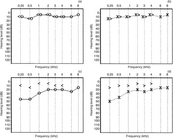
Audiograms were performed under conditions of rest and maximal contraction of the tensor tympani muscle (Figure 1). During contraction, a marked reduction in air conduction was observed, particularly at low frequencies, with air–bone gaps of 35 and 30 dB at frequencies of 250 and 500 Hz respectively. No change was observed in bone conduction (as assessed by our audiologist).
Auditory thresholds of the right ear (a) and left ear (b) during relaxation, and of the right ear (c) and left ear (d) during maximal contraction of the tensor tympani muscle. <= bone conduction right ear; ○ = air conduction right ear; ×= air conduction left ear; >= bone conduction left ear

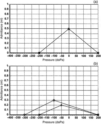
Tympanometric curves are shown in Figure 2. During contraction, there was an increase in impedance, which was fully expected. There was also a shift in tympanometric peak pressure, indicative of an associated change in middle-ear pressure.
Tympanometric curves obtained during relaxation (a) (curves are overlapped) and contraction (b) of the tensor tympani muscle. ○ = right ear; ×= left ear

The diagnosis was discussed with the patient and it was agreed that no therapeutic measures would be employed.
Discussion
The role of the tensor tympani muscle in auditory function has been a subject of controversy. Based on previous studies, Gelfand affirmed that the tensor tympani muscle does not participate in the acoustic reflex.Reference Gelfand4 In his words, ‘in humans the acoustic reflex is a stapedius reflex’. However, tensor tympani muscle contractions have been documented as being part of an alert response to very intense sound. They have also been associated with an anticipatory state; for instance, they have been reported in startle reaction or reflex studies, under conditions where subjects are expecting to hear a sudden, startling noise.Reference Djupesland5
Klockhoff and Anderson recorded changes in middle-ear impedance secondary to tensor tympani muscle contraction in response to a jet of air directed into the eye.Reference Klockhoff and Anderson6 Recently, Bance et al. described a similar response after an identical stimulus.Reference Bance, Makki, Garland, Alian, van Wijhe and Savage1 Rock reported a case of forceful eyelid closure syndrome, in which the tensor tympani muscle contracted simultaneously with vigorous closure of both eyes.Reference Rock2 In that particular case, contraction was long and powerful enough to demonstrate a conductive hearing loss, primarily at lower frequencies.
Contractile activity in the tensor tympani muscle has also been recorded (using electromyography) in association with vocalisationReference Klockhoff7 and swallowing.Reference Klockhoff7, Reference Salen and Zakrisson8 Ingelstedt and Jonson recorded similar muscular activity during belching and yawning (using volume and flow measurements).Reference Ingelstedt and Jonson9 Indeed, in his book A Text-book of the Diseases of the Ear for Students and Practitioners, written in 1909, Politzer had already stated that ‘the deafness which occurs during yawning is brought about by a simultaneous contraction of the tensor tympani’.Reference Politzer10
The importance of the tensor tympani muscle in eustachian tube function is purely speculative. Honjo et al. investigated pressure changes in the middle ear of cats; the authors found that tensor tympani muscle contraction did not result in any pressure rise.Reference Honjo, Ushiro, Haji, Nozoe and Matsui11 To date, there is no evidence of a significant role of the tensor tympani muscle in middle-ear ventilation.
The tensor tympani muscle has been implicated in cases of myoclonus, which may present aloneReference Cohen and Perez12 or in association with neurological diseases such as multiple sclerosis.Reference Zipfel, Kaza and Greene13 In these rare cases, tinnitus is usually the chief complaint, described by the patient as a rumbling or cracking sound. Tenotomy has been used as a treatment for patients with incapacitating tinnitus in whom conservative measures failed.Reference Zipfel, Kaza and Greene13
Voluntary control of the tensor tympani muscle is an extremely rare event. Once again, we quote the words of Politzer, who described voluntary contractions at the beginning of the twentieth century, reporting that ‘during the contractions … the deep tones became deadened and indistinct …’.Reference Politzer10 As audiograms could not be performed at that time, no documentation of this event was obtained. In 1943, Smith became the first author to document the audiometric effects of a purely voluntary contraction, which were similar to those reported here.Reference Smith3 Rock also described a comparable audiogram (as mentioned above); however, contraction in that case was associated with vigorous eye closure.Reference Rock2 Bance et al. reported two cases of voluntary contraction, but only one of them could hold contraction long enough to undergo audiometry in a contraction condition.Reference Bance, Makki, Garland, Alian, van Wijhe and Savage1 Once again, the air–bone gap was evident at lower frequencies. In that particular case, tympanometric curves showed an increase in middle-ear impedance during contraction, but there was no displacement of the pressure peak. In the case presented here, we observed not only a decrease in compliance, but also a reduction in middle-ear pressure, even when no eustachian tube activity was detected.
The tensor tympani muscle was described by Ramírez et al. as being a masticatory muscle within the middle ear.Reference Ramírez, Ballesteros and Sandoval14 This seems to be an adequate definition, as the tensor tympani muscle shares innervation with masticatory muscles (temporal, masseter and pterygoid muscles). Based on this definition, it is proposed that the tensor tympani muscle functions in a co-active manner, additionally attenuating sounds produced during the mastication process and velopharyngeal movements. In this sense, the tensor tympani muscle may be a functional link between temporomandibular joint disorders and ear symptoms such as otalgia, tinnitus, hypoacusis, and aural pressure or fullness.
Even when the tensor tympani muscle is not directly involved in auditory function, the impact of its contraction on sound conduction through the tympano-ossicular system is remarkable. However, the unusual, but strong, powerful contraction described here may not mirror its physiological role in normal subjects. In other words, the regular, ordinary muscular activity of the tensor tympani muscle is probably weaker than that observed in the present case. Nevertheless, a better understanding of the potential audiometric effects of tensor tympani muscle contraction could aid the diagnosis of hearing disorders.
• Tensor tympani muscle contraction increases tympano-ossicular system stiffness
• Its voluntary contraction is an unusual event
• Very few papers have documented its audiometric effects
• The most remarkable effect is an air–bone gap, particularly at lower frequencies
• Tensor tympani muscle function has been related to attenuation of sounds produced during mastication
Appendix 1. Supplementary video material
A short video-otoscopy recording demonstrating voluntary contraction of the tensor tympani muscle is available on The Journal of Laryngology & Otology website, at http://journals.cambridge.org/10.1017/S0022215113003149.