INTRODUCTION
Leeches belong to the phylum Annelida, class Hirudinea (Sawyer, Reference Sawyer1986). The latter class includes leeches living in fresh and marine waters. There is a lot of published work in respect to freshwater leeches, such as, Becker & Katz (Reference Becker and Katz1965), Daniels & Freeman (Reference Daniels and Freeman1976), Bur (Reference Bur1994), Sket & Trontelj (Reference Sket and Trontelj2008), to name just a few, both because they are much easier to collect and study and they are used for medicinal purposes. In contrast, the literature is somewhat scarce regarding leeches of the marine environment. Important information on marine leeches can be accessed through the fundamental work of Sawyer (Reference Sawyer1986) and more recent work, such as, Utevsky et al. (Reference Utevsky, Utevsky, Schiaparelli and Trontelj2007) and Utevsky (Reference Utevsky and Adrianov2008), to name some of them.
Marine leeches belong to the families Ozobranchidae and Piscicolidae (Sket & Trontelj, Reference Sket and Trontelj2008) with representatives by somewhat 25 described genera and they occur as ectoparasites, mainly, on fish. In Europe, members of this family are known from reports since 1850 (Meyer, Reference Meyer1940).
Marine leeches have been described from both cultured and wild fish, including groupers, Epinephelus coioides Hamilton (Cruz-Lacierda et al., Reference Cruz-Lacierda, Toledo, Tan-Fermin and Burreson2000) in the Philippines by Zeylanicobdella arugamensis De Silva and by Piscicola sp. (Bondad-Reantaso, Reference Bondad-Reantaso, Langdon, Enriquez and Sukimin1992), rays and skates, Torpedo marmorata Risso and Raja clavata Linnaeus by Pontobdella muricata Linnaeus, scorpion fish, Scorpaena porcus Linnaeus and Scorpaena scrofa Linnaeus by Trachelobdella lubrica Grube, 1840 in the Sea of Marmara and the Dardanelles, Turkey (Erguven & Candan, Reference Erguven and Candan1992; Saglam et al., Reference Saglam, Orguz, Celik, Doyuk and Usta2003), species of Serranidae, Priacanthidae and Pomacentridae by Trachelobdella lubrica (Sawyer, Reference Sawyer1986; Williams et al., Reference Williams, Bunkley-Williams and Burreson1994), yellow fin bream, Acanthopagrus australis (Gunther) by Austrobdella bilobata Ingram (Roubal, Reference Roubal1986), to name some of them.
Skates and rays are a common fisheries product in the Mediterranean, caught and consumed usually locally because of the rapid development and build up of ammonia taste and smell that starts from the skin. That is why they are usually marketed fresh after the removal of their skin. Catches of R. clavata and Raja species in the Aegean and Ionian Seas in Greece during the period of 2002–2010 amounted to approximately 500 t per year, dropping from a maximum of approximately 1400 t in 1994 (FAO, 2013).
Reports from local fishermen in the area of Lesvos Island, Greece of an occasionally-seen worm-like ectoparasite on the skin surface of skates prompted research on ectoparasites of skates caught in the area for the assessment of the occurrence of parasites and their identification.
This paper reports on the findings of the parasitological macroscopic and histological investigation of ectoparasites of skates caught off the island of Lesvos at the north-eastern Aegean Sea in Greece.
MATERIALS AND METHODS
Sampling
Sampling concerned only skate species caught by the commercial trawlers that included Raja clavata Linnaeus, Raja radula Delaroche, 1809, Raja miraletus Linnaeus, Dipturus oxyrinchus Linnaeus and Dasyatis pastinaca Linnaeus. The survey was performed over a 21 month period from May 2010 to February 2012 on-board fishing trawlers.
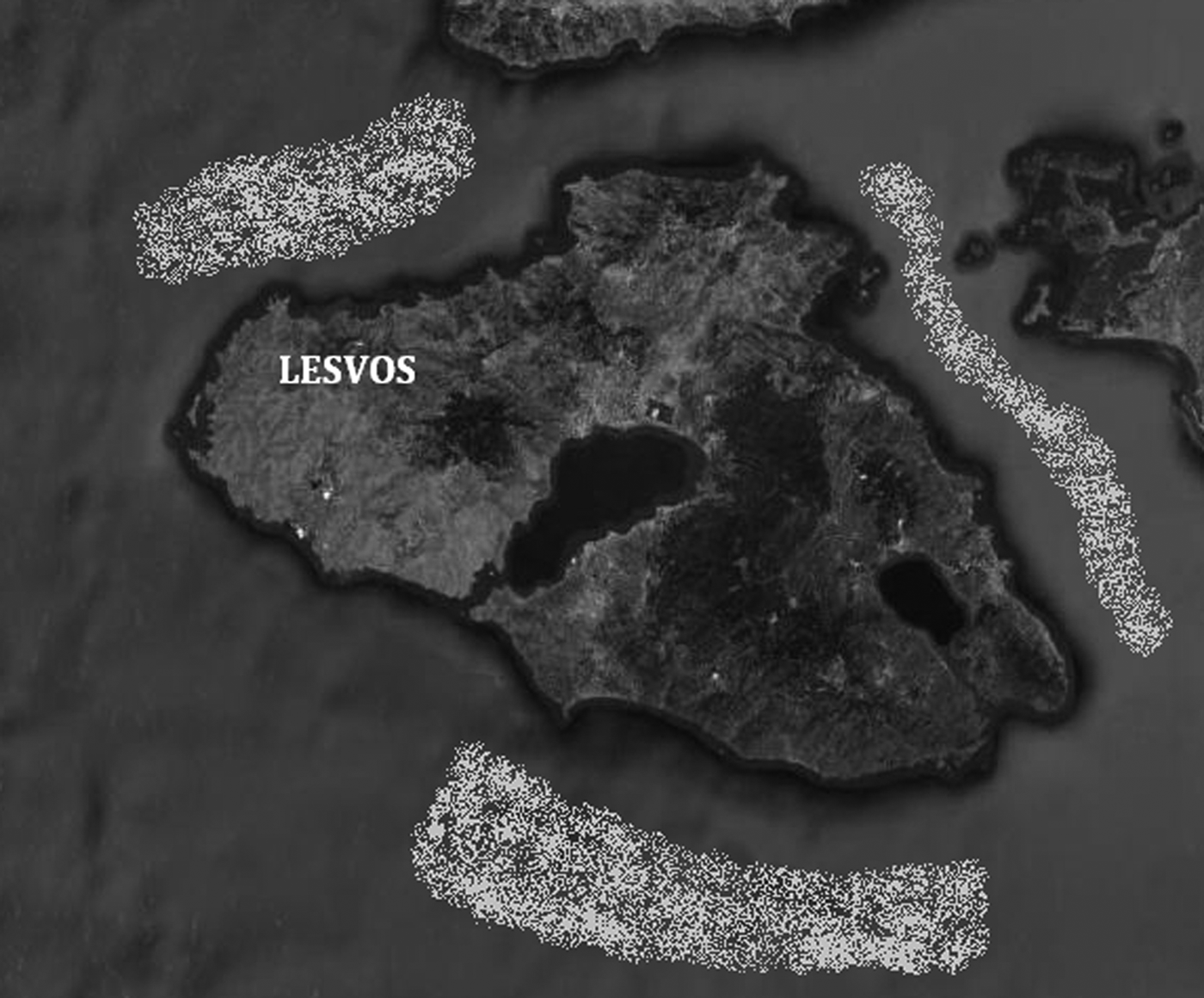
The area of sampling was off the coast of Lesvos Island, Greece, at the north-eastern Aegean Sea as depicted in Figure 1.
Fishing areas off the coast of Lesvos, Greece.

Fish caught by the trawler, following the routine procedure of commercial fisheries, were placed in polystyrene boxes, covered in ice and preserved in a refrigerator at 0–4oC. This procedure causes a cold shock in live specimens that leads to death in minutes. Sampling procedures initiated between 1 and 2 h, maximum, after fish were placed in the refrigerator and consisted of recording the fish species, the numbers per species caught and the number of fish species that were parasitized externally. Fish caught during fishing that had ectoparasites and/or visible lesions on the skin were dissected in situ and ectoparasites, either detached or attached to the skin, and skin lesions, were placed in 10% phosphate buffered formalin (Drury & Wallington, Reference Drury and Wallington1980) in order to be further processed for histological examination.
Fish and parasites identification
Fish species were identified using the keys of the FAO species identification guide for fishery purposes (Serena, Reference Serena2005). The ectoparasites were identified using the key by Sawyer (Reference Sawyer1986).
Measurements of parasitism
The prevalence, mean intensity and mean abundance of ectoparasites on the fish were defined according to Bush et al. (Reference Bush, Lafferty, Lotz and Shostak1997).
Histology
Formalin-preserved samples from D. pastinaca Linnaeus were processed for histology following the procedure of Drury & Wallington (Reference Drury and Wallington1980). Briefly, samples were dehydrated in a series of alcohol solutions starting from 70% to 100% in a Leica TP 1020 Histokinette and then tissues were embedded in paraffin. Paraffin-embedded tissues were cut in slices 3–5 μm thick using a Leica RM2125 RT microtome. Tissue sections were placed on glass slides and after rehydration were stained with haematoxylin–eosin using a Leica Autostainer XL device.
Stained samples were then observed under light microscope at 4 × , 10× and 40× using an Olympus CH20 microscope.
RESULTS
The ectoparasites observed on the skin of inspected samples were identified, exclusively, as the marine leech Pontobdella muricata Linnaeus (Sawyer, Reference Sawyer1986).
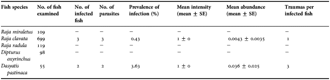
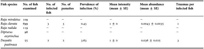
As it is evident in Table 1, Raja clavata Linnaeus represented the major part of the catch, with a percentage of 64.7%, followed by R. radula Delaroche, 1809 at 11%, R. miraletus Linnaeus at 10.1%, Dipturus oxyrinchus Linnaeus at 9% and Dasyatis pastinaca Linnaeus at 5.1%.
Prevalence of Pontobdella muricata (%), mean abundance, mean intensity and trauma lesions in the skate species examined.
S.E: standard error
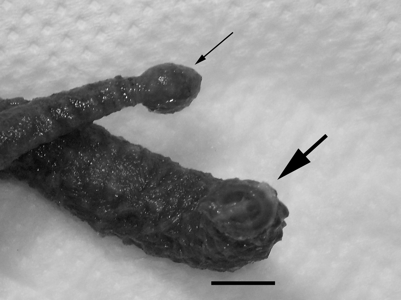
Pontobdella muricata (Figure 2) was observed on the skin of only two fish species, R. clavata and D. pastinaca, with the prevalence of infection being 0.43% and 3.63%, respectively, of the catch these fish species for the examination period.
The marine leech Pontobdella muricata: detail of the oral (thick arrow) and caudal (thin arrow) suckers. Scale bar represents 1 cm.
Mean intensity of the parasitism was 1 ± 0 for both skate species infected, while the mean abundance of the parasite in the total number of the fish species found to be infected, was calculated at 0.0043 ± 0 and 0.036 ± 0 for R. clavata and D. pastinaca, respectively (Table 1).
Macroscopic observation of lesions and parasites
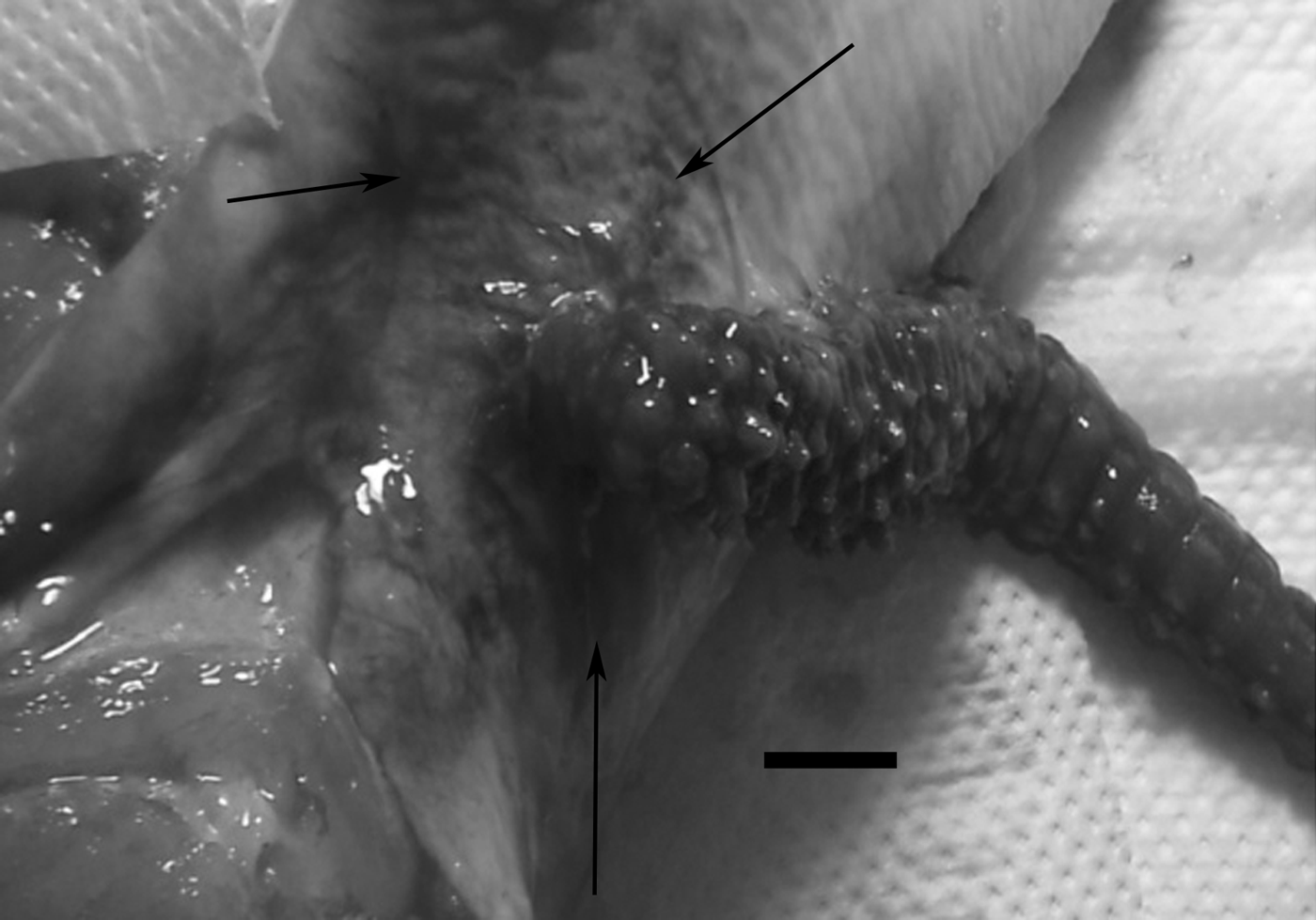
The leeches were observed attached on the skin only. The areas of attachment on R. clavata were the dorsal surface of the body and laterally of the caudal part of the body, as is seen in Figure 3. Attachment of the parasite on D. pastinaca occurred on the dorsal and ventral surface of the body with many lesions occurring very near the edges of both body surfaces. Due to the lack of scales on the skin of D. pastinaca multiple lesions incurred by the parasite were observed, in a way that, as is evident from Table 1, each fish had multiple traumas, in contrast to R. clavata, where the trauma was associated with an attached parasite.
Attachment of Pontobdella muricata on the lateral surface of the body caudal area of Raja clavata. Haemorrhages are evident on the skin (arrows). Scale bar represents 1 cm.
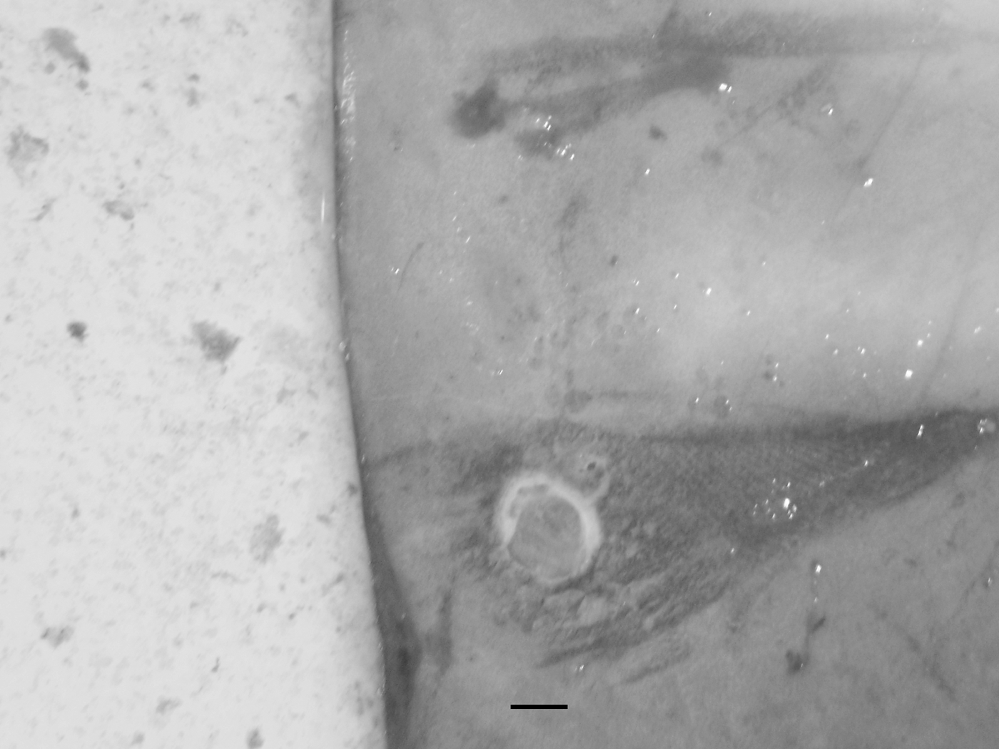
The lesions consisted of roundish wounds where all the skin had disappeared. Connective tissue inside the rim of the lesions in the form of whitish tissue was evident in seemingly older wounds, while the centre of the lesions was red owing to the colour of the exposed muscle beneath, as is evident in Figure 4.
Lesion on the skin of Dasyatis pastinaca after detachment of the leech. Ventral view near the body edge. Scale bar represents 1 cm.
Where the leech was found attached, and especially in R. clavata, there were skin petechias, haemorrhages and swelling (Figure 3). These symptoms were milder in D. pastinaca.
The mean length of the five parasites found in this study was 40 ± 9.06 cm (30–50 cm).
Microscopy
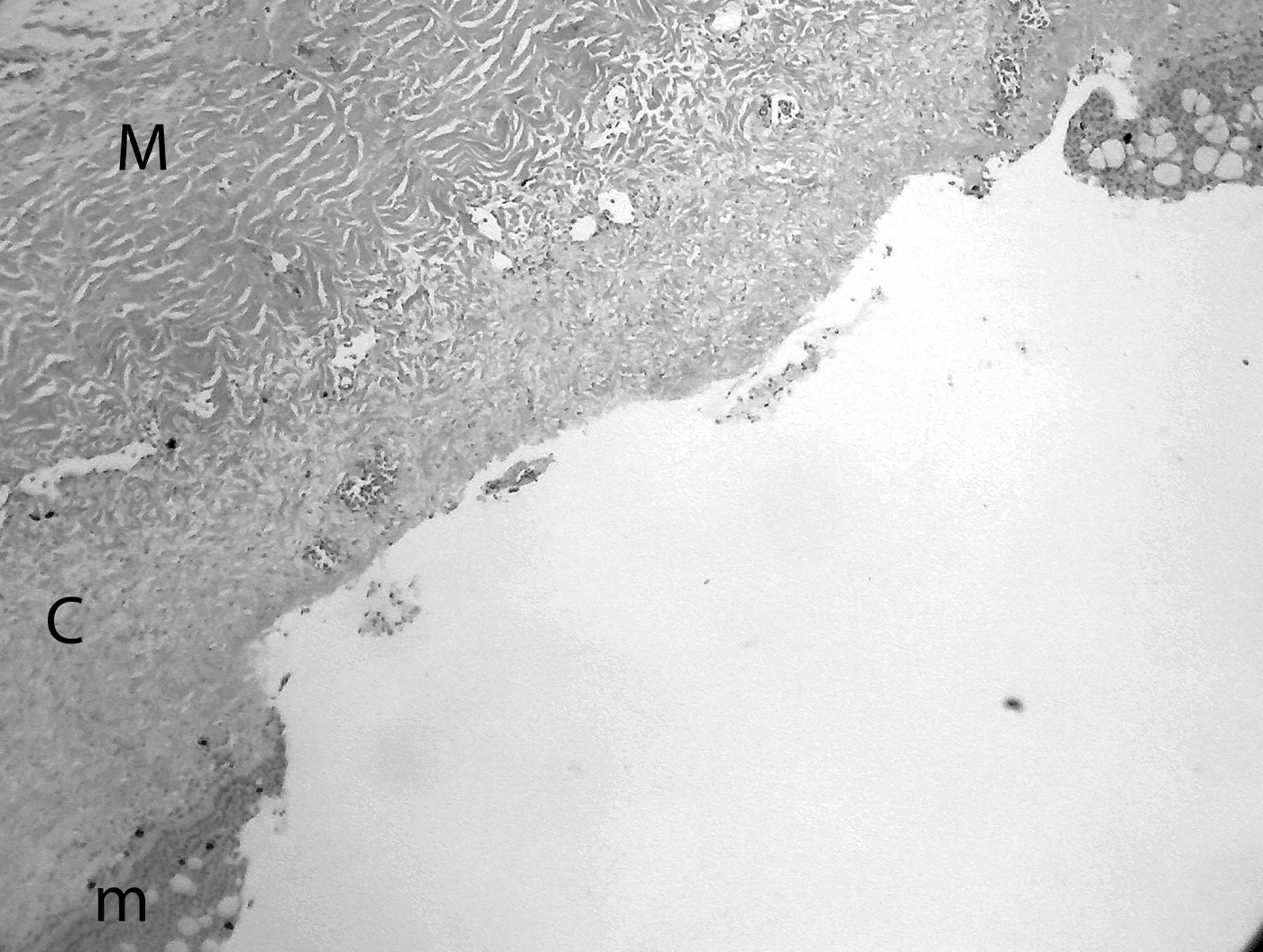
After the detachment of the leech a roundish shallow trauma is seen (Figure 5). The mucosa, the lamina propria and part of the underlying submucosa tissue have disappeared, with no evidence of the lesion reaching the muscle tissue. Interestingly, there was no inflammation on the site of the lesion since no blood leucocytes and macrophages were seen in the area.
Microscopic view of skin lesion of Dasyatis pastinaca after detachment of the leech. M, muscle tissue; C, connective tissue; m, mucosa. Haematoxylin–eosin, 10×.
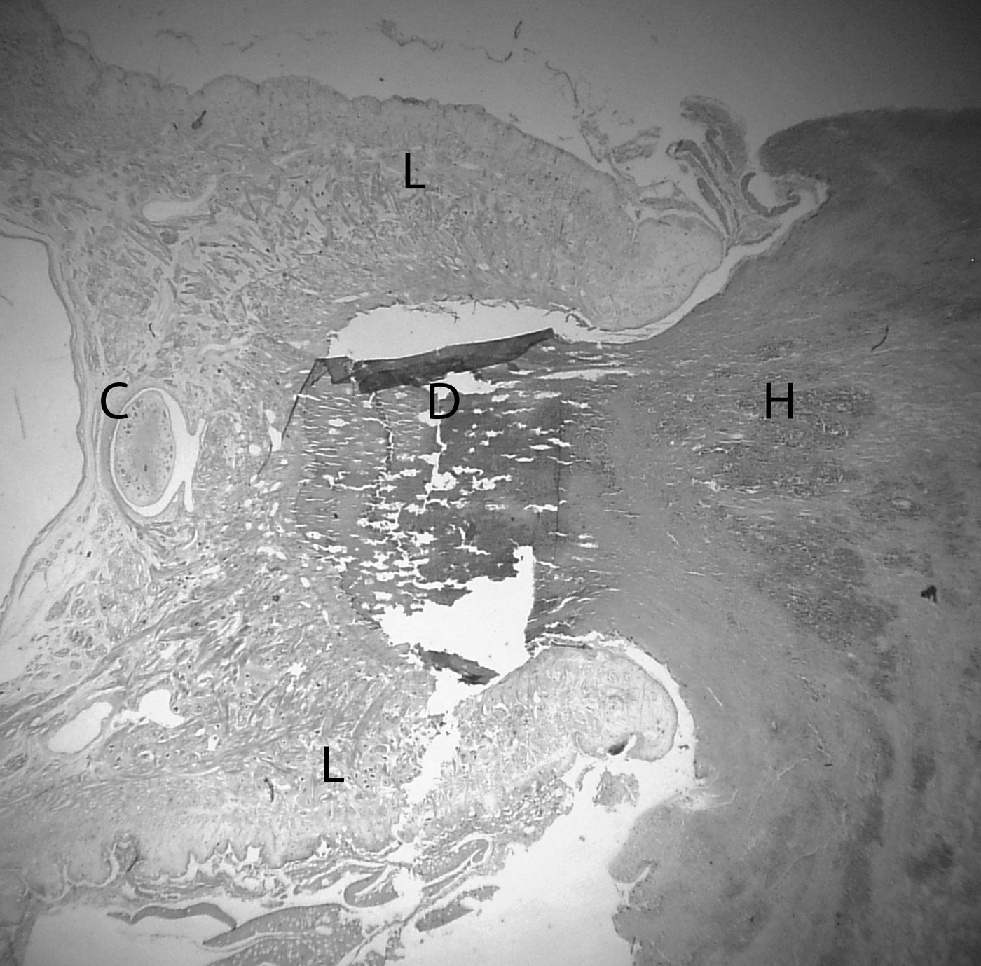
Figure 6 depicts the condition of the fish skin while the parasite is attached. Severe haemorrhage is evident at the part of the skin that is inside the sucker of the leech and in areas of the skin outside it and in the underlying tissue. The part of the fish tissue inside the sucker is necrotized and liquefied.
Microscopic view of the attachment of Pontobdella muricata on the skin of Dasyatis pastinaca. L, leech body (oral sucker); C, cerebral node; D, necrotic and liquefied skin fish tissue; H, haemorrhages. Haematoxylin–eosin, 4×.
Tubules initiating from the sucker of the leech are seen carrying a reddish material to the leech's body—apparently, the liquefied tissues and blood of the fish. The roundish organ at the bottom-centre of the sucker of the leech represents the cerebral node or ganglion.
DISCUSSION
A study of the ectoparasites of skates caught off the coast of Lesvos Island, north-eastern Aegean, Greece, was undertaken in order to assess the parasite species, frequency and fish species affected.
The major catch of skates is represented by the skate Raja clavata Linnaeus, reaching a percentage of 64.7% of the total catch during the investigation period.
Only one ectoparasite species was identified after the inspection of a total of 1080 fish individuals. The ectoparasite was identified as being the marine leech Pontobdella muricata Linnaeus using the key of Sawyer (Reference Sawyer1986). The latter author reported that marine leeches identified as P. muricata have been described from fish in the Mediterranean and Black Seas, and the north-east Atlantic to Spitzbergen and South Greenland. A more recent study (Oktener & Utensky, 2010) reaffirmed the occurrence of P. muricata in the Black Sea. The leech was observed only on skin surfaces, while no other parasites were identified in the gills of examined fish. These findings are in accordance to those reported by Erguven & Candan (Reference Erguven and Candan1992) and by Saglam et al. (Reference Saglam, Orguz, Celik, Doyuk and Usta2003). However, there is a noticeable difference on the prevalence of occurrence of the parasite. While in this study only a 0.43% of R. clavata was found to be infected by the leech, Saglam et al. (Reference Saglam, Orguz, Celik, Doyuk and Usta2003) reported a 100% prevalence of the parasite on the same species. These authors, however, examined only one specimen of R. clavata. A very recent study by Oktener & Utevsky (Reference Oktener and Utevsky2010) calculated a prevalence of 16.6% of P. muricata found on R. clavata in the Black Sea, after examination of 12 specimens. Literature on P. muricata prevalence on fish is very scarce making it difficult to compare our results and highlights the need for more research on the subject. Research on other marine leeches and fish species reported a prevalence of 0.4% of an unidentified marine leech of grouper cultured in floating cages (Leong & Wong, 1988), while Cruz-Lacierda et al. (Reference Cruz-Lacierda, Toledo, Tan-Fermin and Burreson2000) reported a prevalence of 83% and 17% in cultured young and adult grouper Epinephelus coioides Hamilton, respectively.
This study identified a new fish species that is parasitized by P. muricata. Dasyatis pastinaca Linnaeus, which represented the 5.1% of the total catch, was found to be infected by the marine leech with a prevalence of 3.63%. To our knowledge, this is the first report of such parasitism on this fish species.
The macroscopic observations of local haemorrhages, petechias and swelling were evident in the skin around the attachment of the leech only of R. clavata. No such lesions were seen around the attachment sites of P. muricata on D. pastinaca. The latter species seems to develop a much milder reaction to the local lesions. Similar macroscopic lesions have been reported by Saglam et al. (Reference Saglam, Orguz, Celik, Doyuk and Usta2003) from P. muricata on the skin of R. clavata and by Roubal (Reference Roubal1986) from Austrobdella bilobata Ingram on the skin of Acanthopagrus australis (Gunther).
Histological examination was performed on tissue samples isolated from D. pastinaca. The samples included tissues from lesions during the attachment and after detachment of the leech. The latter lesion included the disappearance of mucosa, basal layer of lamina propria and part of the submucosa, and the lack of an inflammatory response. Apart from the lack of inflammatory response, the histological picture of the lesion is similar to that reported by Roubal (Reference Roubal1986). With the leech attached, a severe haemorrhaging condition is evident in the tissue being inside, adjacent and beneath the attachment site. The liquefied tissue inside the sucker corresponds well to the lesions left behind after detachment of the leech.
CONCLUSION
A parasitological examination of skates caught off the coast of Lesvos in the north-eastern Aegean, Greece, revealed the presence of the marine leech Pontobdella muricata Linnaeus attached on the skin of fish. The prevalence of the ectoparasite was 0.43% and 3.63% on the skates Raja clavata Linnaeus and Dasyatis pastinaca Linnaeus, respectively, with the latter skate species being described for the first time as a host of this leech. Pontobdella muricata is feeding on blood and tissues of the host causing, locally, haemorrhages and tissue necrosis and, when detached, a roundish lesion is left behind, with disappearance of the upper layers of the skin tissue with a mild inflammatory response in D. pastinaca.
ACKNOWLEDGEMENT
We wish to thank the local fishermen's association of Lesvos for providing access to fishing boats and material for investigation.
FINANCIAL SUPPORT
The research was supported by funding from The Department of Marine Sciences, University of The Agean, Greece.