Introduction
Initial fish development is a highly dynamic process, with ontogenetic changes resulting in morphofunctional changes of the main body systems, a consequence of the fast morphological and physiological evolution of fish embryos and larvae during their development (Osse et al., Reference Osse, Van Den Boogart, Van Snik and Van Der Sluys1997).
In fish, the larval stage can last from days to months. During this period, the larva doubles in length and increases in weight up to 100 times. Progressive differentiation of adult characteristics takes place through a process of morphogenesis up to the juvenile stage, when the larvae looks like the adults (Nakatani et al., Reference Nakatani, Agostinho, Baumgartner, Bialetzki, Sanches and Cavicchioli2001). Thus, several teleostei fish undergo great changes in body shape, which directly affect the growth and survival of young specimens (Gisbert et al., Reference Gisbert, Williot and Castelló-Orvay2000).
Fish larvae are morphologically different from the adults and have different ecological demands in regards to habitat, feeding, and behavior (Leis & Trnski, 1989 apud Sanches et al., Reference Sanches, Baumgartner, Bialetzki, Subviertom, Gomes, Nakatani and Barbosa2001). Thus, studies on fish ecology cannot be considered adequate without previous knowledge of the initial development of the species (Sanches et al., Reference Sanches, Baumgartner, Bialetzki, Subviertom, Gomes, Nakatani and Barbosa2001). Furthermore, the great similarity between the larvae of different species and the lack of comparative literature make identification studies difficult.
The catfish Zungaro jahu, commonly known as ‘jaú’, is a vulnerable species in danger of extinction due to loss of habitat. It can be found in the Paraná-Paraguay and Uruguay River basins in South America and is one of the largest neotropical fish, reaching up to 150 kg and 144 cm in length (Agostinho et al., Reference Agostinho, Gomes, Suzuki, Júlio, Carolsfield, Harvey, Ross and Baer2004). It reaches first maturation with approximately 70 cm total length, migrates during the reproductive period, has free-floating eggs, and spawns between the months of December and February. Information on its biology and ecology are scarce and the majority of data available is related to nutrition and parasitology (Agostinho et al., Reference Agostinho, Gomes, Suzuki, Júlio, Carolsfield, Harvey, Ross and Baer2004).
Larval ontogenesis studies under controlled conditions enable the monitoring of the morphological changes taking place, providing information on environmental demands and feeding needs during the initial phases of development. Therefore, studies aimed at understanding the development of larval body systems are amongst the most useful for larvae and juvenile husbandry techniques, which in turn increases the chances of survival and, consequently, success in incubation, larviculture, and juvenile production.
Thus, the aim of this study was to observe and describe the ontogeny of the digestive and sensory systems and of other major organs in Z. jahu larvae from hatching to total yolk absorption through stereomicroscopy, light microscopy (LM), and scanning electron microscopy analysis (SEM).
Materials and methods
All procedures were performed according to the ethical principles established by the Brazilian College of Animal Research (Colégio Brasileiro de Experimentação Animal – COBEA) and the study approved by the Ethics Committee in the Use of Animals (Comissão de Ética no Uso de Animais – CEUA, protocol no. 10348/16) of the UNESP – Universidade Estadual Paulista, Jaboticabal-SP, Brazil.
Broodstock of the species Z. jahu Ihering, 1898 (= Paulicea luetkeni Steindachner, 1875) from the fishery Usina Hidrelétrica Engenheiro Souza Dias (Companhia Energética do Estado de São Paulo, Castilho-SP, Brazil) were subjected to induced reproduction and dry fertilization, according to the techniques by Woynarovich & Horváth (Reference Woynarovich and Horváth1983).
The females (n = 5) received two injections of common carp (Cyprinus carpio) pituitary extract (0.8 and 5.0 mg/kg body weight) in the coelomic cavity at 12-h intervals. The males (n = 8) received a single dose (2.5 mg/kg body weight) concomitantly to the females’ second dose.
Approximately 12 h after the effect of the hormone, the gametes were obtained by manual craniocaudal massage of the abdomen and subsequently mixed and hydrated.
Embryos were placed in 60-litre incubators with continuous water flow, where they remained for the whole study period. Artemia nauplii and plankton were provided from the second day after hatching, and ‘lambari’ (Astyanax altiparanae) and ‘curimbatá’ (Prochilodus lineatus) larvae from the third day post-hatching.
The following physical-chemical characteristics of the hatcheries water were recorded: temperature and dissolved oxygen (mg/l), by YSI 550 A equipment, pH, conductivity (μS/cm), by YSI 63 equipment, and alkalinity (mg/l) ammonia (μg/l) by Goltermann et al. (Reference Goltermann, Clymo and Ohnstad1978) method.
Samples were collected at the time of hatching (time zero) and then every 4 h until total yolk absorption. At each sampling time, 10 larvae were fixed in Karnovsky solution (2.5% glutaraldehyde + 2.5% paraformaldehyde) for 24 h, washed in 0.1 M sodium cacodylate buffer (pH 7.4), and stored in 70% alcohol for further analysis according to the following methodologies.
Stereomicroscopy and morphometry
Larvae were observed and photomicrographed using the LEICA DFC 280a photomicrography equipment attached to a LEICA MZ8 stereomicroscope. Larval total length was obtained using the program IM 50-LEICA.
Light microscopy (LM)
Samples were processed for embedding in historesin (kit Leica Historesin®) according to the manufacturer's recommendations. Samples were cut into 4-μm sections and every fifth section mounted onto slides (semi-serially) and stained with hematoxylin–phloxin (HP) or periodic acid Shiff (PAS) (Tolosa et al., Reference Tolosa, Behmer and Freitas-Neto2003). Slide analysis and photodocumentation were carried out using a LEICA DM 2500 photomicroscope.
Scanning electron microscopy (SEM)
After being washed in sodium cacodylate buffer, the larvae were post-fixed in 1% osmium tetroxide solution for 2 h, washed again in the same buffer, dehydrated in increasing graded ethanol, dried to critical point with liquid CO2 (BAL-TEC dryer), mounted onto copper stands, coated with palladium-gold, and observed and electron micrographed using a scanning electron microscope (JEOL-JSM 5410).
Results
The mean values for the physical–chemical water variables were: temperature = 28.75 ± 0.59°C; pH = 8.18 ± 0.035; dissolved oxygen = 7.33 ± 0.21 mg/l; alkalinity = 19.00 ± 1.73 mg/l; conductivity = 32 ± 2 μS/cm and ammonia = 12.47 ± 1.79 μg/l.
Z. jahu larvae hatched 13 h after fertilization with mean total length of 3.79 ± 0.11 mm. Total yolk absorption was observed at 60 hours post-hatching (hph) and mean total length was 7.05 ± 0.33 mm.
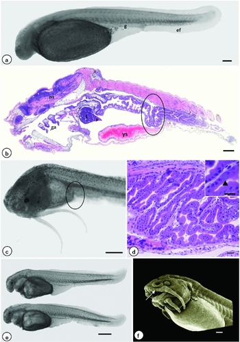
At hatching, the larvae were transparent with pigmentation on the yolk sac and on the cephalic and ventral regions (Fig. 1 a), which increased with larval development. A highly developed embryonic fin covering the dorsal and ventral regions of the body was also observed (Fig. 1 a).
Photomicrography (a–e) and electron micrography (f) of Z. jahu larvae. (a) Hatching – primitive gut; (b, c) 36 hph – first digestive tract fold (circles); (d) 60 hph – detail of the gut wall, mucus-producing cell (arrowhead); (e) 12 hph – opening of the oral cavity; (f) 20 hph – emergence of teeth, taste buds on lip borders and inside the mouth (arrow). Abbreviations: ga = gill arches, l = liver, g = gut, ef = embryonic fin, p = pancreas, ns = nervous system, ys = yolk sac. Bars: (a, e) = 500 μm; (b, f) = 100 μm; (c) = 500 μm; (d) detail = 30 μm. Staining: (b) = hematoxylin–phloxin (HP); (d) = periodic acid Shiff (PAS).

Digestive system
The digestive system was divided into cephalic gut (oral cavity and pharynx), anterior gut (esophagus and stomach, when present), midgut (intestine per se), and hindgut (rectum and anus).
At hatching, a straight and simple tube was observed in the ventral region of the larvae, corresponding to the outline of the primitive gut (Fig. 1 a). This tube was lined with a single layer of prismatic cells throughout its extension and had no connection to the oral cavity. At 8 hph, the gut began to show small invaginations and, by 36 hph, the first digestive tract fold could be seen past the proximal region of the midgut (Fig. 1 b, c). From 24 to 60 hph, there was an increase in the number of invaginations, leading to multiple folds in the midgut. After complete yolk absorption, the midgut had a simple prismatic lining epithelium with striated borders and mucus-producing cells (Fig. 1 d).
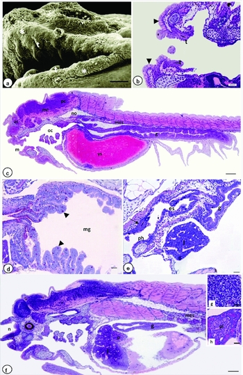
By 12 hph, the oral cavity was opened (Fig. 1 e) and, from 16 hph, the oral cleft began to elongate. From 20 hph, the lining epithelium of the oral cleft was elevated due to the emergence of teeth and a high number of taste buds was observed on the labial borders and inside the mouth (Figs 1 f and 2 a). By 24 hph, small teeth at the early stages of development could be seen in the maxilla and mandible (Figure 2 a, b) and a large number of mucus-producing cells were present on the labial borders (Fig. 2 b).
Electron micrography (a) and photomicrography (b–h) of Z. jahu larvae. (a, b) 24 hph – teeth in the maxilla and mandible, taste buds (*), mucus-producing cells (arrowhead); (c) 24 hph – opening of the esophagus; (d) 28 hph – mucous-producing cells (arrowheads, stained pink); (e) 28 hph – pharyngeal teeth (arrow); (f) 20 hph – liver and pancreas; (g) 60 hph – detail of the liver; (h) 24 hph – pancreatic islet. Abbreviations: m = mouth, ac = anterior cerebrum, mc = mid cerebrum, oc = oral cavity, pc = posterior cerebrum, t = teeth, es = esophagus, l = liver, ph = pharynx, g = gut, mg = midgut, pi = pancreatic islet, n = nares, no = notochord, p = pancreas, mes = mesonephros, ys = yolk sac. Bars: (a) = 50 μm; (b, c, e) = 100 μm; (d) = 30 μm; (g, h) = 20 μm. Staining: (b, c, e–h) = hematoxylin–phloxin (HP); (d) = periodic acid Shiff (PAS).

At 24 hph, the cephalic gut communicated with the anterior gut through the esophagus opening (Fig. 2 c). At 28 hph, the differentiation of the proximal region of the midgut began with an increase of its lumen and the number of mucus-producing cells increased as differentiation progressed (Fig. 2 d). The esophagus, lined by a single layer of goblet cells, showed some mucus-producing cells. The pharynx, also composed of goblet cells, did not show significant alterations in its structure. At 28 hph, the first pharyngeal teeth could be seen (Fig. 2 e). Anal opening occurred at 16 hph.
The liver appeared at 16 hph and became more evident at 20 hph, occupying all the area anterior to the yolk sac and behind the pericardial cavity (Fig. 2 f). The liver stained positive to PAS at 37 hph, indicating glycogen storage. There was an increase in the number of hepatocytes and in the quantity of hepatic glycogen with time (Fig. 2 g).
The pancreas, located between the liver and the yolk sac, was also visualized at 16 hph. It initially displayed few zymogenic granules (Fig. 2 f) that increased significantly in number until total yolk absorption. The first pancreatic islet was observed at 24 hph (Fig. 2 h).
Reabsorption of the yolk sac took place through the yolk syncytial layer; however, the presence of food inside the digestive tube of the larvae before complete yolk absorption indicates an overlapping of endogenous and exogenous feeding.
Gill arches
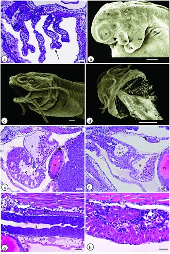
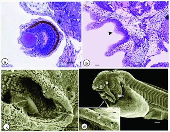
The outline of the gill arches in Z. jahu was present soon after hatching. The start of chondrogenesis and blood supply occurred at 20 hph, and by 24 hph the gills were branching out (Fig. 3 a). At 8 hph, the branchiostegal membrane began developing (Fig. 3 b) and was totally formed and covering the gill arches by 28 hph (Fig. 3 c). The gill arches were well developed after complete yolk absorption (Fig. 3 d).
Electron micrography (b, c, d) and photomicrography (a, e–h) of Z. jahu larvae. (a) 24 hph – gills branching out (arrow); (b) 8 hph – branchiostegal membrane, nasal fold (arrowhead); (c) 28 hph – branchiostegal membrane covering the gill arches, presence of the first two pairs of barbels; (d) 60 hph – gill arches; (e) 28 hph – heart, detail of the atrium and ventricle with trabeculae and atrioventricular orifice (*); (f) 32 hph – heart, detail of the ventricle-bulbus orifice (•); (g) 16 hph – mesonephros; (h) 60 hph – mesonephros with evident lumen. Abbreviations: a = atrium, ga = gill arches, I b = first pair of barbels, II b = second pair of barbels, g = gut, bm = branchiostegal membrane, n = nares, e = eye, mes = mesonephros, v = ventricle, ys = yolk sac. Bars: (a, e, f, h) = 30 μm; (b, c) = 100 μm; (d) = 500 μm; (g) = 50 μm. Staining: (a, e–h) = hematoxylin–phloxin (HP).

Heart
A rudimentary heart was present at hatching and located in the pericardial cavity, anterior to the yolk sac and abdominal cavity. At 24 hph, there were two heart chambers (atrium and ventricle). The atrium had a thin wall while the ventricular myocardium was composed of several cell layers and, from 28 hph, the trabeculae started to develop (Fig. 3 e). By 32 hph, blood flowed through the heart chambers, coming in by the sinoatrial orifice in the atrium, flowing through the atrioventricular orifice and into the ventricle, and then reaching the bulbus arteriosus by the ventricle-bulbus orifice (Fig. 3 f) before leaving the heart by the ventral aorta to be oxygenated at the gill arches.
Pronephros/mesonephros
The pronephros is temporary structure and was present soon after hatching. With the development, the pronephros transforms in a mesonephros. It was shaped as two straight lateral tubules composed of simple cuboidal epithelium. It was located above the gut and followed its entire extension. The cranial region became convoluted at 16 hph, while the caudal region remained straight (Fig. 3 g). As larval development progressed, the mesonephros became more convoluted and its lumen more evident (Fig. 3 h). The mesonephros tubules were joined and opened into the urogenital papilla, together with the intestine.
Thyroid
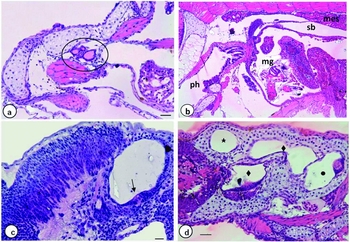
The first thyroid follicles were observed from 56 hph. They were located above the ventral aorta and consisted of a single layer of cuboidal cells, with colloid in their interior (Fig. 4 a).
Photomicrography of Z. jahu larvae. (a) 56 hph–thyroid follicles (circle); (b) 24 hph – swim bladder; (c) 8 hph – inner ear, detail of neuromast (arrow); (d) 60 hph – inner ear with anterior (⋆), lateral (♦), and posterior canals (●). Abbreviations: mg = midgut, ph = pharynx, mes = mesonephros, sb = swim bladder. Bars: (a, c) = 30 μm; (b) = 50 μm; (d) = 20 μm. Staining: hematoxylin–phloxin (HP).

Swim bladder
With the opening of the esophagus at 24 hph, the swim bladder developed between the stomach and pronephros (Fig. 4 b). It was composed of a thin layer of squamous epithelium, with a pneumatic duct connecting it to the esophagus. It did not show significant alterations throughout larval development.
Encephalon and sensory system
Encephalon
The differentiation of the neural tube regions into prosencephalon, mesencephalon, and rhombencephalon occurred concomitantly to somitogenesis. At 24 hph, there was the differentiation of the telencephalon and diencephalon regions of the anterior portion of the cerebrum, the mid ventral and dorsal portions of the cerebrum, and the development of a prominent transverse cerebellum that separated the mid and posterior cerebrum (Fig. 2 c).
Inner ear
The inner ear developed from the otic vesicle. At 8 hph, structures similar to free neuromasts began to form inside the ear (Fig. 4 c) and were well developed by 24 hph, when the anterior and posterior canals were observed. At total yolk absorption, the inner ear already showed well-defined anterior and posterior canals (Fig. 4 d).
Eyes
During somitogenesis, the optic vesicle was already being developed. At 8 hph, retina pigmentation was observed. At the end of yolk absorption, this pigmentation was intense, the eye was well developed with nuclear layers (internal and external) connected through the thick plexiform layer (Fig. 5 a), while the nucleus of the cones and rod receptors began to differentiate into two layers.
Photomicrography (a, b) and electron micrography (c, d) of Z. jahu larvae. (a) 60 hph – eye; (b) 0 hph – nares with cilia (arrowhead); (c) 48 hph – division of the nares (arrow), sensory buds (*); (d) 16 hph – primordium of the third pair of barbels (arrow); (d, inset) 24 hph – detail of the barbel highlighting the sacciform papillae on the ventral surface (arrowhead). Abbreviations: bm = branchiostegal membrane, n = nares, e = eye. Bars: (a) = 30 µm; (b, c) detail = 50 μm; (d) = 100 μm. Staining: (a, b) = hematoxylin–phloxin (HP).

Nares
At hatching, it was observed the initial development of the nares, with olfactory cells containing small cilia (Fig. 5 b). At 8 hph, the nares were more evident and the nasal fold began to form (Fig. 3 b). After 16 hph, the olfactory organs were well developed, with strands of cilia on the sensory plaque. At 28 hph, there was a protuberance in the interior of the nares and the start of lamellae formation that began to divide and had several sensory buds around it by 48 hph (Fig. 5 c). At 60 hph, the nares were lined by a ciliated pseudostratified prismatic epithelium.
Neuromasts
Neuromasts were observed mainly in the cephalic region at 44 hph. These structures were characterized by a group of sensitive cells with a protruding gelatinous cupula and a layer of supporting cells over the basal lamina.
Barbels
The primordium of the first pair of barbels (maxillary barbels) was observed soon after hatching (Fig. 4 f). At 4 hph, the primordium of the second pair of barbels appeared (first mentonian pair) and began to elongate. At 16 hph, the primordium of the third pair of barbels (second mentonian pair) could be seen (Fig. 5 c). At 28 hph, the first two pairs of barbels were elongated and sharp, whereas the maxillary barbels were longer than the first mentonian pair (Fig. 3 c) and the third pair of barbels began to sharpen by the last analyzed time. By 24 hph, several papillae could be seen on the ventral surface of the barbels, where tasting buds developed (Fig. 5 c, detail). Under LM, the barbels showed a layer of epithelial cells and three cell layers, two longitudinal and external and one transversal.
Discussion
The degree of differentiation of newly hatched larvae varies among species according to egg size (Blaxter, Reference Blaxter, Hoar and Randall1969). Species with small eggs and high fecundity rates have faster embryonic development and poorly developed larvae at hatching, as observed in Z. jahu and in the majority of migratory species. Conversely, species with large eggs and low fecundity rates have slower embryonic development and well developed larvae at hatching (Sato et al., Reference Sato, Fenerich-Verani, Nuñer, Godinho, Verani, Godinho, Godinho, Godinho and Godinho2003).
In fish, the development of the body structures and the start of exogenous feeding are important events for larval survival (Balon, Reference Balon, Simenstad and Cailliet1986). The opening of the mouth in Z. jahu was observed at 12 hph, close to the time reported by Maciel et al. (Reference Maciel, Lanna, Maciel Juniorm, Donzele, Neves and Menin2010) in Brycon orbignyanus (9 hph). Total yolk absorption occurred at 60 hph, as also observed by Landines et al. (Reference Landines, Senhorini, Sanabria and Urbinati2003) in larvae of spotted sorubim (P. coruscans). Yolk absorption occurred through the yolk syncytial layer, in agreement with reports by Gomes et al. (Reference Gomes, Scarpelli, Arantes, Sato, Bazzoli and Rizzo2007) in the larvae of three species of trahiras (Erythrinidae sp.).
The transition from endogenous to exogenous feeding is a critical stage in early development, often characterized by high mortality rates (Gordon and Hecht, Reference Gordon and Hecht2002). In the majority of fish species, larvae hatch with a simple and undifferentiated digestive tract and later develop digestive organs (Govoni et al., Reference Govoni, Boehlert and Watanabe1986). This was observed in the Z. jahu larvae, which had a rudimentary digestive tube at hatching that became more developed by the time of total yolk absorption and could then be divided into cephalic gut, anterior gut, midgut, and hindgut.
Stomach and gastric glands development could not be seen during the period analyzed. According to Watanabe and Kiron (Reference Watanabe and Kiron1994), to compensate for the lack of gastric enzymes, the fish depend on their ability to select food, mechanical digestion, and pancreatic and gut enzymes that can act in an alkaline environment. According to Gonzáles et al. (Reference Gonzáles, Flores, Domínguez and Valle2002), the efficiency of food digestion and absorption in the gut is due to the presence of folds and microvilli. Nevertheless, the mature liver and pancreas aid food digestion by producing digestive secretions that digest proteins, fats, and sugars.
A few mucus-producing cells were observed in the esophagus, just past its connection to the midgut. The early presence of these cells in the larvae of Z. jahu indicates that the larvae are morphologically prepared to protect themselves against food abrasiveness (Galvão et al., Reference Galvão, Fenerich-Verani, Yamanaka and Oliveira1997). The pharyngeal teeth in carnivore fish aid in the apprehension of food by preventing the scape of prey (Rodrigues et al., Reference Rodrigues, Navarro and Menin2006). In Z. jahu, these teeth were present at 28 hph, soon after the opening of the esophagus, when the larvae began exogenous feeding.
Some authors have reported that hepatocytes store glycogen during the larval stage (Hamlin et al., Reference Hamlin, Hunt Von Herbing and Kling2000; Micale et al., Reference Micale, Garaffo, Genovese, Spedicato and Muglia2006) and, according to Hamlin et al. (Reference Hamlin, Hunt Von Herbing and Kling2000), glycogen storage in the liver can be considered as the start of hepatocyte function, which remains throughout the larval and juvenile stages. In Z. jahu larvae, there was an increase in hepatic glycogen as development progressed, suggesting an improvement in hepatic function.
Similarly to reports by Sampaio et al. (Reference Sampaio, Prado, Yoshimi, Bazzoli and Rizzo2015) in larvae of B. orthoataenia, S. brasiliensis, L. abtusidens, and P. argenteus, the pancreas in Z. jahu had an endocrine and exocrine portion before total yolk absorption. The endocrine portion is composed of Langerhans islets (Gonzáles et al., Reference Gonzáles, Flores, Domínguez and Valle2002) and their presence suggests that the larvae are able to efficiently assimilate sugars. Well-differentiated liver and pancreas can be seen in larvae that begin exogenous feeding soon after hatching and become functional before total yolk absorption, as in Paralabrax maculofasciatus (Peña et al., Reference Peña, Dumas, Villalejo-Fuerte and Ortíz-Galindo2003), Amphirion percula (Gordon and Hecht, Reference Gordon and Hecht2002), A. lupus (Falk-Petersen and Hansen, Reference Falk-Petersen and Hansen2001), and Melanogrammus aeglefinus (Hamlin et al., Reference Hamlin, Hunt Von Herbing and Kling2000).
The gills are vital structures for fish health, as they are the main site for gas exchange as well as being involved in osmoregulation, acid-base balance, and excretion of nitrogen compounds (Maciel et al., Reference Maciel, Lanna, Maciel Juniorm, Donzele, Neves and Menin2010). The outline of the gill arches in Z. jahu was observed soon after hatching and was well developed by the time of total yolk absorption. The development of heart in Z. jahu was similar to that observed in zebrafish (Danio rerio), appearing during somitogenesis and showing thickening only of the ventricular myocardium during larval ontogenesis (Hu et al., Reference Hu, Sedmera, Post and Clark2000). According to Hu et al. (Reference Hu, Sedmera, Post and Clark2000), the heart is the first definite organ to develop and become functional during embryogenesis.
In Z. jahu larvae, the sensory system developed concomitantly to the digestive system. The eyes, inner ear, and the nares were the first sensory structures to appear, while the taste buds were the last and could only be identified after the opening of the mouth and elongation of the barbels. Taste buds can be found on the epithelium of the lips, oropharyngeal cavity, barbels, head, and occasionally throughout the whole body (Hansen et al., Reference Hansen, Reutter and Zeiske2002).
The development of the sensory organs is important for larval survival and is closely related to the onset of exogenous feeding, scape from predators, and vertical migration in the water column (Matsuoka, Reference Matsuoka2001). Kawamura et al. (Reference Kawamura, Masuma, Tezuka, Koiso, Jinbo and Namba2003) suggest that the development of sensory organs accompanies behavioral changes with important implications to ecology and fish farming, as high mortality rates in larvae under farming conditions could be related to the stress caused by high agitation of the water, aeration, and exaggerated flow, amongst other environmental disturbances that affect chemical and mechanical sensitivity at the different stages of the life cycle.
Total yolk absorption in Z. jahu was fast (60 hph), a characteristic of predatory species such as B. amazonicus (Neumann, Reference Neumann2008), P. fasciatum (Pérez et al., Reference Pérez, Bocanegra and Orbe2001), and B. gouldingi (Faustino et al., Reference Faustino, Makino, Neumann and Nakaghi2015). The development pattern of Z. jahu prioritized the formation of sensory and feeding structures that enable piscivorous expression at the start of development, before the depletion of endogenous energy reserve, thus ensuring fast growth and increasing the chances of larval survival.
Acknowledgements
The authors would like to thank Mr Orandi Mateus for his help in processing the materials, the Companhia Energética de São Paulo (CESP), Mr René Belmont and Mr Roberto Soares Ferreira for sample collection, and CNPq and CAPES for the financial support provided.