Introduction
Supracricoid laryngectomy with cricohyoidoepiglottopexy is an organ-preserving surgical procedure used to treat patients with laryngeal cancer. This procedure was first proposed by Majer and Rieder in 1959.Reference Majer and Rieder1 By removing approximately three-quarters of the larynx, including both vocal folds, supracricoid laryngectomy with cricohyoidoepiglottopexy allows patients to continue breathing via the natural airway, without a tracheal stoma, and preserves swallowing and vocal function.Reference Laccourreye, Laccourreye, Weinstein, Menard and Brasnu2–Reference Nakayama, Okamoto, Miyamoto, Takeda, Yokobori and Masaki4 Interaction between the arytenoids and the epiglottis has been reported to be responsible for this post-operative function.Reference Saito, Kimura, Imagawa, Nito, Tayama and Shiotani5, Reference Hayashi, Hirose, Tayama, Imagawa, Nakayama and Seino6 However, the post-operative neoglottis tends to be variable and therefore difficult to predict in terms of anatomy and function.
The neoglottis is usually evaluated using laryngoscopy, stroboscopy and high-speed endoscopic imaging. However, these modalities provide only two-dimensional images from above. In contrast, three-dimensional imaging is an ideal method of evaluating the altered features of the neoglottis, following supracricoid laryngectomy with cricohyoidoepiglottopexy.
We have previously reported that three-dimensional models of the neoglottis can be reconstructed utilising data obtained from multidetector-row computed tomography (CT).Reference Seino, Nakayama, Okamoto and Hayashi7
The current study used this method to examine a larger group of patients, in order to evaluate the three-dimensional morphology of the neoglottis following supracricoid laryngectomy with cricohyoidoepiglottopexy.
Patients and methods
Patients
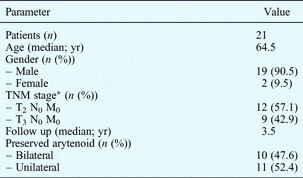
We retrospectively analysed three-dimensional images reconstructed from multidetector-row CT data for 21 patients (mean age, 64.5 years; age range, 53–74 years). These patients had undergone supracricoid laryngectomy with cricohyoidoepiglottopexy between November 1997 and February 2007. All patients had shown a complete response for more than one year post-operatively, and were living with a natural airway and satisfactory laryngeal function. Clinical data for these 21 patients are summarised in Table I.
Patient clinical data

*Including radiation failure. Yr = years; TNM = tumour-node-metastasis
The ipsilateral arytenoid was resected when the cancer had invaded the posterior part of the larynx (i.e. the paraglottic space or the arytenoid cartilage); in such cases, the corniculate cartilage was preserved to reduce the neoglottic gap. Both arytenoids were preserved when the cancer was confined to the anterior part of the larynx.
Methods
A 16-channel multidetector-row CT (Light Speed Ultra 16; GE Healthcare UK, Little Chalfont, UK) was utilised for regular patient follow up and evaluation. Computed tomography scanning was performed under the following conditions: (1) X-ray tube voltage, 120 kV; (2) X-ray tube current, 250 mA; (3) slice thickness, 1.25 mm; and (4) slice time, 0.6 seconds.
The patient was instructed to lie on the CT table in a supine position with the chin relaxed; a head band was then applied to the patient's forehead to restrict head and neck movement and thus minimise imaging errors.
Scanning was usually performed using the plane along the hyoid bone as a horizontal landmark. Scanning was performed from the oropharynx down to the level of the cricoid cartilage, during both respiration and phonation. In order to obtain data for both these states, the patient's radiation dose was increased by 20 to 30 per cent, compared with conventional CT scanning. This fact was explained to each patient, and the procedure was only performed with the patient's full consent.
Three-dimensional images were processed using Digital Imaging and Communication in Medicine (DICOM) files, and analysed using Intage Realia Pro (version 1.1.2.421) and Volume Player (Cybernet Systems, Tokyo, Japan) software programs installed on a Windows personal computer. Three-dimensional images of the cricoid, arytenoid cartilages and airway configuration were created using the volume rendering method (Figure 1).
Three-dimensional images of typical neoglottic morphology, from patients with bilateral and unilateral arytenoids.

Arytenoid movement
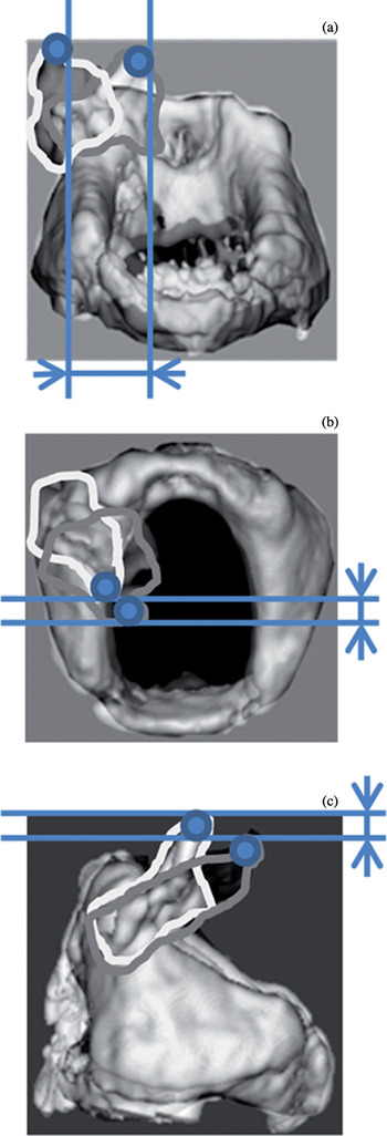
This was analysed based on the movement of a designated point located at the pinnacle of the arytenoid cartilage, during respiration and phonation. Motion of the designated point was measured using (1) a single linear distance; and (2) distances along the transverse (i.e. lateral to median), sagittal (i.e. anterior to posterior) and coronal (i.e. superior to inferior) axes (Table II and Figure 2); all axes were defined based on the inferior horizontal plane of the cricoid cartilage. As the movement of the arytenoid along the cricoid slope is relatively complex, the single linear measurement was used to quantitatively simplify the imaging data, in order to facilitate comparisons involving single and bilateral arytenoids. Measurements along the three axes were used to compensate for the simplicity of linear measurement. Movement along these three axes was measured based on the perpendicular axial source from the inferior horizontal plane of the cricoid. Arytenoid movement was analysed in patients with single and bilateral arytenoids.
Diagrams illustrating measurement parameters for (a) transverse (i.e. lateral to median), (b) sagittal (i.e. anterior to posterior) and (c) coronal (i.e. superior to inferior) arytenoid movement.

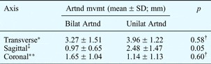
3D Movement of unilateral and bilateral arytenoids

*Lateral to median movement; ‡anterior to posterior movement; **superior to inferior movement; see Figure 2 for illustration. †Not significant. 3D = three-dimensional; artnd = arytenoid; mvmt = movement; SD = standard deviation; bilat = bilateral; unilat = unilateral
Minimum neoglottic gap cross-sectional area. During phonation, the minimum cross-sectional area of the neoglottic gap reflects narrowing of the air stream, and is considered to indicate the status of laryngeal function. This parameter was calculated using three-dimensional data, with reference to a plane parallel to the inferior horizontal plane of the cricoid (as this was the only prominent horizontal plane in the area of interest). Moreover, the minimum neoglottic cross-sectional area is considered a substitute parameter which enables reasonable analysis of neoglottic function, despite the variable amount of soft tissue which surrounds the arytenoid(s) and cricoid cartilages post-operatively. When multiple air stream pathways were present, the sum of the minimal cross-sectional areas of all streams was analysed.
This study was approved by the Institutional Review Board of Kitasato University Hospital (B ethics 09-20). All patients provided written, informed consent.
Results
Arytenoid movement
The mean arytenoid movement, measured as a single linear distance, was 3.98 mm for bilateral arytenoids (being the mean distance of the two arytenoids in motion) and 5.15 mm for single arytenoids (Figure 3). This distance was slightly larger in patients with one single arytenoid, compared with patients with bilateral arytenoids; however, this difference did not reach statistical significance (p = 0.44).
Measurement points for linear movement calculation. Mean ± standard deviation movement distances were 3.98 ± 1.46 mm for bilateral arytenoids (n = 10) and 5.15 ± 1.09 mm for unilateral arytenoids (n = 11); this difference was not significant (p = 0.42).

Table II shows the mean distances of arytenoid movement along the three axes described above. The mean movement measurements for bilateral arytenoids represent the mean distance of the two arytenoids in motion. Mean movement along the transverse axis (i.e. lateral to median) was 3.27 mm for bilateral arytenoids and 3.96 mm for unilateral arytenoids (p = 0.58; not significant). Mean movement along the sagittal axis (i.e. anterior to posterior) was 0.97 mm for bilateral arytenoids and 2.48 mm for unilateral arytenoids (p < 0.05). Mean movement along the coronal axis (i.e. superior to inferior) was 1.65 mm for bilateral arytenoids and 1.14 mm for unilateral arytenoids (p = 0.60; not significant). While the mean movement distance along the transverse and coronal axes were similar for bilateral and unilateral arytenoids, the mean movement along the sagittal axis was greater for unilateral arytenoids than bilateral arytenoids.
Minimum neoglottic gap cross-sectional area
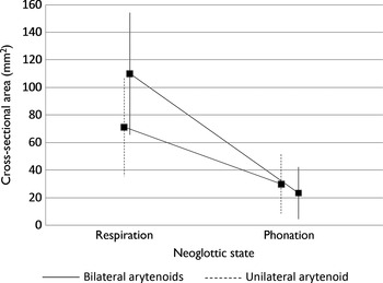
Results of the mean minimum neoglottic gap cross-sectional area are shown in Figure 4. During respiration, the mean cross-sectional area was 109.9 mm2 for bilateral arytenoids and 71.2 mm2 for unilateral arytenoids. During phonation, the mean cross-sectional area was 23.3 mm2 for bilateral arytenoids and 29.9 mm2 for unilateral arytenoids. The neoglottic gap during respiration was wider in patients with bilateral arytenoids, compared with patients with unilateral arytenoids; however, both groups had a similar neoglottic gap during phonation.
Mean minimum neoglottic gap cross-sectional area for patients with bilateral (n = 10) and unilateral (n = 11) arytenoids, during respiration and phonation. Outliers indicate ±1 standard deviation. p < 0.05, bilateral vs unilateral respiration results.

Discussion
Following supracricoid laryngectomy with cricohyoidoepiglottopexy, it has been reported that a sufficient degree of efficient arytenoid movement (of at least one remaining arytenoid) is important in order to maintain good laryngeal function.Reference Nakayama, Hirose, Okamoto, Miyamoto, Yokobori and Takeda8 Following resection of both vocal folds, the remaining arytenoids play a major role in neoglottic closure, and thus in post-operative laryngeal function.Reference Weinstein, Laccourreye, Ruiz, Dooley, Chalian and Mirza9
Analysis of neoglottic morphology and movement is crucial in order to elucidate function following supracricoid laryngectomy with cricohyoidoepiglottopexy. Due to the relatively variable and unstable vibration patterns of the neoglottis, high-speed digital imaging has been reported to provide better information on neoglottic morphology, compared with laryngostroboscopic evaluation.Reference Makeieff, Giovanni and Guerrier10, Reference Granqvist and Lindestad11
However, both these methods allow only two-dimensional observation. Because of the altered configuration of the neoglottis, three-dimensional evaluation is superior to two-dimensional observation methods.Reference Bruno, Napolitano, Sciuto, Giordani, Garaci and Floris12 The present study used our previously reported three-dimensional reconstruction techniques to further explore the three-dimensional morphology of the neoglottis, in additional cases.Reference Seino, Nakayama, Okamoto and Hayashi7
Mean linear distance measurements demonstrated sufficient movement in both bilateral and unilateral arytenoids. Although there was no statistically significant difference, unilateral arytenoids exhibited slightly greater mobility than bilateral arytenoids. When comparing mean arytenoid movement along the three axes described above, the greatest movement distance was seen along the transverse (i.e. lateral to median) axis, for both bilateral and unilateral arytenoids. Unilateral arytenoids exhibited greater movement along the sagittal (i.e. anterior to posterior) axis, compared with bilateral arytenoids (p < 0.05). In addition to transverse movement, anterior shifting of the unilateral arytenoid plays an important role in compensating for the inability to perform neoglottic closure. The clinical importance of anterior shifting has been noted previously.Reference Seino, Nakayama, Okamoto and Hayashi7 In this study, we were able to present quantitative evidence supporting this fact; this evidence could only be obtained by three-dimensional analysis.
In addition, analysis of mean neoglottic gap cross-sectional area demonstrated that the neoglottic gap during respiration was wider for bilateral arytenoids than for unilateral arytenoids. However, both groups had similar narrowing during phonation. The air stream pattern varied between cases with single versus multiple air streams, due to variability in neoglottic structure and mucosa pliability. These results contribute to our understanding of neoglottic function in patients with a single arytenoid. Again, accurate estimation of smallest neoglottic gap cross-sectional area could only be obtained via three-dimensional imaging.
These data provide evidence that the unilateral arytenoid is capable of achieving sufficient neoglottic narrowing to compensate for the resected contralateral arytenoid.
• After supracricoid laryngectomy with cricohyoidoepiglottopexy, three-dimensional computed tomography was used to assess the neoglottis
• Unilateral arytenoids showed more sagittal movement than bilateral arytenoids; movement in other directions was comparable
• Anterior shift of the unilateral arytenoid is important to compensate for lack of neoglottic closure
• Three-dimensional analysis was useful for this comparison
It has previously been reported that there is no difference between patients with one and two arytenoids as regards voice function one year after the initial surgery.Reference Yüce, Cağli, Bayram, Karasu, Sati and Güney13 Our results support these findings. In patients with a unilateral arytenoid, well balanced interaction between the residual arytenoid mucosa on the resected side, and favourable motion of the remaining arytenoid, are crucial for good laryngeal function.
Our data on linear arytenoid movement, movement along three axes and minimum neoglottic gap cross-sectional area could not have been obtained without three-dimensional CT imaging. Other useful CT techniques for such patients include transverse imaging with multiplanar reconstructions, use of static reference points (e.g. adjacent to a vertebral body), and measurement of cross-sectional area by multiplanar reformatting.
Further research is needed to fully elucidate the morphological and functional aspects of the neoglottis, following supracricoid laryngectomy.
Acknowledgement
This study was supported by a Grant-in-Aid for Young Scientists (B) from the Ministry of Education, Culture, Sports, Science and Technology of Japan (grant number 20791225: 2010–2011).