I. INTRODUCTION
Fe–Cr–Mo alloys are widely used in power plants, as a corrosion resistant alloy, in pressurized water reactors Reference Greeff, Louw and Swart1 and are specially designed to be used in oil industries at temperatures ranging from 500 °C to 600 °C. Reference Klueh2–Reference Mohapatra, Panda and Mitra4 Nowadays Fe–9% Cr–1% Mo is the most common alloy in use due to a combination of properties such as toughness, creep resistance, and corrosion resistance at high temperatures. Reference ElMassalami, de Sousa, Areiza, Rabello and Elzubair5,Reference Kim, Park and Lee6 However, in some refineries with higher acidity levels, strong corrosion processes have been observed, specially in processes related to naphthenic acid corrosion. Increase of molybdenum content has been suggested as a possible solution to this problem, since the addition of this alloying element results in a better corrosion resistance. Reference Wu, Jing, Zheng, Yao and Ke7
Precipitation of undesired phases like carbides and intermetallic compounds in Cr/Mo-rich stainless steels tends to deteriorate important properties such as corrosion resistance, ductility, and creep strength. While difficult to avoid, the appearance of these precipitates should be at least controlled for industrial applications of these alloys. Due to its ferritic BCC structure, a Fe–Cr–Mo steel exhibits magnetic properties that can be exploited to obtain information about changes on its microstructure and appearance of deleterious phases.
Mössbauer spectroscopy has been used to investigate the formation of precipitates present in the matrix of metallic alloys. Kuzmann et al., Reference Kuzmann, Bene, Domonkos, Hegedus, Nagy and Vertes8 in a pioneering work, used Mössbauer spectroscopy to study the crystalline structure of various MC-type carbides (M7C3, M6C, and M23C6) formed in the course of Fe–Cr alloys production. Vardavoulias and Papadimitriou Reference Vardavoulias and Papadimitrou9 used Mössbauer spectroscopy to study isolated iron–chromium carbides extracted from 29% Cr ferritic stainless steels. The hyperfine parameters of M7C3 and M23C6 were obtained in a more accurate way by Schaaf et al. Reference Schaaf, Kramer, Wiesen and Gonser10 All iron carbides presented paramagnetic response at room temperature (RT) and their Mössbauer spectra comprises singlets or doublets.
Intermetallic compounds such as σ, μ, and χ phases can also precipitate in Fe–Cr–Mo alloys. These phases lack a definite stoichiometry and have tetragonal (σ), rhombohedral (μ), and α-Mn (χ) structure. They can be formed in alloys in which at least one constituting element is a transition metal. While σ- and μ-phases are also found in binary Fe–Cr and Fe–Mo alloys, χ-phase appears only in ternary Fe–Cr–Mo alloys. It has been shown that σ-phase, Reference Cieślak, Reissner, Steiner and Dubiel11,Reference Cieślak, Dubiel and Reissner12 μ-phase, Reference Cieślak, Przewoznik and Dubiel13 and χ-phase Reference Kimball, Phillips, Nevitt and Preston14 have paramagnetic or antiferromagnetic response at RT and their Mössbauer spectra comprises doublets.
Analysis of the magnetic properties of ferritic steels obtained by VSM measurements have been used to evaluate the microstructure and the formation of precipitates in the matrix of Fe–Cr–Mo alloys. Reference Byeon and Kwun15 According to Moorthy et al. Reference Moorthy, Vaidyanathan, Raj, Jayakumar and Kashyap16 and Mohapatra et al., Reference Mohapatra, Panda, Gunjan, Bandyopadhyay, Mitra and Ghosh17 it is possible to obtain relations between changes in microstructure and magnetic hysteresis parameters as coercive field, remanent magnetization and saturation magnetization.
In this work, we study the magnetic properties of Fe–9% Cr–X% Mo (X = 1, 5, 7, and 9 wt%) alloys in the as-received condition and thermally aged at 650 °C for up to 10,000 min. Microstructure and corrosion properties of these very same alloys have been studied by Gomes da Silva et al. Reference Gomes da Silva, Herculano, Urcezino, Araújo, de Abreu and de Lima Neto18 Thermodynamical simulations using the software Thermocalc TM predicted the precipitation of μ and χ phases in aged E1–E3 alloys, wt% percentages depending upon aging temperature. The presence of small amounts of M23C6 and M6C carbides was also predicted. Only M23C6 was predicted to precipitate in the P9 alloy. Characterization by scanning electron microscopy and X-ray dispersive energy spectroscopy confirmed the presence of a combination of these precipitates in samples aged at various temperatures for various amounts of time. All precipitates formed or likely to form (M23C6 and M6C carbides and μ and χ intermetallic phases) are paramagnetic and their Mössbauer signature and magnetic response are expected to be indistinguishable regardless of type and relative amounts of precipitates.
The goal of the present work is to study the influence of precipitates on the magnetic properties of these alloys using XRD, transmission Mössbauer spectroscopy (TMS), and VSM measurements, and to show that these techniques may be used as alternative to the usual microscopy-based techniques for this purpose.
II. EXPERIMENTAL PROCEDURE
Model Fe–Cr–Mo alloys were fabricated with higher Mo content than that found in commercial P9 alloys (ASTM A213 grade T91 alloy with composition Fe–9% Cr–1% Mo). The alloys were produced in an induction furnace from commercial ferro-alloys containing 60–65 wt% Mo and 52–56% Cr wt%. The ingots were then cut, and small portions were hot rolled in multiple reductions to achieve a thickness on the order of 5–10 mm.
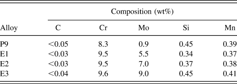
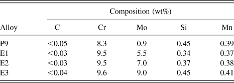
Four alloys (P9, E1, E2, and E3) were analyzed by XRD, TMS, and VSM measurements. The P9 alloy was analyzed in the as-received condition (not solution treated) to maintain their original characteristics and to simulate the typical use of P9 in the petrochemical industry. It serves as a comparison study with the E1, E2, and E3 alloys. E1, E2, and E3 alloys were cast and hot-forged with approximately the same Cr content (9 wt%) and different Mo contents (5, 7, and 9 wt%, respectively). Table I shows the chemical composition of the alloys obtained by optical emission spectroscopy.
Composition of alloys P9, E1, E2, and E3, in wt% obtained by optical emission spectroscopy.
The alloys were solution treated at 1100 °C for 20 min to dissolve any minority phases (except for the P9 alloy). Some of the solution-treated samples were subjected to thermal aging for 10, 100, 1000 and 10,000 min at 650 °C to induce the formation or precipitates. Samples were cut and ground (using SiC paper up to grit 1200) into 100 μm thin foils for XRD and TMS measurements and into 3 × 3 mm (height × diameter) cylinders for VSM measurements.
RT XRD measurements were carried out in a X’Pert Pro MPD diffractometer from PANalytical B.V. (Eindhoven, The Netherlands) using a Co tube (Kα Co = 1.7889 Å) with 2θ ranging from 40° to 120°. TMS measurements were performed at RT using a constant acceleration Mössbauer spectrometer with a 57Co-Rh source. All the isomer shifts were measured with respect to the centroid of the α-Fe spectrum at RT. The spectra were then analyzed using fitting routines based on a least square algorithm. VSM measurements were performed at RT in a Lakeshore 7404 model VSM with a sensitivity of 0.4 μemu and 2.17 T of maximum magnetic field.
III. RESULTS AND DISCUSSION
A. X-ray diffraction
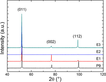
XRD patterns of the solution-treated samples are shown in Fig. 1 and indicate that the crystalline structure is BCC, similar to that of α-Fe. The most intense peak is at approximately 2θ = 52° and refers to the family of planes {011}. The variation in the relative intensities of the other peaks is due to texture effects.
XRD patterns of solution-treated E1–E3 and as-received P9 alloys.

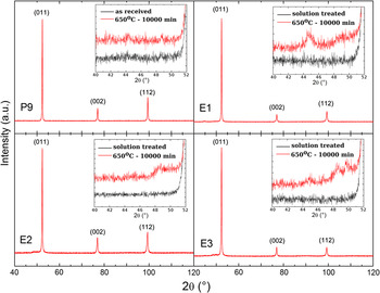
Figure 2 shows XRD patterns of samples aged at 650 °C for 10,000 min. The insets show a detailed view (on the region between 40° and 52°) of the diffractograms of solution-treated and aged samples. It can been seen that the aged samples show minor peaks in their patterns which are due to the presence of precipitates identified in Ref. Reference Gomes da Silva, Herculano, Urcezino, Araújo, de Abreu and de Lima Neto18. There are no such peaks in the solution-treated XRD patterns. It was not possible to identify the crystal structure of the precipitates due to the low intensity of the diffraction peaks associated with the precipitates.
XRD patterns of alloys aged at 650 °C for 10,000 min. The insets show a comparison between XRD patterns of aged and solution-treated alloys in the range from 40° to 52°.

B. Transmission Mössbauer spectroscopy
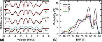
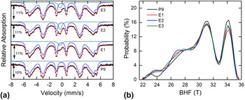
Mössbauer spectra and respective hyperfine magnetic field (BHF) distributions of solution-treated samples are shown in Fig. 3. Spectra consist of six-line patterns consistent with ferromagnetic iron sites typical of the BCC structure of α-iron. Isomer shifts (δ) and quadrupole splitting (∆) were found to be the same as for α-Fe while BHF distributions ranged from 18 to 36 T. BHF values on pure iron sites (sites without alloying elements in the immediate surroundings) are amplified in relation to the expected α-Fe field value of 33 T. This amplification is due to effects associated to more distant coordination spheres and is related to the variation of iron magnetic moment with the concentration of alloying elements. Reference Marcus and Schwartz19
(a) Mössbauer spectra and (b) hyperfine magnetic field distributions of solution-treated E1–E3 and as-received P9 alloys.
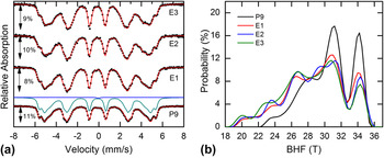
The spectrum of the P9 alloy consists of both a sextet (six-lined ferromagnetic subspectrum) and a central doublet (two-lined paramagnetic subspectrum), while the spectra of other alloys consist of the ferromagnetic sextet only. Peaks at about −5.9 mm/s and +5.2 mm/s refer to lines 1 and 6 of a sextet associated with the iron sites with no alloying element (Cr and/or Mo) in its environment, and present the higher BHF value of all possible iron sites. Note that these peaks are sharper and more intense in the P9 alloy spectrum, which indicates that sites of this type are more abundant in this alloy than in the other ones. This is due to the lowest Mo concentration in the P9 alloy. In terms of a Mössbauer spectrum, these alloying element atoms are comparable to impurities that work to decrease the hyperfine magnetic field on a given iron site in their neighborhood. In the context of this work, the terms alloying elements and impurities will be interchangeable. Moreover, the small (relative area of less than 1%) paramagnetic subspectrum found in the P9 spectrum is associated to the presence of precipitates, since this sample was analyzed in the as-received condition unlike the others which were solution treated.
Figure 3(b) shows the respective BHF distributions of each Mössbauer spectrum shown in Fig. 3(a). Hyperfine magnetic field distributions are highly sensitive to the distribution of alloying elements throughout the matrix. The rightmost peaks (centered at about 34 T) are attributed to sites with no alloying elements in the near neighborhood of a given iron site. These peaks correspond to the outermost sextet mentioned in the paragraph above. The peaks located to the left are attributed to sites with one or more impurity atoms in the same neighborhood. For the P9 alloy, the BHF values are mostly distributed over two reasonably well-defined values with the higher probabilities associated with sites with no impurity and with one or more impurity atoms. As the Mo content increases, the intensities of the two outermost peaks decrease and the probabilities redistribute among other BHF values, causing an increase in the probabilities of the sites with medium and lower BHF. This result indicates an increasing number of sites containing more diverse positional configurations of the alloying element atoms in the E1, E2, and E3 alloys than those in the P9 alloy. Further increase in Cr-equivalent content leads to a random distribution of alloying elements within the BCC matrix as was shown in Ref. Reference Vasconcelos, Tavares, Reis and Abreu20 where Mössbauer results on Fe–Cr–Mo alloys with Cr-equivalent of 21.5% and higher yielded symmetrical Gaussian-like BHF distributions about 28–30 T.
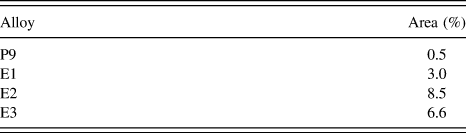
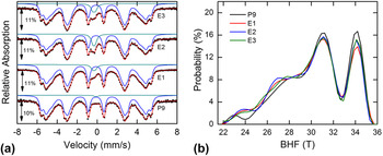
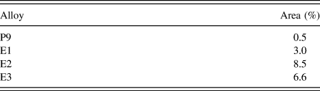
Figure 4 shows the Mössbauer spectra and the respective BHF distributions of the samples aged at 650 °C for 10,000 min. The presence of precipitates in all alloys is evidenced by the Mössbauer subspectra in the form of doublets. These doublets indicate the presence of Fe-bearing paramagnetic phases formed during the aging process. It can be seen in Table II that the doublet’s relative area increases from sample P9 to E2, which indicates that the volume of precipitates increases with Mo content. This pattern is not followed by the E3 sample, in which a slight decrease in the doublet’s relative area is observed. This is probably due to the relative increase of alloying elements (Cr and/or Mo) in the composition of the precipitates, since the intensity of Mössbauer spectra is related only to the iron content in a compound. The areas of the doublets found on the unaged and aged P9 alloy spectra are very similar indicating that the aging process does not induce the formation of a substantial amount of precipitates in the P9 alloy.
(a) Mössbauer spectra and (b) hyperfine magnetic field distributions of alloys aged at 650 °C for 10,000 min.
Relative areas of paramagnetic doublets on Mössbauer spectra of alloys aged at 650 °C for 10,000 min.
The formation of precipitates also affects BHF distributions as can be seen in Fig. 4(b). Precipitation of other phases in the E1–E3 alloys removes alloying elements from the BCC matrix changing the respective BHF distribution. In fact, BHF distributions obtained from the aged E1, E2, and E3 alloys become very similar to the one obtained from either the aged or unaged P9 alloy. This finding suggests that the excess of Mo in the E1–E3 alloys as compared to the commercial P9 alloy moves to the precipitates formed under aging.
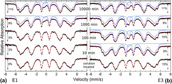
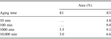
To study the kinetics of precipitates formation, Mössbauer spectra of E1 and E3 alloys (lower and higher Mo content) aged at 650 °C for various times were collected and are shown in Fig. 5. Paramagnetic contributions appear on E1 spectra [Fig. 5(a)] after aging for 1000 min and on E3 spectra [Fig. 5(b)] as soon as 10 min. Table III shows the doublets’ relative areas in these spectra. Again, the increase in doublet area is related to the relative fraction of iron in the precipitates. The slight decrease observed in the samples aged for 10,000 min is probably due to the increase of Mo and/or Cr content in the precipitates along with a decrease in the Fe content.
Mössbauer spectra of (a) E1 and (b) E3 alloys aged at 650 °C for various times.

Relative areas of paramagnetic doublets on Mössbauer spectra of alloys E1 and E3 aged at 650 °C for various times.
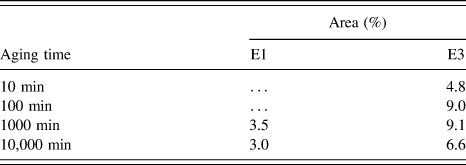
These results confirm that the presence of Cr and Mo leads to the formation of precipitates and that the volume of precipitate is a function of Mo content in the alloy. They also suggest that the concentration of Mo also determines the kinetics of precipitation. The relative area of the paramagnetic doublet is at the most 9% of the total spectrum (see Tables II and III), which hinders the ability of the fitting procedure to determine the doublets’ hyperfine parameters with a certainty level required to identify the types of precipitates present in each sample. Nevertheless, as was discussed in the introduction, all possible precipitates (M23C6 and M6C carbides and μ and χ intermetallic phases) are paramagnetic, which is consistent with the doublets found.
C. Vibrating sample magnetometry
As mentioned before, it was not possible to identify the types of precipitates by XRD and TMS measurements due to their small relative fraction. However, the RT magnetic response of all possible precipitates is nonferromagnetic as shown in the literature and confirmed by TMS. Therefore, irregardless of type and composition of the mix of precipitates, their magnetic behavior contrasts the strong ferromagnetic nature of the BCC matrix, which serves the purpose of this work.
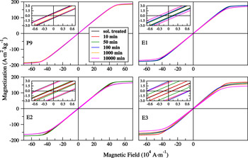
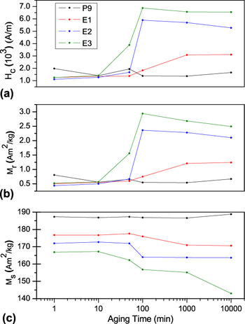
Figure 6 shows magnetic hysteresis cycles of the alloys as a function of Mo composition and aging time. Magnetic properties were extracted from these data. Figure 7 shows values of coercive field, remanent magnetization and saturation magnetization of the alloys aged at 650 °C as a function of aging time. Results shown for 1 min are those of as-received P9 and as-solution-treated E1, E2, and E3 alloys.
Magnetic hysteresis cycles of alloys as a function of Mo composition and aging time.

(a) Coercive force, (b) remanent magnetization, and (c) saturation magnetization as function of aging time for samples aged at 650 °C.

Coercive field and remanent magnetization of all alloys [Figs. 7(a) and 7(b), respectively] show very similar general behavior. Magnetic data of the P9 alloy show a slight decrease in coercive field and remanent magnetization between 1 min and 10 min. There is no significant change in coercive field and remanent magnetization from 10 min to 10,000 indicating that the aging process does not induce the formation of a substantial amount of precipitates in the P9 alloy.
As opposed to the P9 case, magnetic data of the E1–E3 alloys show a considerable increase of coercive field and remanent magnetization as function of aging time which can be associated with the formation of precipitates. This increase is more noticeable at 1000, 100, and 50 min for E1, E2, and E3 alloys, respectively, which suggests that the onset of precipitation is a function of Mo content. Moreover, for the same aging temperature, coercive field, and remanent magnetization are consistently more intense as the Mo content increases. These results are in agreement with the TME results that show (Fig. 5 and Table III) an increasingly larger relative doublet area (associated with the presence of precipitates) as Mo content increases.
The data then suggest that the presence of molybdenum favors the formation of precipitates which contribute directly to the increase of the magnetic hardness (measured by coercive field) through the process of pinning the magnetic domain walls. In addition, because the pinning of domain walls affects their movement during the demagnetization process, the remanent magnetization follows a similar behavior. Reference Jiles and Atherton21–Reference Bertotti23
Figure 7(c) shows saturation magnetization as a function of aging time at 650 °C. It is clear from the figure that the value of saturation, for each aging time, is an inverse function of the Mo content in the alloys. In fact, the presence of a certain amount of Mo tends to cause a proportional decrease in net magnetic moment of the material. The hyperfine magnetic field distributions shown in Fig. 3 reinforce this result.
Values of saturation magnetization of E1–E3 alloys experience a consistent decrease as a function of aging time, in a similar, but inverted, fashion observed for coercive field and remanent magnetization. As shown in previous sections, aging at 650 °C induces the formation of precipitates (doublets in the Mössbauer spectra) which produce two competing effects. The first effect is to remove alloying elements from the BCC matrix making it more ferromagnetic (with consequent increase in saturation magnetization) as can be seen comparing BHF distributions of Figs. 3 and 4. The other effect is the appearance of demagnetizing fields created by paramagnetic precipitates that contribute to decreasing the saturation values in the proportion of the relative amount of these phases. Because saturation values decrease as the relative amount of precipitates increases, it is clear that latter effect is stronger the former.
IV. CONCLUSIONS
This work has shown that the techniques of VSM and TMS can be useful to study the influence of precipitates in the magnetic properties of Fe–Cr–Mo alloys. The results show that the increase in the Mo content and aging time contributes to the formation of precipitates in the matrix.
Mössbauer spectra of solution-treated samples fitted by BHF distribution presented only sextets (associated to ferromagnetic phases) while spectra of the 650 °C aged samples showed doublets as well. The doublets are associated with the presence of paramagnetic phases while their relative areas are related to the relative Fe content in the precipitates. BHF distributions are strongly affected by the concentration of alloying elements (impurities) and the presence of precipitates. The intensity of the peaks associated with sites with none or one impurity atom in the near neighborhood decrease with the increase in the Mo content causing the distributions to be more homogeneous in the aged samples than in the solution-treated ones.
It was not possible to identify the types of precipitates by XRD and Mössbauer spectroscopy measurements due to their small relative fraction. However, all possible precipitates present nonferromagnetic response and thus have a similar effect in the alloys magnetic properties. The increase in coercive field and remanent magnetization confirms the formation of paramagnetic precipitates in the ferritic matrix. As the precipitates are paramagnetic, they act as pinning centers for the motion of magnetic domain walls. The saturation was reduced with the formation of precipitates as these phases create demagnetizing fields that decrease the signal during the magnetization process. Although the precipitation of paramagnetic phases yielded more strongly ferromagnetic BCC matrices, the effect of demagnetizing field created by the precipitated was stronger which resulted in a net decrease on saturation.
The combinations of XRD, Mössbauer spectroscopy, and VSM techniques used in this work were shown to be very useful to understand the mechanisms through which the formation of precipitates affects the magnetic properties of the alloys and may be used as complement to the usual microscopy-based techniques for this purpose.
ACKNOWLEDGMENTS
The authors would like to acknowledge the Brazilian research agencies FUNCAP, CNPq, and CAPES for financial support. The authors are thankful to Prof. M. Beres and Prof. M.J. Gomes da Silva for the insightful discussions and suggestions.