Introduction
The nematode family Philometridae consists of three subfamilies, Alineminae, Neophilometroidinae and Philometrinae; the latter contains nine genera, Buckleyella Rasheed 1963, Clavinema Yamaguti, 1935, Dentiphilometra Moravec et Wang, 2002, Nilonema Khalil, 1960, Paraphilometroides Moravec et Shaharom-Harrison, 1989, Philometra Costa, 1845, Philometroides, Yamaguti, 1935, Rumai Travassos, 1960 and Spirophilometra Parukhin, 1971 (Moravec, Reference Moravec2006). Philometrid nematodes are known to infect various body organs and cavities of the fish host, such as body muscles, gonads, abdominal cavity, buccal cavities, eyes, subcutaneous tissues and other body tissues. Nematodes of this group are known to have a tremendous impact on fisheries industries, affecting fish reproduction, marketability and aquaculture potential of infected host fishes (Moravec, Reference Moravec2004, Reference Moravec2006; Quiazon et al., Reference Quiazon, Yoshinaga and Ogawa2008a, Reference Quiazon, Yoshinaga and Ogawab, Reference Quiazon, Yoshinaga and Ogawa2010; Portes Santos & Moravec, Reference Portes Santos and Moravec2009). In spite of their economic importance, very few taxonomic studies have been carried out on this parasite group, especially in so far little-explored countries such as the Philippine Archipelago. Currently, 142 philometrid species are known to infect the body cavities and various body tissues of fish worldwide, including 29 species infecting body musculature and subcutaneous tissues (Moravec, Reference Moravec2006; González-Solís et al., Reference González-Solís, Moravec and Tuz Paredes2007; Moravec et al., Reference Moravec, Santana-Piñeros, González-Solís and Torres-Huerta2007, Reference Moravec, Crosby, de Buron, González-Solís and Roumillat2008a, Reference Moravec, Montoya-Mendoza and Salgado-Maldonadob, Reference Moravec, Charo-Karisa and Jirku˙2009, Reference Moravec, Bakenhaster and Fajer-Avila2010; Quiazon et al., Reference Quiazon, Yoshinaga and Ogawa2008a, Reference Quiazon, Yoshinaga and Ogawab, Reference Quiazon, Yoshinaga and Ogawa2010; Moravec & Bakenhaster, Reference Moravec and Bakenhaster2010).
In the municipality of Casiguran, located in the Aurora Province, the Philippine Archipelago, heavy infections of unknown nematodes, seen in the body cavity, subcutaneous tissue near the bases of the pectoral and pelvic fins and surrounding body tissues in the hypaxial musculature of the loach goby, Rhyacichthys aspro (Valenciennes), have been reported by local communities. The fish is known to be amphidromous as it inhabits both freshwater and saltwater environments at different life stages. The fish presumed to be infected were caught by local fishermen in a freshwater environment. There have been no previous attempts to examine the nematode infection in this fish. As a result, preliminary examinations of fish revealed a very high prevalence (100%) and intensity (1–37 worms) of the nematode infection. The limited knowledge on the diversity of nematode parasites in fishes and the concern of local communities about the possibility of human infection prompted the authors to study closely the taxonomy of these nematodes in view of food safety. A detailed study of their morphology shows that these nematodes represent a new genus and species, as described below.
Materials and methods
Several specimens of non-gravid, subgravid and gravid female nematodes have been collected from the body cavity, subcutaneous tissues near the bases of pectoral and pelvic fins, and surrounding body tissues in the hypaxial body musculature of R. aspro. The fish samples were caught from the freshwater environment in the upper Bianuan River, at an elevation of 71 m above sea level, in the municipality of Casiguran, Aurora Province, Philippines (16° 13.778'N; 122° 0.967'E) in April 2011. The collected nematodes were washed in a physiological saline solution, followed by fixing in 70% ethanol. For morphological examination, both light microscopy (LM) and scanning electron microscopy (SEM) were used. For light microscopic examination, the 70% ethanol-fixed samples were cleared in glycerine, followed by mounting of the whole individual nematodes on glass slides. Furthermore, some morphological features difficult to study by LM were examined using SEM. Briefly, the 70% ethanol-fixed specimens were transferred to 4% formaldehyde solution and then post-fixed in 1% osmium tetroxide (in phosphate buffer), dehydrated through a graded acetone series, critical point-dried and sputter-coated with gold. Finally, the specimens were observed under a scanning electron microscope (JEOL JSM-7401F) at an accelerating voltage of 4 kV (gentle beam, low mode). Drawings of the nematodes were made with the aid of a Zeiss microscope drawing attachment. Type specimens of the new nematode species were deposited at the Meguro Parasitological Museum, Tokyo (MPM collection no. 20 789) and at the Helminthological Collection of the Institute of Parasitology, Biology Centre of the ASCR, in České Budĕjovice (collection number N-978). All measurements are in micrometres unless otherwise stated. The fish name follows FishBase (Froese & Pauly, Reference Froese and Pauly2011).
Dentirumai n. gen
Diagnosis: Dracunculoidea, Philometridae, Philometrinae. Body of female elongate, with somewhat narrowed and blunt anterior end, conical and sharply pointed posterior end. Cuticle smooth. Cephalic end with apical outgrowths. Cephalic papillae small, arranged in two circles. Oral aperture circular, with sclerotized oral ring armed with numerous minute circumoral teeth. Oesophagus cylindrical at its anterior part, feebly swollen near mouth, and markedly expanded at posterior part. Small ventriculus present. Vagina and vulva absent. Male unknown. Parasites of body cavity and tissues of perciform fishes.
Taxonomic summary
Type and the only species. Dentirumai philippinensis n. sp.
Etymology. The generic name comprised of two words: denti ( = having teeth) and rumai, which relates to the name of the closely related genus Rumai Travassos, 1960
Dentirumai philippinensis n. sp
Description. Male: unknown.
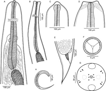
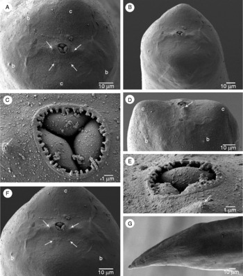
Female (17 gravid specimens; measurements of 25 subgravid specimens in parentheses) (figs 1 and 2). Body of fixed non-gravid and subgravid specimens whitish; fully gravid specimens light yellowish, with smooth cuticle; anterior end somewhat narrowed, blunt, posterior end conical and sharply pointed. Body length 24.38–75.25 (11.87–44.20) mm, maximum width at oesophageal region 250–816 (170–380); maximum width/length ratio 1:69–259 (1:59–170). Cephalic end of body 105–180 (70–126) wide, truncated in lateral view, rounded in dorsoventral view, bearing two conspicuously large (one dorsal and one ventral), rounded anterior projections 10–25 (5–15) high. Oral aperture circular, surrounded by narrow sclerotized ring armed with distinct numerous (33) minute teeth on its inner surface. Bottom of mouth formed by surfaces of three lobular sectors of oesophagus. Mouth surrounded by 14 small cephalic papillae arranged in two circles and a pair of slightly outlined lateral amphids; eight papillae of external circle form two dorsolateral and two ventrolateral pairs, each pair consisting of one circular and one elongate-oval papilla in apical view; internal pair consists of four small, submedian single papillae and two small, lateral papillae situated near oral aperture. Tissue of anterior cephalic projections appears to be somewhat sclerotized, hyaline. Oesophagus relatively short, 600–925 (500–694) long (representing 1–3 (2–5)% of body length) and 55–109 (38–68) at maximum width, with feebly developed anterior inflation 36–54 (18–60) long and 38–45 (20–50) wide; it consists of shorter and narrower anterior muscular portion and much longer and wider posterior muscular-glandular portion. Nerve ring encircles muscular oesophagus 200–300 (150–290) from anterior extremity. Small ventriculus 27–52 (12–55) long and 50–70 (22–58) wide present. Oesophagus opens into intestine through valve. Intestine light-coloured, relatively narrow anteriorly; posterior end of intestine atrophied, forming narrow ligament 45–80 (30–150) long attached to body wall short distance anterior to posterior extremity. Posterior end of body conical. Ovaries narrow, long, reflexed, situated near anterior and posterior ends of body. Uterus amphidelphic, in gravid specimens occupying major part of body, being filled with first-stage larvae, developing embryos and eggs. Vagina and vulva absent. Anterior end of larvae blunt, posterior end sharply pointed; larvae 218–279 long, with maximum width 12–18.
Dentirumai philippinensis n. gen., n. sp., female. (A) Anterior end of gravid specimen, lateral view; (B) anterior end of smallest subgravid specimen, dorsoventral view; (C, D) cephalic end of gravid specimen, lateral and dorsoventral views; (E) posterior end of gravid specimen, lateral view; (F) oral aperture armed with teeth, apical view; (G) cephalic end of gravid specimen, apical view; (H) larva from uterus, lateral view.

Dentirumai philippinensis n. gen., n. sp., SEM micrographs of gravid female. (A) Region of oral aperture and cephalic papillae, apical view (arrows indicate papillae of internal circle); (B) cephalic end, sublateral view; (C) oral aperture, apical view (arrow indicates lateral papilla of internal circle); (D) cephalic end, dorsoventral view (arrow indicates lateral papilla of internal circle); (E) oral aperture, sublateral view (arrow indicates lateral papilla of internal circle); (F) cephalic end, sublateral view (larger magnification) (arrows indicate papillae of internal circle); (G) caudal end, lateral view. Abbreviations: b, submedian pair of papillae of external circle; c, cephalic projection.

Taxonomic summary
Type host. Loach goby, Rhyacichthys aspro (Rhyacichthyidae, Perciformes; total body length 90–160 mm; standard length 80–135 mm; body weight 20–50 g); local name – Pilingan.
Sites of infection. Body cavity, subcutaneous tissues near the bases of pectoral and pelvic fins, and surrounding body tissues in the hypaxial musculature.
Type locality. Bianuan River, Municipality of Casiguran, Aurora Province, Philippines (16° 13.778′N, 122° 0.967′E; 71 m above sea level; collected in April 2011).
Prevalence and intensity. 100% (55 fish examined); 1–37 (mean 6) worms per fish.
Deposition of types. Female holotype and paratypes deposited in the Meguro Parasitological Museum (MPM collection number 20 789); female paratypes deposited also in the Helminthological Collection of the Institute of Parasitology, Biology Centre of the ASCR, České Budĕjovice (collection number N-978).
Etymology. The specific name of this worm relates to the type locality where the type host was collected, i.e. Philippines.
Diagnosis. Within the family Philometridae, only subfamilies Philometrinae and Neophilometroidinae are reported to include genera characterized by the presence of peribuccal teeth in gravid females (i.e. Dentiphilometra Moravec et Wang, 2002 for the former and Neophilometroides Moravec, Salgado-Maldonado et Aguilar-Aguilar, 2002 for the latter subfamily) (Moravec & Wang, Reference Moravec and Wang2002; Moravec et al., Reference Moravec, Salgado-Maldonado and Aguilar-Aguilar2002). Dentirumai also possesses peribuccal teeth. The presence of a buccal capsule and embossed cuticle separates Neophilometroidinae from Philometrinae. In contrast to Dentiphilometra of the subfamily Philometrinae, the general morphology of the type species, D. philippinensis, is very similar to that of the only species of Rumai, R. rumai Travassos, 1960; therefore, the new genus Dentirumai is assigned to the subfamily Philometrinae.
Discussion
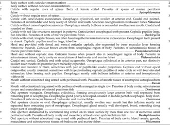
According to the key to dracunculoid nematodes given by Moravec (Reference Moravec2006), the present nematodes belong to the subfamily Philometrinae, family Philometridae. The philometrid nematodes collected from R. aspro are morphologically similar to the genus Rumai. Comparison with the only known species of Rumai, R. rumai, revealed similar morphological features, such as: (a) body surface without cuticular ornamentations; (b) caudal end conical; (c) cephalic end with apical outgrowths; (d) oesophagus cylindrical at its anterior part which is feebly swollen at the anterior end near the mouth; and (e) posterior part of the oesophagus markedly expanded (Moravec, Reference Moravec2006; Portes Santos & Moravec, Reference Portes Santos and Moravec2009). However, some morphological differences were found between the current specimens and that of R. rumai, including the colour of live specimens and shape of the posterior end. Live gravid R. rumai females have a red-coloured body (yellowish after fixation) while non-gravid females are white. In contrast, the current specimens of non-gravid and subgravid females from R. aspro have a whitish cream body colour, whereas fully gravid females have light yellowish body colouration. In addition, R. rumai has a conical posterior end compared to the conical and sharply pointed posterior end in the current philometrid specimens. Based on these differences, the present philometrids could represent a separate species of Rumai. However, they differ considerably from Rumai in possessing a sclerotized ring in the oral aperture armed with numerous minute teeth on its inner surface. Currently, in the subfamily Philometrinae, only the genus Dentiphilometra has been reported to have an oral aperture demarcated by a sclerotized ring armed on its inner surface by numerous small peribuccal teeth. A comparison of the present philometrid specimens with the type species of the genus Dentiphilometra, D. monopteri Moravec et Wang, 2002, showed entirely different morphological features in the latter, which include the following: (a) caudal end almost rounded or blunt in lateral view bearing two conspicuous lateral, papilla-like projections that are sometimes unequal in size; (b) cephalic end without apical outgrowths; (c) oesophagus with bulbous inflation at the anterior end; and (d) minute teeth on the inner surface of the oral aperture arranged in more than one row. Since the present nematodes cannot be assigned to any of the existing philometrid genera, a new genus Dentirumai is proposed to accommodate them. A modified key to genera of the Philometrinae is provided below (table 1).
Key to genera of Philometrinae (updated from Moravec, Reference Moravec2006)

Dentirumai philippinensis was collected during the dry month of April 2011 in R. aspro from the Bianuan River. Its biology, particularly the life history, transmission and seasonal maturation cycle remain unknown. Because the fish host inhabits both freshwater and saltwater environments, it is also unclear where the fish acquired the infection. To date, this is the first record of a philometrid in fish belonging to the perciform family Rhyacichthyidae. Of many described nominal species of philometrids, only Philometra robusta Moravec, Möller et Heeger, 1992, a parasite of the map puffer, Arothron mappa (Lesson), has so far been recorded from the Philippines (Moravec et al., Reference Moravec, Möller and Heeger1992). Schmidt & Kuntz (Reference Schmidt and Kuntz1969) also reported Philometra lateolabracis Yamaguti, 1935 from fishes of Palawan (see also Arthur & Lumanlan-Mayo, Reference Arthur and Lumanlan-Mayo1997), but with respect to the paper of Quiazon et al. (Reference Quiazon, Yoshinaga and Ogawa2008a), this identification was evidently wrong and, therefore, these nematodes should be designated as Philometra sp. Consequently, D. philippinensis is the second nominal philometrid species reported from the Philippine Archipelago. Hence, discovery of many other philometrids can be expected in this region.
Portes Santos & Moravec (Reference Portes Santos and Moravec2009) commented on the relationship between tissue-dwelling philometrid nematodes (R. rumai) and Nilonema senticosum (Baylis, 1927) and the high mortality of young Arapaima gigas (Schinz) in the conditions of an aquaculture system in the Mexiana Island, Brazil. There have been several other reports showing effects of philometrid nematodes infecting host fishes (Moravec & Genc, Reference Moravec and Genc2004; Clarke et al., Reference Clarke, Dove and Conover2006; Quiazon et al., Reference Quiazon, Yoshinaga and Ogawa2008a, b). Given that R. aspro is a source of protein for the local communities, the presence of D. philippinensis in the host tissues could result in a reduction of fish marketability and acceptance. The pathogenicity and biology of this nematode are still unclear. It would be desirable to know where the infection commenced and if there are any pathological effects of these nematodes on their fish hosts. With the trend in molecular taxonomy, it would also be interesting to contribute the molecular taxonomy of this group of nematodes to supplement the conventional taxonomy, which could help in the reclassification of this nematode group, as suggested by Moravec (Reference Moravec2006) and Quiazon et al. (Reference Quiazon, Yoshinaga and Ogawa2008b).
Acknowledgements
Thanks are greatly extended to the Commission on Higher Education, Zonal Research Center, Region 3, Program Cluster Implementer, Philippines and CLSU CHED ZRC PCI for funding and supporting the research. Thanks are also due to the staff of the Laboratory of Electron Microscopy of the Institute of Parasitology, BC ASCR, in České Budĕjovice for their technical assistance and to Blanka Škoríková from the same institute for help with illustrations. The main author would also like to thank his colleagues (Gemerlyn G. Garcia, Geraldine C. Sanchez, Rowena R. Sazon, Glenda D. Abad and Mark Joseph R. Rafael) and to his student (Mc Anthony Estrada) for their assistance and help. This study was partly supported by the research projects of the Institute of Parasitology, ASCR (Z60220518 and LC522).