Introduction
Comamonas testosteroni (formerly Pseudomonas testosteroni) is a Gram-negative bacterium. It has the ability to utilize various steroids (as well as many other aromatic compounds) as sole carbon and energy source through a complex metabolic pathway involving many steps catalysed by steroid-inducible enzymes (Leppik, Reference Leppik1983). 3α-Hydroxysteroid dehydrogenase/carbonyl reductase (3α-HSD/CR), a steroid-inducible enzyme, plays an important role in steroid degradation and has served as an indicator for enhanced steroid metabolism (Xiong et al., Reference Xiong, Martin and Maser2003a, Reference Xiong, Martin and Maserb). Three genes, repressor A, repressor B and activator involved in hsdA gene (3α-HSD/CR encoding gene) regulation, have been isolated (Xiong et al., Reference Xiong, Martin and Maser2003a, Reference Xiong, Martin and Maserb). Characterization of the activator gene was reported previously by Dai et al. (Reference Dai, Zhou, Xiong, Wu and Pan2005, Reference Dai, Pan, Xiong, Wu and Zhou2006). It was estimated that the complete degradation of the steroids into CO2 and H2O requires more than 20 enzyme reactions. However, enzymes involved in steroid metabolism are still a mystery, so far. Two steroid degradation gene clusters were identified in C. testosteroni TA441 (Horinouchi et al., Reference Horinouchi, Hayashi, Yamamoto and Kudo2003, Reference Horinouchi, Kurita, Yamamoto, Hatori, Hayashi and Kudo2004a, Reference Horinouchi, Hayashi and Kudob, Reference Horinouchi, Hayashi, Koshino, Kurita and Kudo2005, Reference Horinouchi, Hayashi, Koshino and Kudo2006). Recently, a LuxR-type testosterone-inducible regulator (teiR) was found by transposon mutagenesis and its domain distribution [N-terminal autoinducer binding domain and C-terminal helix-turn-helix (HTH) DNA binding domain] resembled TraR, a specific transcriptional regulator in Agrobacterium tumefaciens (Pruneda-Paz et al., Reference Pruneda-Paz, Linares, Cabrera and Raimondi2004). Northern blot analysis revealed that the teiR gene was required for full expression of Δ1-dehydrogenase and 3α-dehydrogenase. Moreover, when teiR gene was provided to the teiR-disrupted strain in trans, the transcription levels of these genes were restored (Horinouchi et al., Reference Horinouchi, Kurita, Yamamoto, Hatori, Hayashi and Kudo2004a; Pruneda-Paz et al., Reference Pruneda-Paz, Linares, Cabrera and Raimondi2004).
In the present work, the teiR gene was cloned from C. testosteroni, and ligated into pK18 and pKtac2 plasmids. Co-transformation with the hsdA gene into Escherichia coli was investigated to investigate the relationship between the teiR and hsdA genes.
Materials and methods
Bacterial strains and plasmids
Host strains E. coli HB101, C. testosteroni ATCC11996, plasmids pK18 (kanamycin resistance) and p6 (ampicillin resistance, a 5.257 kb EcoRI fragment was cloned into pUC18, including hsdA, repressor A and repressor B genes) were used for cloning and gene expression.
Growth and growth conditions
E. coli was grown at 37°C in Luria–Bertani (LB) medium. C. testosteroni ATCC11996 was grown at 30°C in LB medium with or without 60 μg/ml ampicillin (Ap) and 30 μg/ml kanamycin (Km).
Restriction enzymes and other reagents
Restriction enzymes, T4 ligase, shrimp alkaline phosphatase and PCR Fragment Recovery Kit were purchased from TaKaRa (Dalian, Liaoning, China). Biowest agarose and MediBEST Plasmid Purification Kit were obtained from Qiagen (Shanghai, China). Ampicillin and kanamycin were from AMERSOCO (Solon, Ohio, USA).
DNA manipulations and teiR gene cloning
The preparation of recombinant DNA was carried out according to Sambrook and Russell (Reference Sambrook and Russell2002). To generate pKteiR100, forward (5′-GCTCTAGAATGTGCCCATATTTCGACACC-3′) and reverse (5′-CCCAAGCTTTCACTTGTTCCCCAG-3′) primers were used in a polymerase chain reaction (PCR) with C. testosteroni chromosomal DNA as the template. The resulting fragment was digested with XbaI and HindIII, then introduced into pK18 downstream from the lacZ promoter. The plasmid pKtac2-teiR was generated by producing a PCR fragment with forward (5′-GCTCTAGAATGTGCCCATATTTCGACACC-3′) and reverse (5′- TCGGGGTACCTCACTTGTTCCCCAGCCA-3′) primers with C. testosteroni chromosomal DNA template. The amplified fragment was digested with XbaI and KpnI and inserted in pKtac2 downstream from the tac promoter. The PCR procedure was: 5 min at 94°C, followed by 30 cycles of 94°C for 45 s, 60°C for 45 s and 72°C for 80 s, plus a final extension step at 72°C for 10 min in a Bio-Rad® thermal cycler (Beijing, China).
Transformation of bacteria
All constructs were verified by restriction enzyme analysis of the resulting band patterns or sequence confirmation. Ligated constructs were transferred into competent E. coli cells. Double co-transformations were performed by exploiting the kanamycin resistance gene of pK18 and the ampicillin resistance gene of pUC18. In this case, both antibiotics were added to the culture medium. Plasmid isolation and agarose gel electrophoresis were performed to prove successful double transformations.
Protein extraction
After overnight culture, a 3 ml sample of bacteria was centrifuged at 13 000 rpm for 30 s and the pellet suspended in 300 μl phosphate-buffered saline with 3 μl lysozyme (5 mg/ml). The suspension was frozen and thawed 3 times (−20°C and room temperature, 30 min each), then incubated for 15 min at 37°C after adding 2 μl DNase I (5 mg/ml) and 100 μl B buffer containing 100 mmol/l sodium phosphate, 10 mmol/l Tris-Cl, 8 mol/l urea, pH 8.0. Finally, the suspension was centrifuged at 13 000 rpm for 20 min. The protein concentration in the supernatant was determined by the bicinchoninic acid (BCA) method. The extracted protein was diluted to 1 mg/ml and stored at −20°C.
ELISA assay
To quantify the expression of 3α-HSD/CR and TeiR, an ELISA assay was conducted, and respective antibodies against 3α-HSD/CR and TeiR were prepared in rabbits. The ELISA plate was coated with protein extracts and incubated at 37°C for 30 min. After washing, each antibody (1:2000 dilution) was added and incubated for 30 min, before the addition of the secondary antibody (1:20 000 dilution). The further procedure was carried out according to Ausubel et al. (Reference Ausubel, Brent and Kingston1998).
Results
teiR gene amplification
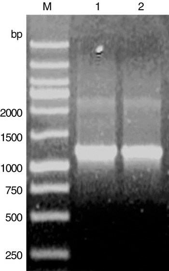
As shown in Fig. 1, the molecular mass of the resulting bands following PCR amplification was identical to the previously reported mass of the teiR gene (Pruneda-Paz et al., Reference Pruneda-Paz, Linares, Cabrera and Raimondi2004), indicating the specificity of the PCR amplification. After sequence confirmation, the resulting bands were purified using the PCR Fragment Recovery Kit (TaKaRa).
PCR amplification of teiR gene from C. testosteroni. Lanes: M, wide range DNA marker (TaKaRa); 1 and 2, the resultant bands.

Plasmids pKteiR100 and pKtac2-teiR construction
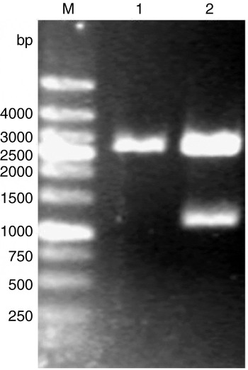
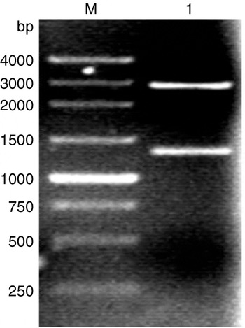
The purified amplified fragments were digested with restriction enzymes and ligated into pK18 and pKtac2. The ligated constructs were transferred into competent E. coli cells. All constructs were verified by restriction enzyme analysis of the resulting band patterns. Results are shown in Figs 2 and 3. As expected, two bands were clearly observed on 1% agarose gels.
pKteiR100 verified by restriction enzyme analysis with XbaI and HindIII. Lanes: M, 250 bp DNA marker (TaKaRa); 1, pK18 digestion with XbaI and HindIII; 2, pKteiR100 digestion with XbaI and HindIII.

pKtac2-teiR verified by restriction enzyme analysis with XbaI and KpnI. Lanes: M, DNA marker D (Sangon); 1, pKtac2-teiR digestion with XbaI and KpnI.

Expression of recombinant plasmids in E. coli
TeiR expression by transformed E. coli
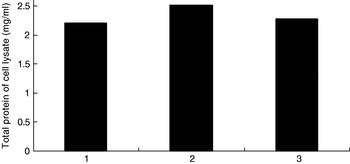
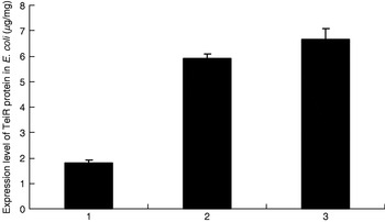
Recombinant plasmids pKteiR100 and pKtac2-teiR were transformed into competent E. coli cells with plasmid pK18 as a control. The host protein was extracted and, after its concentration was determined, was used for tests of TeiR expression level by ELISA. The total protein concentration after pKteiR100 and pKtac2-teiR were transformed into E. coli, were estimated to be up to 2.51 mg/ml and 2.27 mg/ml, respectively, in contrast to pK18-transformed cells (2.20 mg/ml) (Fig. 4). There was no significant difference in TeiR protein expression level between pKteiR100 (6.65 μg/mg) and pKtac2-teiR (5.93 μg/mg) transformed cells (Fig. 5). However, TeiR protein expression levels of E. coli transformed with the two recombinant plasmids were significantly different from that of E. coli transformed with pK18 at the P=0.01 level.
Total protein concentration of E. coli after transformation with recombinant plasmids. 1, pK18; 2, pKteiR100; 3, pKtac2-teiR.

TeiR expression level by E. coli after transformation with recombinant plasmids. 1, pK18; 2, pKteiR100; 3, pKtac2-teiR. Each value is the average of data from three independent experiments (error bars indicate standard deviations).

TeiR and 3α-HSD/CR expression of E. coli co-transformed with recombinant plasmids and plasmid p6
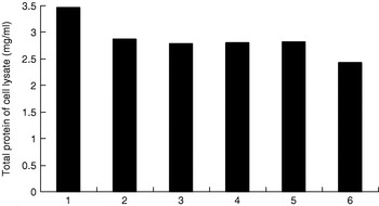
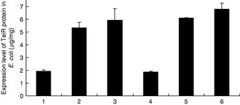
In order to investigate the relationship between TeiR and 3α-HSD/CR, recombinant plasmids pKteiR100, pKtac2-teiR and pK18 were co-transformed into E. coli with plasmid p6. p6 contained the hsdA gene (3α-HSD/CR encoding gene), which served as an indicator of enhanced steroid metabolism and an adaptation to steroids. The host E. coli was cultured overnight at 37°C in LB medium or LB medium plus 1 mmol/l isopropyl-β-d-thiogalactoside (IPTG) induction, and total protein was extracted for gene expression analysis. The total protein concentration values of pK18 and p6, pKteiR100 and p6, and pKtac2-teiR and p6 were 3.46, 2.87 and 2.79 mg/ml, respectively; and 2.80, 2.82 and 2.43 mg/ml, respectively, after induction with 1 mmol/l IPTG (Fig. 6). After co-transformation, the TeiR protein expression levels of E. coli transformed by pKteiR100 and p6 and pKtac2-teiR and p6 could be up to 5.33 μg/mg and 5.94 μg/mg, respectively, and up to 6.10 μg/mg and 6.81 μg/mg after induction with 1 mmol/l IPTG (Fig. 7). Whether induced with 1 mmol/l IPTG or not, no significant difference was found in the TeiR protein expression levels between pKteiR100 and pKtac2-teiR. However, TeiR protein expression levels of E. coli co-transformed with the two recombinant plasmids and p6 were significantly different from that of E. coli transformed with pK18 and p6, at the P=0.01 level.
Total protein concentration of E. coli after co-transformation with recombinant plasmids and p6. 1, pK18 and p6 plasmids; 2, pKteiR100 and p6 plasmids; 3, pKtac2-teiR and p6 plasmids; 4, pK18 and p6 plasmids induced with 1 mmol/l IPTG; 5, pKteiR100 and p6 plasmids induced with 1 mmol/l IPTG; 6, pKtac2-teiR and p6 plasmids induced with 1 mmol/l IPTG.

TeiR expression level of E. coli after co-transformation with recombinant plasmids and p6. 1, pK18 and p6 plasmids; 2, pKteiR100 and p6 plasmids; 3, pKtac2-teiR and p6 plasmids; 4, pK18 and p6 plasmids induced with 1 mmol/l IPTG; 5, pKteiR100 and p6 plasmids induced with 1 mmol/l IPTG; 6, pKtac2-teiR and p6 plasmids induced with 1 mmol/l IPTG. Each value is the average of data from three independent experiments (error bars indicate standard deviations).

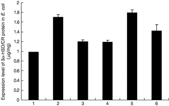
After pKteiR100 and pKtac2-teiR co-transformation into E. coli with plasmid p6, increased levels of 3α-HSD/CR protein expression were observed compared to E. coli co-transformed with pK18 and p6: 1.71, 1.20 and 0.99 μg/mg, respectively (P<0.01) (Fig. 8), and up to 1.80, 1.42 and 1.19 μg/mg, respectively, after induction with 1 mmol/l IPTG (P<0.05). These differences indicated a specific role of TeiR in the regulation of 3α-HSD/CR expression and a mechanism of the teiR gene.
3α-HSD/CR expression level of E. coli after co-transformation with recombinant plasmids and p6. 1, pK18 and p6 plasmids; 2, pKteiR100 and p6 plasmids; 3, pKtac2-teiR and p6 plasmids; 4, pK18 and p6 plasmids induced with 1 mmol/l IPTG; 5, pKteiR100 and p6 plasmids induced with 1 mmol/l IPTG; 6, pKtac2-teiR and p6 plasmids induced with 1 mmol/l IPTG. Each value is the average of data from three independent experiments (error bars indicate standard deviations).

Discussion
Comamonas testosteroni has the ability to utilize various steroids as a sole carbon and energy source through a complex metabolic pathway catalysed by steroid-inducible enzymes. 3α-HSD/CR, a steroid-inducible enzyme, plays an important role in steroid degradation. Interestingly, steroid-inducible enzymes of C. testosteroni are not constitutively expressed, consequently limiting the wide use of this organism in environmental protection. With the development of metabolic engineering, target-oriented improvement of C. testosteroni is achievable.
LuxR-type HTH proteins occur widely in bacteria. The DNA-binding HTH domain is usually located in the C-terminal part. The N-terminal part can contain an autoinducer binding domain or a response regulatory domain. Most luxR-type regulators act as transcription activators, but some can be repressors or have a dual role for different sites. LuxR-type HTH regulators control a wide range of activities in various biological processes (Egland and Greenberg, Reference Egland and Greenberg2001; Ducros et al., Reference Ducros, Lewis and Verma2001; Vannini et al., Reference Vannini, Volpari and Gargioli2002; Pristovsek et al., Reference Pristovsek, Sengupta and Lohr2003). Pruneda-Paz et al. (Reference Pruneda-Paz, Linares, Cabrera and Raimondi2004) reported that testosterone-inducible regulator (teiR) was almost identical to TraR, a specific transcription regulator in Agrobacterium tumefaciens, and was necessary for full expression of Δ1-dehydrogenase and 3α-dehydrogenase. In the present work, the teiR gene was introduced into pK18 and pKtac2 to yield pKteiR100 and pKtac2-teiR. After co-transformation into E. coli, 3α-HSD/CR expression level was increased. It was shown that the teiR gene could act as an activator for hsdA gene expression. Construction of a knock-in mutant of the tac promoter upstream of the teiR gene in C. testosteroni by homologous integration would greatly increase the expression of 3α-HSD/CR and TeiR, and finally result in several-fold greater steroid degradation ability compared to the wild type of C. testosteroni.
The tac promoter, produced by combining the −10 (bases 197–203) region of the lacUV5 and the −35 region (bases 175–180) of the trp promoter, is about 11 times more efficient than the lacUV5 promoter and about 3 times more than the trp promoter. Plasmids into which a foreign gene was ligated downstream from the tac promoter showed an efficient expression level, but down mutation or deletion of the full tac promoter was observed in the host cells, especially in E. coli (Otto et al., Reference Otto, Niagro, Su and Rawlings1995; Dai et al., Reference Dai, Zhou, Xiong, Wu and Pan2005, Reference Dai, Pan, Xiong, Wu and Zhou2006). In this paper, the teiR gene was introduced into plasmid pKtac2 to yield pKtac2-teiR downstream from the tac promoter. However, after sequence confirmation, neither down mutation nor deletion of tac promoter was found in E. coli (data not shown). Our results indicate that the tac promoter was more efficient than the lacZ promoter in E. coli (Figs 5 and 7). But after co-transformation into E. coli, the high expression of TeiR protein did not mean the high expression level of 3α-HSD/CR protein. The expression of 3α-HSD/CR protein after pKtac2-teiR co-transformation into E. coli with p6, was lower than that of pKteiR100 co-transformation. This could possibly be due to an inhibitory effect of TeiR protein on the growth of E. coli. Overall, we cloned the teiR gene and obtained heterologous expression in E. coli to reveal the relationship between the teiR gene and hsdA (3α-HSD/CR encoding gene). We discovered that the teiR gene could act as an activator in regulating 3α-HSD/CR expression. Our results also suggest the existence of more complex regulation mechanisms in 3α-HSD/CR expression.
Acknowledgements
This work was supported by the Natural Science Foundation of Fujian Province (2008J0041) and Key Science and Technology Financing Projects of National Education Department (207056, JA06011), and was partly supported by the Focused Project of Science and Technology of Fujian Province (2003N028).