Pulmonary vein stenosis in children is a challenging condition refractory to surgical and transcatheter treatments. Although stent placement is a relatively effective option, stent placement in pulmonary vein is a technically challenging procedure. The caliber narrowing of the intraparenchymal pulmonary vein and the slippery nature of the pulmonary vein wall tend to extrude the stent toward the left atrium. Reference Mullins1 The implantation of larger stents has reportedly improved survival and pulmonary vein patency. Reference Balasubramanian, Marshall and Gauvreau2,Reference Kurita, Baba and Kondo3 However, the use of larger stents is likely to complicate with extrusion of stents during procedure. In this report, we introduce a novel “half-uncovered technique” to secure a larger stent for pulmonary vein stenosis.
Case series
Patient 1
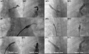
A 3-year-old boy with the diagnosis of pulmonary vein stenosis after total anomalous pulmonary venous return repair was referred to our hospital. The patient had already undergone surgical release for pulmonary vein obstruction four times at the previous hospital. After referral, pulmonary vein stenosis progressed as expected which was refractory to percutaneous transluminal angioplasty. The left lower pulmonary vein had eventually occluded and lethal pulmonary haemorrhage occurred. We elected to place larger stents for each lesion except for the left lower pulmonary vein to release the pulmonary congestion. As the stenosis was near the orifice of the pulmonary vein, extrusion of stents into the left atrium was likely to occur (Fig 1 – a1, a2, d1, and d2). To prevent extrusion, we planned to place Omnilink Elite stents 8/19 mm and 10/19 mm (Abbott Vascular, Santa Clara, CA, USA) for the left and right upper pulmonary vein using the half-uncovered technique. After confirming the location and distance between the pulmonary vein orifice and the stenotic lesion by pulmonary venous angiography, we advanced the stent applying back-loading technique using a 0.035” stiff guide wire (Amplatz Extra Stiff Whisker, 260 cm, Cook Medical, Bloomington, IN, USA) and a 7Fr long sheath (Parent Cross, 55 cm, Medikit, Tokyo, Japan). Subsequently, we inflated the stent half-uncovered by the sheath at the center of the stenotic lesion with nominal pressure, not pushing or pulling the sheath (Fig 1 – b, e1, and e2). After the distal side of the stent was sufficiently apposed to the pulmonary vein wall, it was totally uncovered and inflated. We successfully placed stents at the left and right upper pulmonary veins (Fig 1 – c1, c2, f1, and f2).
Stent placement for left and right upper PVs using “half-uncovered technique”. ( a1,2 ) Left upper pulmonary venous angiography before stenting. The stenosis was near the orifice of the pulmonary vein. ( b ) Inflation of the stent half-covered by sheath (frontal view). After the distal side of the stent was sufficiently apposed to the PV wall, the stent was totally uncovered and inflated. ( c1,2 ) The secured stent at the stenotic site. The stenosis completely resolved. ( d1,2 ) Right upper pulmonary venous angiography before stenting. ( e1,2 ) Inflation of the stent half-covered by sheath. ( f1,2 ) The stent was precisely placed and released the stenosis. PV, pulmonary vein.

Patient 2
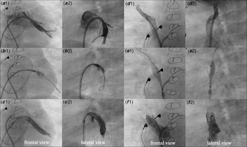
A 3-year-old girl diagnosed with total anomalous pulmonary venous return was referred to our hospital aged 1 month and underwent the surgical repair immediately after referral. Pulmonary vein stenosis was revealed 1 month after the surgery. Despite surgical release for pulmonary vein obstruction, pulmonary vein stenosis recurred within a short time. The effect of PTA was also limited. We intended to place the larger stents for each lesion. The stenosis was near the orifice of the pulmonary vein (Fig 2 – a1, a2, d1, and d2). We placed an Omnilink Elite stent 10/19 mm for the right upper pulmonary vein and 8/19 mm for the left lower pulmonary vein, using the half-uncovered technique (Fig 2 – b1, b2, e1, e2, and Supplementary Video S1). Both stents were precisely placed at the planned sites (Fig 2 – c1, c2, f1, and f2). Computed tomography 1 year after stenting indicated that all stents were patent with mild intimal proliferation, which did not require re-dilation.
Stent placement for left lower and right upper PV using “half-uncovered technique”. ( a1,2 ) Left lower pulmonary venous angiography before stenting. The orifice of the PV was near the right upper PV. ( b1,2 ) Inflation of the stent half-covered by sheath. Similarly, after the distal side of the stent was sufficiently adhered to the PV wall, the stent was uncovered and fully inflated. ( c1,2 ) The secured stent at the stenotic site. ( d1,2 ) Right upper pulmonary venous angiography before stenting. ( e1,2 ) Inflation of the stent half-covered by the sheath. ( f1,2 ) The stent was precisely placed and released the stenosis. PV, pulmonary vein.

Discussion
Postoperative pulmonary vein stenosis is common after repair of total anomalous pulmonary venous return, with an incidence of 14 to 21%. Reference Seale, Uemura and Webber4,Reference White, Ho and Faerber5 Despite multiple surgical pulmonary vein obstruction releases and catheter treatments including balloon angioplasty and stent implantation, the outcome remains poor and the mortality is extremely high (46–80%). Reference Seale, Uemura and Webber4,Reference Caldarone, Najm and Kadletz6 Countermeasures have been reported to improve the outcomes of stent implantation. Balasubramanian et al. reported that using large stents >7 mm in diameter improved patency and reduced the re-intervention rate. Reference Balasubramanian, Marshall and Gauvreau2 Kurita et al also reported that implanted stents with diameters ≧8 mm had a longer freedom from in-stent stenosis. Reference Kurita, Baba and Kondo3 The half-uncovered technique will be helpful to place larger stents percutaneously without extrusion due to caliber narrowing and the slippery pulmonary vein wall. To our best knowledge, this is the first report to address this technique. Drug-eluting stents are reportedly able to prevent internal proliferation and have superior patency to that of bare metal stents of the same size. Reference Furukawa, Kishiro and Fukunaga7–Reference Khan, Qureshi and Justino9 However, the diameter of drug-eluting stents is relatively small because they are designed for coronary arteries. We believe the larger stent size is more important for the prognosis, especially in medium-sized vessels.
The difference between this technique and the existing method is to secure the distal end of the stent beforehand. Fixing the distal end under long sheath support is more likely to prevent stent migration. To secure the stent in position is crucial in pulmonary vein stenting. If the stent is partially extruded into the left atrium, the following approach and post-dilatation become challenging. In this technique, the stent size and initial wire position are critical. In each lesion, the stent size was at least 1.1 times larger than the reference vessel diameter. To secure the distal end of the stent on the lesion, sufficient stent size is crucial. Although vascular injury is an expected complication when using larger stents, there was no vascular tear or rupture as long as the stent size was maximally 3.5 times larger than the minimal lumen diameter and 1.4 times larger than the reference diameter in these lesions (Supplementary File S2). The wire should be also positioned deep inside the main branch. With inappropriate wire positioning, especially in narrower side branch, extrusion of stents, or vascular injury is likely to occur. Moreover, this technique may not be suitable for the right lower pulmonary vein because the access route through the interatrial septum from the inferior vena cava is too acute that will make the sheath bounce off and be ejected during the procedure. Hence, we did not apply this technique to the right lower pulmonary vein in both patients. The route from the superior vena cava may resolve the difficulty of the access route to the right lower pulmonary vein. Reference Mullins1 In these cases, all lesions were discrete-type stenoses. Although a long sheath supports well to prevent the stent from extrusion, stent extrusion may occur in long-segmental lesions because small distal space of the stent and the gap between the stent size and the reference diameter cause a strong force to push out the stent.
Conclusion
Implantation of larger stents for pulmonary vein stenosis is challenging, although it alleviates the issue of re-stenosis and reduces re-intervention. The half-uncovered technique is a promising method of securing larger stents for such narrow and slippery pulmonary vein lesions.
Supplementary material
To view supplementary material for this article, please visit https://doi.org/10.1017/S1047951121004790
Acknowledgements
Special thanks are due to Yoshinori Miyahara, Suguru Tarui, Nobuo Oyama, Takeshi Shimizu, and Kota Nagaoka for their assistance and comments that were very helpful in completing the procedures in the patients addressed in this manuscript.
Financial support
This study received no specific grant from any funding agency in the public, commercial, or not-for-profit sectors.
Conflicts of interest
None.
Ethical standards
The authors assert that all procedures contributing to this work comply with the ethical standards of the relevant national guidelines on human experimentation and with the Helsinki Declaration of 1975, as revised in 2008.