





Impact statement
This work contributes to the growing field of biodesign by demonstrating how biologically derived materials can be shaped through growth-guided fabrication to create functional, scalable and accessible technologies.
First, this research advances the development of hydrogel microneedles, an emerging class of non-invasive transdermal devices with the potential to transform both drug delivery and diagnostics. Unlike conventional injections, hydrogel microneedles can deliver therapeutics across the skin without pain or specialized training while maintaining injection-like delivery kinetics. In addition, their ability to absorb interstitial fluid enables minimally invasive sampling of biomarkers, offering an alternative to blood-based testing. By exploring bacterial cellulose (BC) as a hydrogel microneedle material, this work introduces a biocompatible, sustainable option with intrinsic swelling and mechanical properties well suited for these dual functions.
Second, the work introduces biolithography as a novel fabrication strategy for shaping BC with micrometric precision. This one-pot, low-energy approach replaces complex, resource-intensive manufacturing steps using a design-guided biological growth. The method is low-cost, scalable and adaptable to large-area production, positioning it as a valuable platform not only for microneedles but also for a broader range of biofabricated materials and devices.
Finally, the investigation of both in situ biomineralization and ex situ mineralization strategies demonstrates how the properties of living-derived materials can be tuned after growth. By modulating the mechanical behaviour of BC structures, this work expands the design space for functional biofabricated systems.
Together, these contributions support a shift toward more sustainable, patient-friendly and design-driven approaches in healthcare technologies, while offering transferable tools and concepts for the wider biodesign and biofabrication communities.
Introduction
Microneedles are an emerging non-invasive drug delivery platform with the potential to transform how therapeutics and diagnostics are administered (Larrañeta et al. Reference Larrañeta, Lutton, Woolfson and Donnelly2020). By enabling transdermal delivery with injection-like pharmacokinetics – while avoiding pain, invasiveness and the need for trained professionals – microneedles offer a patient-friendly alternative for decentralized, self-administered care.
Among the various microneedle systems developed to date, hydrogel microneedles are particularly compelling due to their dual functionality (Donnelly et al. Reference Donnelly, McCrudden, Alkilani, Larrañeta, McAlister, Courtenay, Kearney, Singh, McCarthy, Kett, Caffarel-Salvador, Al-Zahrani and Woolfson2014). In addition to penetrating the skin and delivering therapeutic agents, hydrogel microneedles can absorb substantial volumes of interstitial fluid, enabling their use for minimally invasive diagnostics. Their swelling-based mechanism also allows for the integration of drug reservoirs or patches positioned above the needle array, enabling controlled and sustained release.
Bacterial cellulose (BC) is a biopolymer whose production enables a unique fabrication strategy referred to here as biolithography (Roig-Sánchez et al. Reference Roig-Sánchez, Fernández-Sánchez, Laromaine and Roig-Serra2019). By guiding cellulose growth using predefined, oxygen-permeable polydimethylsiloxane (PDMS) molds floating on a fermentation medium, bacteria can fabricate BC with complex microstructured geometries and micrometric resolution. This growth-driven process enables the one-pot fabrication of BC-based hydrogel microneedles without the need for subtractive manufacturing or harsh processing steps.
Leveraging this approach, this work explores a simple, low-cost and scalable strategy to fabricate hydrogel microneedles from BC. To tailor the mechanical properties required for effective skin penetration, different biomineralization strategies (Hong et al. Reference Hong, Wang, Jia, Huang, Gao and Wan2006) are applied and evaluated through qualitative functional assays, including penetration, rehydration and drug delivery.
Objectives
The objectives of this demo are to:
-
1. Demonstrate the feasibility of manufacturing BC-based hydrogel microneedles using a biolithography approach with consistent geometric fidelity.
-
2. Compare in situ biomineralization and ex situ mineralization strategies to modulate the mechanical properties of BC-based microneedles, assessed qualitatively through handling, penetration and SEM characterization.
-
3. Evaluate the functional performance of the fabricated microneedles through penetration testing on Parafilm M layers, rehydration assays and model drug diffusion (rhodamine B) visualized in real time on agarose gel.
Relevance
This demo showcases a growth-driven, material-led fabrication strategy that enables the molding of BC at micrometric resolution in a scalable and resource-efficient manner. While hydrogel microneedles are used as a demonstrative application, the broader contribution lies in presenting biolithography as a versatile biodesign platform for shaping living-derived materials.
BC offers intrinsic advantages – biocompatibility, high water uptake and tunable mechanics – that align naturally with hydrogel-based systems. By positioning fabrication as a collaboration with biological growth rather than a purely top-down process, this work contributes to emerging biodesign practices that prioritize sustainability, material agency and low-energy manufacturing.
Technology setup
The fabrication process begins with the digital design of microneedle master molds using TinkerCad. Different microneedle parameters were selected based on literature-reported dimensions for comparison and optimization purposes (pyramidal vs conical geometry, 1000 µm height and 400 µm base vs 600 µm height and 300 µm base). Master molds were produced via 3D printing and used to generate negative PDMS molds (10:1 elastomer-to-curing-agent ratio) through standard micromolding.
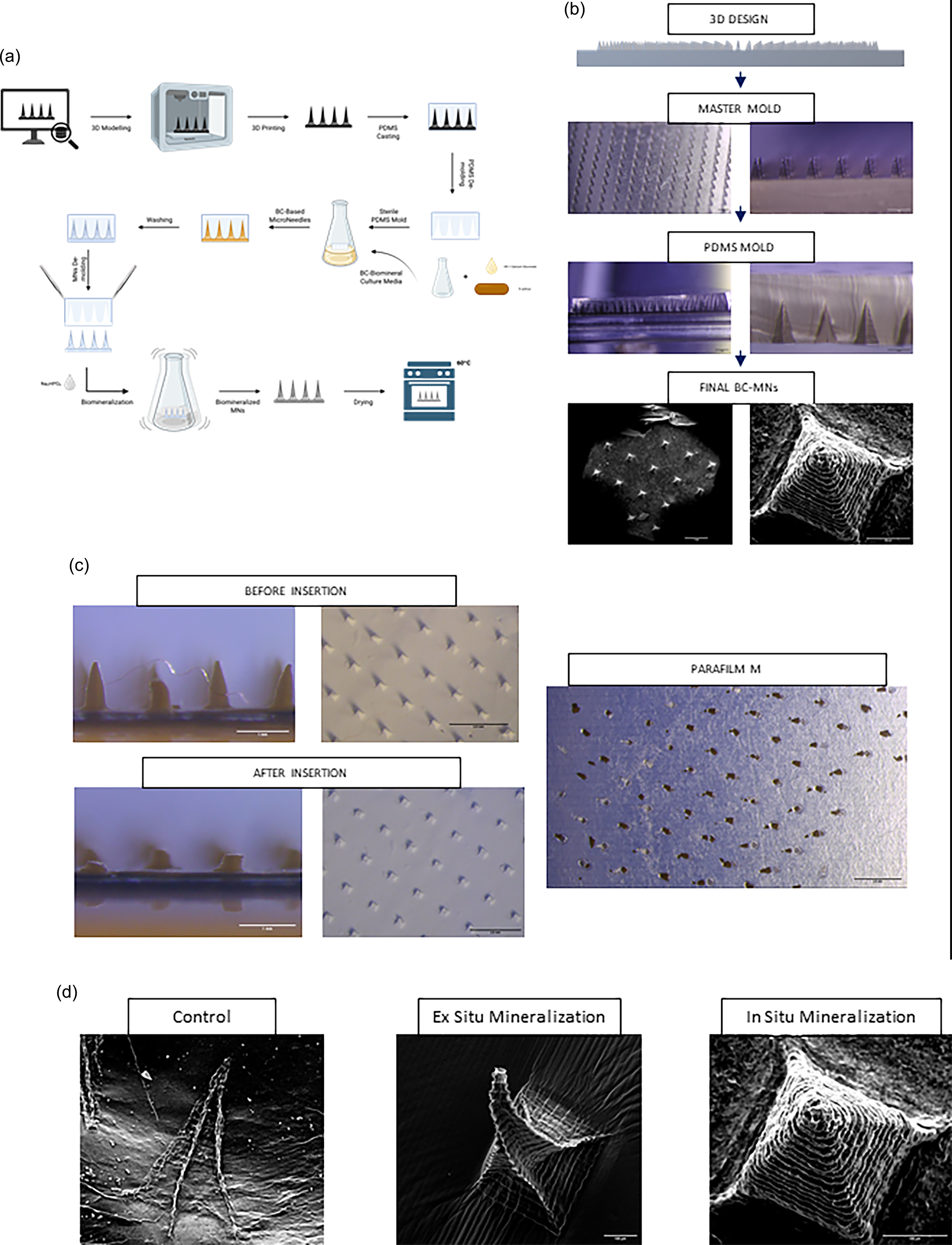
PDMS molds were rendered hydrophilic via plasma treatment and sterilized by autoclaving. The molds were then placed floating on top of BC fermentation media. To enable and enhance biomineralization during synthesis, the fermentation medium was modified, introducing calcium ions directly into the material during growth (In Situ Biomineralization). Due to PDMS oxygen permeability, BC synthesized at the air–liquid interface, conforming to the mold geometry during growth. After synthesis, BC microneedles were cleaned, demolded, mineralized using post-growth mineralization protocols and dried. These final structures were used for characterization, functional testing and demonstration. A schematic overview of the process is provided in Figure 1a.
(a) Schematic of the biolithography fabrication process: growth-guided molding of bacterial cellulose (BC) using floating oxygen-permeable PDMS templates. (b) Optical and SEM images across fabrication stages: (i) TinkerCAD design of the master mold; (ii) optical image of the 3D-printed master mold; (iii) optical image of PDMS replicas; (iv) SEM images of final BC-based microneedles. (c) Optical stereoscope images of penetration testing using in situ biomineralized BC microneedles: arrays before insertion, after insertion and resulting puncture holes in Parafilm M. (d) Comparative SEM images of the three microneedle variants: native BC, ex situ mineralized BC and in situ biomineralized BC microneedles.

Figure 1. Long description
Panel A: The diagram shows the biolithography fabrication process involving growth-guided molding of bacterial cellulose using floating oxygen-permeable PDMS templates. It includes steps like 3D modeling, 3D printing, PDMS casting, and bacterial cellulose growth and mineralization. Panel B: The images display the fabrication stages of microneedles. The first image shows a TinkerCAD design of the master mold. The second image is an optical image of the 3D-printed master mold. The third image is an optical image of PDMS replicas. The fourth image shows SEM images of the final BC-based microneedles. Panel C: The optical stereoscope images depict penetration testing using in situ biomineralized BC microneedles. The images show arrays before insertion, after insertion, and the resulting puncture holes in Parafilm M. Panel D: The comparative SEM images show three microneedle variants: native BC, ex situ mineralized BC, and in situ biomineralized BC microneedles.
Demo setup and interaction
The demo is structured as a guided, hands-on exploration of growth-driven fabrication, positioning BC as an active design material shaped through biological synthesis rather than conventional manufacturing Appendix 1.
The demonstration is structured as a physical, multi-station setup that guides attendees through each stage of the fabrication pipeline. It begins at a digital design station that displays the parametric TinkerCAD models used to generate the 3D-printed master molds, making the design-to-fabrication pipeline tangible from the outset (1). Attendees then progress through mold preparation (3D-printed masters and PDMS replicas) (2), live one-pot BC microneedle growth systems incubated at 30°C (3) and a comparative sample station where native, in situ biomineralized (calcium-supplemented growth medium) and ex situ mineralized (post-synthesis mineralization baths) microneedles can be handled and visually compared side by side (4). Physical samples at each stage of fabrication illustrate the full transition from digital concept to biologically formed, functionally tested microstructures.
Participants interact with pre-prepared BC constructs in both wet and processed states, observing how material properties evolve through post-growth treatments. Comparative samples demonstrate how structural and mechanical characteristics can be tuned through controlled modifications. A visual overview of the process is provided in Figure 1b.
The demo concludes with qualitative functional demonstrations at three interactive stations (5): (A) a penetration station, where microneedles are applied to Parafilm M layers to compare native, in situ biomineralized and ex-situ mineralized insertion capability Figure 1c; (B) a rehydration station, where dried microneedle arrays are submerged to observe swelling kinetics; and (C) a drug delivery station, where microneedles pre-loaded with a model dye (rhodamine B) are applied to an agarose gel, enabling real-time visualization of transdermal diffusion. BC-based Microneedles are evaluated comparatively across stations, with optical stereoscope images on display illustrating structural differences between native, in situ and ex situ variants Figure 1d. These stations provide intuitive, hands-on insight into how growth-driven fabrication and material processing jointly determine functional performance.
The live BC growth component requires standard microbiology incubation facilities (30°C static incubation in sealed containers). In the event that live cultures cannot be maintained on-site, pre-grown and processed samples covering all fabrication stages will be available and time-lapse video documentation will replicate the growth observation experience without interruption to the demonstration flow. In addition, in order for participants to observe the different Master Molds, PDMS Molds and final BC-Based Microneedle structures, a very basic optical microscope or stereoscope would be needed. Finally a computer screen would be nice, in order for participants to play with TinkerCad and to show videos if and when needed.
Conclusion
This demo presents biolithography as a growth-driven fabrication strategy that enables the precise shaping of BC through design-guided biological synthesis. By integrating digital modeling, biolithography and material transformation into an interactive workflow, the project highlights how living-derived materials can function as active partners in fabrication.
Beyond the specific case study presented, the work demonstrates a scalable, accessible and materially expressive approach to designing functional microstructures. It contributes to ongoing conversations in biodesign by shifting focus from fabrication as imposition to fabrication as a collaboration with biological growth.
Supplementary material
The supplementary material for this article can be found at https://doi.org/10.1017/S2977905726100572
Data availability statement
All data can be made available upon request.
Acknowledgements
The authors gratefully acknowledge the collaboration and support of Mariano Campoy-Quiles. We also thank the SEM Service at ICMAB.
Author contributions
Conceptualization: G.W.M; A.R. Methodology: G.W.M; M.T; L.A.P; A.T.N Data curation: G.W.M; M.T. Data visualization: G.W.M; M.T. Writing original draft: G.W.M; A.R. All authors approved the final submitted draft.
Financial support
This work was supported by the Spanish Ministry of Science and Innovation through PID2024-157637OB-I00 project and the “Severo Ochoa” Programme for the Center of Excellence in R&D (CEX2023001263-S). This work is part of the G.W. doctoral thesis, carried out within the Materials Science PhD Program at the Autonomous University of Barcelona. This project has received funding from the European Union’s Horizon Europe research and innovation programme under the Marie Skłodowska-Curie grant agreement No 10181337 awarded to G.W. and A.R
Competing interests
None.
Ethical standards
The research meets all ethical guidelines, including adherence to the legal requirements of the study country.


 Open access

