Introduction
In cadaveric studies, incudal osteomas have been found to be present in up to 1.6 per cent of the population.Reference Arensburg, Belkin and Wolf1 They are usually small and solitary, and are found on the medial surface of the incus.Reference Arensburg, Belkin and Wolf1
Incudal osteomas very rarely become symptomatic. To date, only four cases of symptomatic osteomas of the incus have been described in the English-language literature.Reference Jang and Cho2
Case report
A 51-year-old woman presented with an 18-month history of progressive, left-sided hearing loss. There were no other otological symptoms, and no history of childhood ear infections or surgery.
Otoscopy revealed normal tympanic membranes and ear canals bilaterally.
Tuning fork tests were consistent with a left-sided, conductive hearing loss.
Pure tone audiometry showed a 70 dB mixed hearing loss, with a large conductive component, and a 10 dB reduction in the bone conduction thresholds at 2 kHz.
Tympanometry showed a type A pattern on the right, but a type As pattern on the left, with reduced tympanic membrane compliance on that side.
A diagnosis of left-sided otosclerosis was made, and a left stapedectomy was planned.
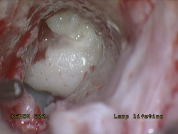
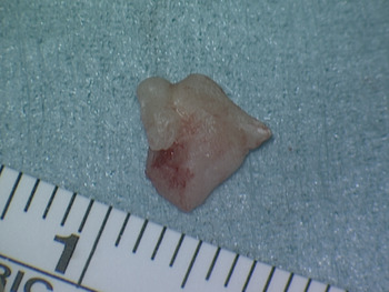
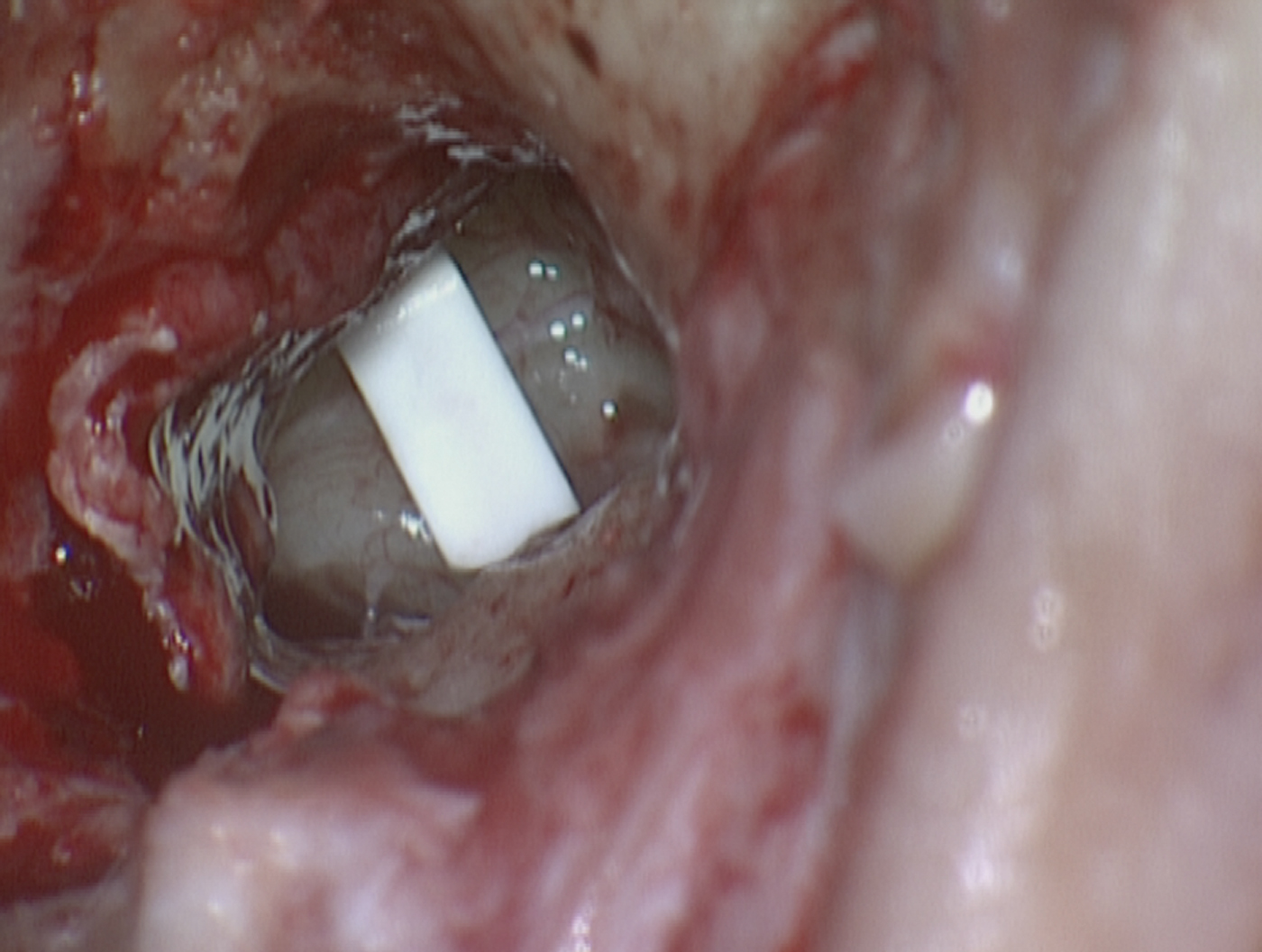
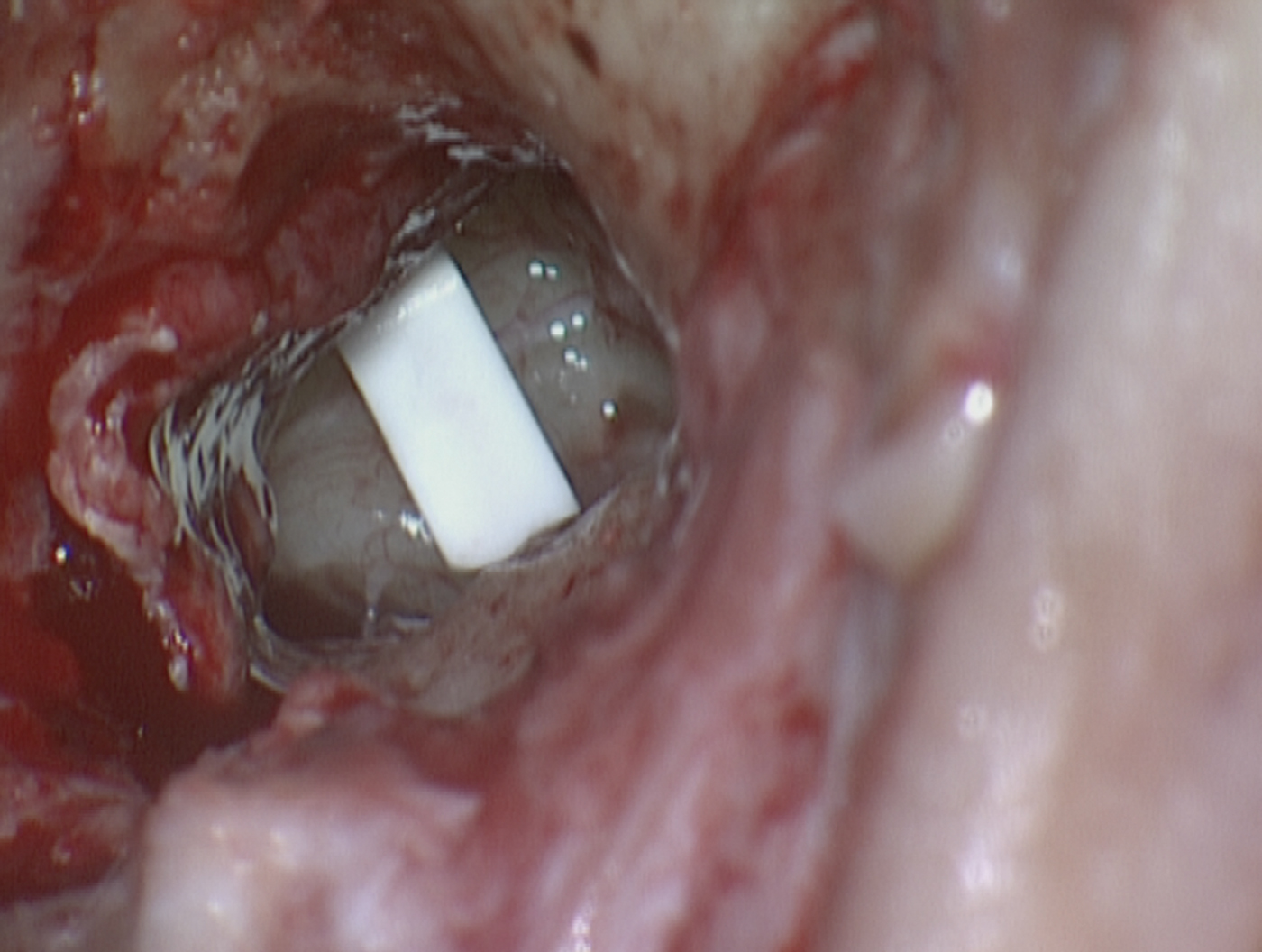
Surgery utilised a permeatal approach, in the standard fashion. After curettage of the postero-superior bony annulus, the stapes superstructure was visible. The exposed long process of the incus appeared normal. Although intact, the ossicular chain was found to be immobile. In order to establish which part of the ossicular chain was fixed, the incudostapedial joint was divided. This demonstrated a mobile stapes, with fixation of the malleus and incus in the attic region. An atticotomy was performed, revealing a bony, 6 × 6 × 4 mm mass arising from the incus, extending onto the malleus head, and fixing the malleus and incus in the attic (Figures 1 and 2). After dividing the neck of the malleus, the atticotomy needed to be extended towards the additus to enable the bony mass to be removed. The ossicular chain was reconstructed using a Wehr's partial ossicular replacement prosthesis (Figure 3).
Operating microscopy photograph showing the osteoma arising from the incus and attached to the malleus head. The malleus neck has been divided.

The excised incudal osteoma, measuring 6 × 6 × 4 mm.

Operating microscopy photograph showing Wehr's partial ossicular replacement prosthesis in situ.
There were no immediate post-operative complications, and the patient was discharged the following day.
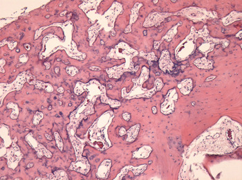
Histological examination of the excised mass showed it to be a benign, nodular lesion composed of densely ossified, sclerotic bone with prominent cement lines (Figure 4). These findings were consistent with a diagnosis of osteoma of the incus.
Photomicrograph of the incudal osteoma. (H&E; ×5)

Discussion
This case highlights a rare cause of unilateral conductive hearing loss. A particular ‘learning point’ from this case is the diagnostic confusion that can occur in the presence of other causes of conductive hearing loss. In this patient, the presence of a Carhart's notch was highly suggestive of otosclerosis (although this finding does occur in other causes of conductive loss), and a stapedectomy was planned accordingly. The final diagnosis was not reached until the time of surgery, when the mobility of the divided ossicular chain was assessed. This reinforces the fact that assessment of the ossicular chain, following division of the incudo-stapedial joint, is an essential step in stapedectomy surgery.
The use of computed tomography (CT) in the diagnosis of otosclerosis is controversial. Proponents of pre-operative CT scanning argue that it is a sensitive means of detecting otosclerosis, it reduces the risk of unexpected intra-operative findings, and it helps to identify individuals at higher risk of post-stapedectomy complications.Reference Lagleyre, Sorrentino, Calmels, Shin, Escudé and Deguine3 However, CT scanning does result in exposure to a significant amount of ionising radiation. Many would argue that this is an unnecessary and unacceptable consequence of an investigation that is not essential in the management of patients with presumed otosclerosis. Finally, the routine use of CT scanning in such patients would increase the cost of their management.
• Symptomatic osteomas of the malleus, incus and stapes are rare
• Such cases often have a normal tympanic membrane
• Presenting symptoms and investigations may mimic otosclerosis
• The diagnosis may be missed if the mobility of the divided ossicular chain is not checked intra-operatively
Conclusion
This case report reinforces the importance of assessing the mobility of the divided ossicular chain during planned stapedectomy. It also serves as a reminder that, in cases with a normal tympanic membrane, otosclerosis is not the only cause of a conductive loss with a Carhart's notch. Although CT scanning would assist the establishment of a pre-operative diagnosis, the authors believe that the ionising radiation exposure and increased cost associated with use of this investigation preclude its routine use in patients with suspected otosclerosis.